Podcast
Questions and Answers
What is the primary function of the mesonephros during development?
What is the primary function of the mesonephros during development?
- It becomes non-functional and disappears.
- It serves as the permanent kidney.
- It differentiates into renal pelvis.
- It has partial function and gives rise to the Wolffian and Müllerian ducts. (correct)
Which structure surrounds the kidney fat and adrenal glands?
Which structure surrounds the kidney fat and adrenal glands?
- Fibrous renal capsule
- Renal sinus
- Pararenal fat
- Renal fascia/Gerota’s fascia (correct)
What is the normal length range of a healthy kidney?
What is the normal length range of a healthy kidney?
- 8-11 cm
- 7-10 cm
- 9-12 cm (correct)
- 6-9 cm
Which of the following accurately describes the relationship between kidney and uterine/vagina anomalies?
Which of the following accurately describes the relationship between kidney and uterine/vagina anomalies?
What is a junctional parenchymal defect in the kidney?
What is a junctional parenchymal defect in the kidney?
What is the funnel-shaped structure that connects major calyces to the ureter?
What is the funnel-shaped structure that connects major calyces to the ureter?
Which layer of the kidney is primarily responsible for filtering wastes to form urine?
Which layer of the kidney is primarily responsible for filtering wastes to form urine?
Which aspect of the kidney vasculature is correct?
Which aspect of the kidney vasculature is correct?
Which of the following conditions is least likely to contribute to postrenal failure?
Which of the following conditions is least likely to contribute to postrenal failure?
What is the expected renal size indicating renal artery stenosis?
What is the expected renal size indicating renal artery stenosis?
What is the primary indicator of renal transplant dysfunction demonstrated by the resistive index (RI)?
What is the primary indicator of renal transplant dysfunction demonstrated by the resistive index (RI)?
Which of the following symptoms is NOT indicative of renal artery stenosis?
Which of the following symptoms is NOT indicative of renal artery stenosis?
Which fluid collection complication would you expect NOT to see 24 hours post-op of a renal transplant?
Which fluid collection complication would you expect NOT to see 24 hours post-op of a renal transplant?
What is the common cause leading to renal vein thrombosis?
What is the common cause leading to renal vein thrombosis?
What is a distinguishing sonographic finding of acute transplant rejection?
What is a distinguishing sonographic finding of acute transplant rejection?
What is the most common cause of renal disease leading to kidney transplantation?
What is the most common cause of renal disease leading to kidney transplantation?
What is the term for the phenomenon that describes diminished function of nephrons in chronic end-stage kidney disease?
What is the term for the phenomenon that describes diminished function of nephrons in chronic end-stage kidney disease?
Where is renal artery stenosis most commonly located?
Where is renal artery stenosis most commonly located?
What is the significance of a renal index (RI) value of 1.0?
What is the significance of a renal index (RI) value of 1.0?
Which condition is associated with unilateral renal agenesis?
Which condition is associated with unilateral renal agenesis?
What differentiates a complete duplex kidney from an incomplete duplex kidney?
What differentiates a complete duplex kidney from an incomplete duplex kidney?
In horseshoe kidney, which pole is more commonly fused?
In horseshoe kidney, which pole is more commonly fused?
What is the most common inherited kidney disease?
What is the most common inherited kidney disease?
Which of the following is an associated condition with ARPKD?
Which of the following is an associated condition with ARPKD?
Which renal cyst characteristics suggest malignancy?
Which renal cyst characteristics suggest malignancy?
What is the most common solid renal mass in adults?
What is the most common solid renal mass in adults?
Which organ is commonly associated with ADPKD besides the kidneys?
Which organ is commonly associated with ADPKD besides the kidneys?
What is a common finding in renal tuberculosis?
What is a common finding in renal tuberculosis?
Which of the following conditions has the highest association with renal cell carcinoma (RCC)?
Which of the following conditions has the highest association with renal cell carcinoma (RCC)?
What distinguishes peripelvic cysts from parapelvic cysts?
What distinguishes peripelvic cysts from parapelvic cysts?
What imaging finding is characteristic of emphysematous pyelonephritis?
What imaging finding is characteristic of emphysematous pyelonephritis?
Flashcards
Kidney development stages
Kidney development stages
The three successive stages of kidney development: Pronephros (non-functional and disappears), Mesonephros (partially functional, gives rise to Wolffian and Mullerian ducts), and Metanephros (permanent kidney).
Wolffian vs. Mullerian ducts
Wolffian vs. Mullerian ducts
The Wolffian ducts develop into male genitalia, while the Mullerian ducts develop into the uterus and vagina in females.
Junctional Parenchymal Defect
Junctional Parenchymal Defect
A junctional parenchymal defect is a triangular, hyperechoic area seen on ultrasound, usually in the upper pole of the right kidney. It's a remnant of fetal lobulation and is considered a normal variant.
Layers of the kidney
Layers of the kidney
Signup and view all the flashcards
Kidney Vasculature
Kidney Vasculature
Signup and view all the flashcards
Renal sinus
Renal sinus
Signup and view all the flashcards
Renal pelvis
Renal pelvis
Signup and view all the flashcards
Minor calyces and medullary pyramids
Minor calyces and medullary pyramids
Signup and view all the flashcards
What is the Renal Index (RI)?
What is the Renal Index (RI)?
Signup and view all the flashcards
What is Unilateral Renal Agenesis?
What is Unilateral Renal Agenesis?
Signup and view all the flashcards
Describe complete vs incomplete duplex kidney.
Describe complete vs incomplete duplex kidney.
Signup and view all the flashcards
Describe a Horseshoe Kidney.
Describe a Horseshoe Kidney.
Signup and view all the flashcards
What is Autosomal Dominant Polycystic Kidney Disease (ADPKD)?
What is Autosomal Dominant Polycystic Kidney Disease (ADPKD)?
Signup and view all the flashcards
What is Autosomal Recessive Polycystic Kidney Disease (ARPKD)?
What is Autosomal Recessive Polycystic Kidney Disease (ARPKD)?
Signup and view all the flashcards
What is Emphysematous Pyelonephritis?
What is Emphysematous Pyelonephritis?
Signup and view all the flashcards
What is an Oncocytoma?
What is an Oncocytoma?
Signup and view all the flashcards
What is an Angiomyolipma (AML)?
What is an Angiomyolipma (AML)?
Signup and view all the flashcards
What is Renal Cell Carcinoma (RCC)?
What is Renal Cell Carcinoma (RCC)?
Signup and view all the flashcards
What is Wilm's Tumor?
What is Wilm's Tumor?
Signup and view all the flashcards
What is Mesoblastic Nephroma?
What is Mesoblastic Nephroma?
Signup and view all the flashcards
Chronic end-stage kidney disease
Chronic end-stage kidney disease
Signup and view all the flashcards
Sonographic findings of chronic kidney disease
Sonographic findings of chronic kidney disease
Signup and view all the flashcards
Postrenal failure
Postrenal failure
Signup and view all the flashcards
Intrinsic renal failure
Intrinsic renal failure
Signup and view all the flashcards
Prerenal failure
Prerenal failure
Signup and view all the flashcards
What is the most common cause of kidney failure leading to transplant?
What is the most common cause of kidney failure leading to transplant?
Signup and view all the flashcards
Why is the left kidney favored for transplant?
Why is the left kidney favored for transplant?
Signup and view all the flashcards
Arterial anastomosis
Arterial anastomosis
Signup and view all the flashcards
Acute Tubular Necrosis (ATN)
Acute Tubular Necrosis (ATN)
Signup and view all the flashcards
Sonographic findings of acute transplant rejection
Sonographic findings of acute transplant rejection
Signup and view all the flashcards
Study Notes
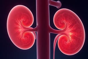
Kidney Development and Anatomy
- Mesoderm forms by the 3rd week of development.
- Three successive kidney pairs develop: pronephros (non-functional and disappears), mesonephros (partial function, forms Wolffian and Müllerian ducts), and metanephros (permanent kidney).
- Wolffian ducts form male genitalia; Müllerian ducts form uterus and vagina.
- Kidney size: typically 9-12 cm long, 5-7 cm wide, and 2-3 cm thick. Length and width should be within 2 cm of each other.
- Kidney layers (outer to inner): fibrous renal capsule, perirenal fat, renal fascia (Gerota's fascia), pararenal fat.
- Renal fascia surrounds kidney, fat, and adrenal glands.
- At the kidney hilum (VAU): vein is anterior, artery is in between vein and ureter, and ureter exits posteriorly.
- Normal renal cortex thickness is greater than 1 cm (10 mm or more).
- Renal sinus contains inner hyperechoic fat, calyces, renal pelvis, connective tissue, vessels, and lymphatics.
- Number of minor calyces equals the number of medullary pyramids.
- Renal pelvis is a funnel-shaped structure connecting major calyces to ureter.
- Kidney vasculature: main renal artery branches to segmental (5), interlobar, arcuate, and interlobular arteries.
- Junctional parenchymal defect: hyperechoic break in cortex, typically at upper pole of right kidney, due to partial fusion of ranunculi (embryonic kidney).
- Column of Bertin: prominent cortical tissue between medullary pyramids; may appear as a mass. Echogenicity is similar to cortex.
Kidney Function and Disease
- Kidney functions: filtering waste for urine, maintaining water/electrolyte/acid balance, hormone release.
- Stages of Chronic Kidney Disease (CKD) are based on glomerular filtration rate (GFR): Normal GFR > 90, end-stage GFR < 15.
- Renal Imaging (RI) evaluates transplant rejection, hydronephrosis, and renal disease. RI equation: (Peak systolic - End diastolic) / Peak systolic; normal RI < 0.7; RI of 1 = diastolic absent.
- Unilateral renal agenesis can be associated with VACTERL and MURCS syndromes.
- Complete duplex kidney: two ureters; incomplete: one ureter.
- Collecting system duplication: usually longer than normal; complete central cortical break within hyper echoic sinus is seen.
- Ectopic ureter: ureter draining upper pole inserts into ectopic bladder location. Common complication: ureterocele.
- Crossed fused renal ectopia: developing kidneys fuse in pelvis and one ascends to its normal position, dragging other across the midline, absence of other kidney, or both ureters connect to both sides of the bladder.
- Horseshoe kidney: lower poles commonly fuse anterior to aorta.
- Autosomal dominant polycystic kidney disease (ADPKD): most common inherited kidney disease; associated with cysts in liver and pancreas (and spleen in rarer cases). Associated with cerebral arterial aneurysms, especially berry aneurysms of the circle of Willis.
- Autosomal recessive polycystic kidney disease (ARPKD): more common in childhood and typically presents at birth; associated with pulmonary hypoplasia, hepatic fibrosis, and portal hypertension.
- Multi-cystic dysplastic kidneys (MCDK): most common abdominal mass in newborns, typically unilateral.
- Renal cysts (complex): hemorrhagic, infected, septate; malignancy suggested by multiple/thick septations, thick calcifications, or mural nodule/solid component.
- Peripelvic cysts are within the renal sinus; parapelvic cysts protrude into the sinus from parenchyma.
- Long-term dialysis increases risk of acquired cystic kidney disease and RCC.
- Von Hippel-Lindau disease: inherited disease presenting in the second and third decade with specific visual impairment and associated tumors (RCC, pheochromocytomas, islet cell tumors, renal and pancreatic cysts).
- Tuberous sclerosis: increased incidence of renal cysts and angiomyolipomas (AML).
- Oncocytomas: benign renal tumor with central scar and spoke-wheel pattern on angiogram.
- Angiomyolipomas (AML): mesenchymal tumor with fat cells, smooth muscle, and thick blood vessels. AML frequently located in the right.
- Renal cell carcinoma (RCC): most common solid renal mass in adults; associated with hematuria, acquired cystic renal disease, Von Hippel Lindau syndrome, tuberous sclerosis, and ADPKD. Males more commonly affected, especially after age 50.
- Wilm's tumor (Nephroblastoma): most common childhood renal tumor; associated with Beckwith-Wiedemann syndrome.
- Mesoblastic nephroma: most common neonatal and infant renal tumor.
- Transitional cell carcinoma (TCC): most common bladder neoplasm.
- Metastatic renal tumors: common sources include lung and breast.
- Hydronephrosis causes: intrinsic (calculi, UPJ obstruction, tumors); extrinsic (neoplasm, trauma, BPH). Hydronephrosis more common on the right side. Most common obstruction location: ureterovesical junction.
- Nephrocalcinosis: renal parenchymal calcium deposits, usually bilateral and diffuse, cortical or medullary, or both.
- Staghorn calculi: large calculi that fill the pelvis and calyces of the kidney.
- Renal infarction: initially difficult to see sonographically; later appears hypoechoic then echogenic.
- Emphysematous pyelonephritis: gas within the kidney, causing reverb or comet-tail artifacts; associated with anaerobic bacteria, often in diabetic patients.
- Xanthogranulomatous pyelonephritis (XGP): chronic pyelonephritis from chronic infections and obstructions. Common cause: Candida.
- Renal tuberculosis: associated with pulmonary infection. Infectious diseases show nonspecific masses, diffusely heterogeneous appearance, and difficult-to-define borders.
- Acute kidney injury (AKI) mechanisms: prerenal (inadequate perfusion), intrinsic (renal damage), and postrenal (obstruction).
- Chronic kidney disease (CKD)/End-stage renal disease: irreversible, nephrons reduced in function, GFR and resorptive capability decrease. Sonographic findings not specific: small, echogenic, shrunken kidneys.
- Hydronephrosis indicates postrenal failure; abnormal RI indicates intrinsic renal disease.
Renal Transplant and Renal Vascular
- Leading cause of kidney transplantation: diabetes.
- Left kidney favored for transplantation due to longer renal vein.
- Cadaveric transplant survival: 7-10 years; living donor: 15-20 years.
- Arterial anastomosis: external or internal iliac artery.
- Immediate post-transplant poor function: acute tubular necrosis (ATN).
- Acute transplant rejection: enlarged kidney, decreased echogenicity, loss of corticomedullary boundary, and increased flow resistance (RI).
- Post-transplant complications: fluid collections (hematomas, urinomas), renal artery kinking/thrombosis, renal vein thrombosis.
- Transplant dysfunction RI: >0.8.
- Renal artery stenosis: most commonly located at origin or first 2 cm; symptoms: uncontrolled or sudden onset hypertension.
- Decreased renal size (<9cm of length) suggests renal artery stenosis.
- Direct evaluation of renal artery stenosis: renal artery velocities and renal artery/aorta ratio (RAR) >3.5.
- Indirect evaluation: intrarenal waveform evaluation (parvus tardus).
- Renal artery thrombosis: causes prerenal failure, acute flank pain, hematuria, and sudden rise in blood pressure.
- Renal artery thrombosis findings: focal hypoechoic areas of infarct, absence of intrarenal arterial flow; renal enlargement.
- Causes of renal vein thrombosis: nephrotic syndrome, hypercoagulability disorders, malignant tumors, extrinsic compression, trauma, transplant rejection.
- Renal vein thrombosis findings: dilated thrombosed renal vein, absence of intrarenal venous flow, enlarged hypoechoic kidney, high resistance renal artery waveform (increased RI).
Studying That Suits You
Use AI to generate personalized quizzes and flashcards to suit your learning preferences.