Podcast
Questions and Answers
What structural feature of the afferent arteriole contributes to maintaining high blood pressure within the glomerulus?
What structural feature of the afferent arteriole contributes to maintaining high blood pressure within the glomerulus?
- It has a smaller diameter than the efferent arteriole.
- It is lined with specialized podocyte cells.
- It has a wider diameter than the efferent arteriole. (correct)
- It directly connects to the peritubular capillaries.
The renal vein carries oxygenated blood with metabolic waste products away from the kidney.
The renal vein carries oxygenated blood with metabolic waste products away from the kidney.
False (B)
What are the three main processes involved in the functioning of the kidney?
What are the three main processes involved in the functioning of the kidney?
Glomerular filtration, tubular reabsorption, and tubular secretion
The network of blood vessels surrounding the renal tubule is known as the ______ capillary network.
The network of blood vessels surrounding the renal tubule is known as the ______ capillary network.
Match the following components of the nephron with their location in the kidney:
Match the following components of the nephron with their location in the kidney:
During which process are useful substances such as glucose and amino acids returned to the bloodstream?
During which process are useful substances such as glucose and amino acids returned to the bloodstream?
The efferent arteriole directly supplies blood to the glomerulus.
The efferent arteriole directly supplies blood to the glomerulus.
What is the function of the renal capsule?
What is the function of the renal capsule?
The ______ is the structural and functional unit of the kidney.
The ______ is the structural and functional unit of the kidney.
What types of waste products are formed during metabolism?
What types of waste products are formed during metabolism?
Flashcards
Renal Artery
Renal Artery
Branch of the aorta that supplies oxygenated blood rich in metabolic waste to the kidney.
Afferent Arterioles
Afferent Arterioles
Smallest arterial branches in the kidney cortex, leading to Bowman's capsules.
Glomerulus
Glomerulus
Capillary network within Bowman's capsule where filtration occurs.
Efferent Arteriole
Efferent Arteriole
Signup and view all the flashcards
Peritubular Capillary Network
Peritubular Capillary Network
Signup and view all the flashcards
Kidney Functions
Kidney Functions
Signup and view all the flashcards
Nephron
Nephron
Signup and view all the flashcards
Afferent Arteriole Function
Afferent Arteriole Function
Signup and view all the flashcards
Bowman's Capsule
Bowman's Capsule
Signup and view all the flashcards
Excretion
Excretion
Signup and view all the flashcards
Study Notes
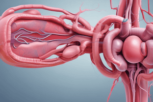
- Each kidney gets its blood supply from a renal artery.
- The renal artery is a branch of the aorta, transporting oxygenated blood loaded with metabolic waste to the kidneys.
- The renal artery enters the kidney at the hilum.
- The artery branches into smaller arteries, extending between the pyramids toward the cortex.
- These further branch into the smallest cortical branches: afferent arterioles.
- One afferent arteriole extends to each Bowman's capsule.
- The afferent arteriole splits into a capillary network, the glomerulus, nestled within the Bowman's capsule.
- These capillaries converge to form an efferent arteriole, carrying blood away from the glomerulus.
- The efferent arteriole branches again into the peritubular capillary network, surrounding the renal tubule.
- Afferent arterioles are wider than efferent arterioles, maintaining high glomerular blood pressure.
- The peritubular capillaries combine into venules and larger veins, forming the renal vein.
- The renal vein carries deoxygenated blood, minus waste, from the kidney to the heart via the inferior vena cava.
- The kidney functioning are divided into three processes: Glomerular filtration, Tubular reabsorption, and Tubular excretion.
- The efferent arteriole has a capillary network on both sides: the glomerulus, and the peritubular capillaries, is called a portal system.
Kidney Structure
- The kidney is encapsulated by a connective tissue membrane, the renal capsule, for protection.
- Directly beneath is the cortex layer.
- The medulla is the lighter-colored, inner region, with grouped tubes.
- Each tube group forms a pyramid, its base faces the cortex.
- The renal papilla is at each pyramid tip.
- Tubes in each renal papilla open into renal calyxes.
- The renal calyxes merge into the ureter region known as the renal pelvis.
Microscopic Internal Structure
- Each kidney is made of about one million nephrons.
- Nephrons are the kidney's structural and functional units.
- 'Structural units' = the building blocks making up the kidney.
- 'Functional units' = independent units performing kidney functions.
Nephron Structure
- Each nephron has two primary parts: the Malpighian body and the renal tubule.
- The Malpighian body, found in the cortex, includes the Bowman's capsule (double-walled, cup-shaped) and the glomerulus (capillary network).
- Glomerular capillaries have single endothelial layer with pores.
- The afferent arteriole transports blood to the glomerulus.
- The efferent arteriole carries blood away from the glomerulus.
- A comes before E - Afferent transports to, Efferent transports away.
Renal Tubule
- The renal tubule, a long and winding tube, lies partly in the cortex and partly in the medulla.
- The tube has three sections: proximal convoluted tubule, loop of Henle, and distal convoluted tubule.
- The proximal convoluted tubule follows the Bowman's capsule and is in the cortex.
- Has a single layer of cuboidal epithelium and is widest, at this part.
- The loop of Henle, situated in the medulla, features a descending limb extending into the medulla, a hairpin loop, and an ascending limb returning to the cortex.
- The distal convoluted tubule is in the cortex, also has a single layer of cuboidal epithelium, receives input from several nephrons' distal tubules.
- Distal convoluted tubules drain into collecting ducts.
- The tubes forming the pyramids and which open into the renal calyx of the renal pelvis, are the ducts of Bellini.
Podocytes
- The Bowman's capsule inner wall has podocytes, specialized cells.
- Podocytes have projections that create filtration slits.
- The cavity between the Bowman's capsule inner and outer walls is the Bowman's capsule cavity.
- The blood in the glomerulus is separated from the Bowman's capsule cavity by: a thin endothelial cell layer with pores & the podocyte layer with filtration slits.
Urinary system in humans
- The urinary system includes two kidneys, two ureters, the bladder, and the urethra.
- Renal arteries and veins are the blood vessels associated with the urinary system.
- Kidneys are in the abdominal cavity, on either side of the vertebral column, just below the diaphragm.
- The renal arteries carry oxygenated blood to the kidneys.
- The renal veins transport purified, deoxygenated blood away.
- A tube, the ureter, extends from each kidney to the bladder.
- The bladder is a thin walled muscular sac storing urine temporarily.
- The urethra transports urine from the bladder to the exterior.
- At the bladder base, a sphincter muscle regulates urine flow to the urethra.
Excretion in Humans
- Metabolic waste diffuses from cells to blood in blood vessels, via tissue fluid.
- Blood transports waste to excretory organs, which remove and release them.
Excretory Organs
- The lungs excrete CO2, water vapor, and heat.
- The kidneys excrete urine consisting of excess water, mineral salts, and nitrogenous wastes; stored in the bladder.
- The liver excretes urea and bile pigments. Urea is transported to the kidneys for excretion in urine. Bile pigments are excreted via feces.
- The skin excretes sweat with excess water, salts, and a bit of urea via the sweat glands.
- CO2 is a product of cellular respiration.
- Excess water comes from cellular respiration, food, and fluids.
- Urea forms in the liver from excess amino acids.
- Uric acid is the end product of nucleic acid metabolism.
- Creatinine comes from creatinine phosphate in cells.
- Bile pigments form in the liver from hemoglobin breakdown.
Metabolism
- Metabolism is all the chemical reactions continuously happening in cells.
- Metabolism produces CO2, excess water, salts, and nitrogenous wastes (i.e., urea, uric acid and creatinine).
- These metabolic wastes must be continuously removed, preventing poisoning and impaired function.
- Excretion is the removal of metabolic wastes from the body
- Egestion is the removal of undigested waste.
- Secretion is the release of useful substances secreted by cells.
Studying That Suits You
Use AI to generate personalized quizzes and flashcards to suit your learning preferences.