Podcast
Questions and Answers
Melyik mechanizmus nem kapcsolódik a kalcium jelátvitelhez?
Melyik mechanizmus nem kapcsolódik a kalcium jelátvitelhez?
- Calmodulin mediált jelátvitel
- Ryanodin receptorok aktiválása
- Na/Ca csere
- RTK jelátvitel (correct)
Milyen szerepe van a kalciumnak az izom összehúzódás során?
Milyen szerepe van a kalciumnak az izom összehúzódás során?
- Csak az energiatermelésben
- Csak a sejthalál folyamatában
- Csak a transzkripciós szabályozásban
- A kalcium transziens irányításában (correct)
Mi nem szerepel a szívi myocyták kalcium homeosztázisának komponensei között?
Mi nem szerepel a szívi myocyták kalcium homeosztázisának komponensei között?
- Na/Ca csere
- T-típusú Ca2+ csatorna
- L-típusú Ca2+ csatorna
- K+-ion pumpa (correct)
Melyik elem nem közvetlenül kapcsolódik a kalcium ciklushoz?
Melyik elem nem közvetlenül kapcsolódik a kalcium ciklushoz?
Melyik állítás nem igaz a kalcium jelátvitelre?
Melyik állítás nem igaz a kalcium jelátvitelre?
Mi jellemzi a fluoreszcens kalcium indikátorokat UV megvilágítás esetén?
Mi jellemzi a fluoreszcens kalcium indikátorokat UV megvilágítás esetén?
Melyik nem része a kalcium indikátorok kiválasztási kritériumainak?
Melyik nem része a kalcium indikátorok kiválasztási kritériumainak?
Mi a Stokes-shift a fluoreszcens anyagok esetén?
Mi a Stokes-shift a fluoreszcens anyagok esetén?
Melyik módszer csökkenti a sejt autofluoreszcenciáját?
Melyik módszer csökkenti a sejt autofluoreszcenciáját?
A Fura-2 indikátor Kd értéke Mg2+ nélkül mi?
A Fura-2 indikátor Kd értéke Mg2+ nélkül mi?
Mi lehet a fluoreszcens indikátorok használatának hátránya?
Mi lehet a fluoreszcens indikátorok használatának hátránya?
Melyik a Fura-2 indikátorral való mérés állapotos jellemzője?
Melyik a Fura-2 indikátorral való mérés állapotos jellemzője?
Melyik megállapítás igaz a kalmodulin struktúrájára?
Melyik megállapítás igaz a kalmodulin struktúrájára?
Mi a szerepe a CaMKIIδ autophosphorylation-nek Thr 286/287 helyszínen?
Mi a szerepe a CaMKIIδ autophosphorylation-nek Thr 286/287 helyszínen?
Melyik CaMK isoform nem található meg a megadott listában?
Melyik CaMK isoform nem található meg a megadott listában?
Mik a CaMKII szerepei a β-adrenoreceptor jelátviteli folyamatában?
Mik a CaMKII szerepei a β-adrenoreceptor jelátviteli folyamatában?
Melyik állítás jellemzi a CaMKIIδ autoinhibitorikus domént?
Melyik állítás jellemzi a CaMKIIδ autoinhibitorikus domént?
Melyik CaMKII isoform van a citoszolban és a membránon?
Melyik CaMKII isoform van a citoszolban és a membránon?
Mi az a Ca2+ 'szivárgás' szerepe a Kardiomiopátiában?
Mi az a Ca2+ 'szivárgás' szerepe a Kardiomiopátiában?
Melyik nem szerepel a CaMKII jelátviteli párhuzamos folyamatban?
Melyik nem szerepel a CaMKII jelátviteli párhuzamos folyamatban?
Mit jelez a CaMKII szerepe a hart-hullám gyakorlatban?
Mit jelez a CaMKII szerepe a hart-hullám gyakorlatban?
Mi a L-típusú kalciumcsatornák (LTCC) fiziológiai szerepe?
Mi a L-típusú kalciumcsatornák (LTCC) fiziológiai szerepe?
Melyik a következő gyógyszerek közül nem inhibitora az L-típusú kalciumcsatornának?
Melyik a következő gyógyszerek közül nem inhibitora az L-típusú kalciumcsatornának?
Hol találhatók főleg a T-típusú kalciumcsatornák?
Hol találhatók főleg a T-típusú kalciumcsatornák?
Melyik állítás nem igaz az LTCC-kre vonatkozóan?
Melyik állítás nem igaz az LTCC-kre vonatkozóan?
Mi a szerepe a kalcium-calmodulin-CaMKII és PKA rendszereknek az LTCC-k szabályozásában?
Mi a szerepe a kalcium-calmodulin-CaMKII és PKA rendszereknek az LTCC-k szabályozásában?
Mi jellemzi a T-típusú kalciumcsatornákat az LTCC-kkal összehasonlítva?
Mi jellemzi a T-típusú kalciumcsatornákat az LTCC-kkal összehasonlítva?
Melyik inhibitor felelős a L-típusú kalciumcsatornák blokkolásáért?
Melyik inhibitor felelős a L-típusú kalciumcsatornák blokkolásáért?
Mi az LTCC gyors aktiválódásának előnye?
Mi az LTCC gyors aktiválódásának előnye?
Melyik ioncsatorna érzékeny a TTX-re és található a patkány kamrai sejtjeiben?
Melyik ioncsatorna érzékeny a TTX-re és található a patkány kamrai sejtjeiben?
Mi jellemzi a Na/Ca cserélőt?
Mi jellemzi a Na/Ca cserélőt?
Melyik gyógyszer csökkentheti a Na+/Ca2+ arányt 1-re?
Melyik gyógyszer csökkentheti a Na+/Ca2+ arányt 1-re?
Melyik mechanizmus felelős a Ca2+ eltávolításáért a citoplazmából?
Melyik mechanizmus felelős a Ca2+ eltávolításáért a citoplazmából?
Milyen fiziológiai szerepe van a Na/Ca cserélőnek a szívben?
Milyen fiziológiai szerepe van a Na/Ca cserélőnek a szívben?
Mi jellemzi a slip mode conductance-t?
Mi jellemzi a slip mode conductance-t?
Milyen hatása van a CaMKIIδB-nek a szívfejlődésre?
Milyen hatása van a CaMKIIδB-nek a szívfejlődésre?
Mi okozhat aritmiát a szívben a Na/Ca csere zavara miatt?
Mi okozhat aritmiát a szívben a Na/Ca csere zavara miatt?
Mi a kalcium szerepe a szívizomsejtek kontraktilitásában?
Mi a kalcium szerepe a szívizomsejtek kontraktilitásában?
Hogyan hat a kalciumciklizálás a szívizomsejtek elektromos aktivitására?
Hogyan hat a kalciumciklizálás a szívizomsejtek elektromos aktivitására?
Melyik állítás jellemző a szív pacemaker aktivitására?
Melyik állítás jellemző a szív pacemaker aktivitására?
Mik azok a korai afterdepolarizációk (EAD)?
Mik azok a korai afterdepolarizációk (EAD)?
Mik a késleltetett afterdepolarizációk (DAD)?
Mik a késleltetett afterdepolarizációk (DAD)?
Mit tervez a „membrán óra” modell a pacemaker aktivitásában?
Mit tervez a „membrán óra” modell a pacemaker aktivitásában?
Milyen hatása van a kalcium túlterhelésének a szívizomsejtekre?
Milyen hatása van a kalcium túlterhelésének a szívizomsejtekre?
Milyen kapcsolat van a Na+ és Ca2+ homeosztázis között a szívizomsejtekben?
Milyen kapcsolat van a Na+ és Ca2+ homeosztázis között a szívizomsejtekben?
Mi történik a kalciumtransziensekkel szívelégtelenség esetén?
Mi történik a kalciumtransziensekkel szívelégtelenség esetén?
Milyen mechanizmusok segítik a szívizomsejtek normális működését?
Milyen mechanizmusok segítik a szívizomsejtek normális működését?
Milyen hatással bír a kalcium érzékenysége a RyR-re?
Milyen hatással bír a kalcium érzékenysége a RyR-re?
Mi jellemző a szív mechanikai diszfunkciójára?
Mi jellemző a szív mechanikai diszfunkciójára?
Flashcards
Fluoreszcencia gerjesztés spektruma
Fluoreszcencia gerjesztés spektruma
A fluoreszcens molekula különböző hullámhosszúságú fényekre adott reakciójának és emissziós képességének grafikus ábrázolása.
Kalcium-indikátorok
Kalcium-indikátorok
Fluoreszcens vegyületek, amelyek kalciumionok jelenlétére reagálnak a fluoreszcencia intenzitás megváltozásával, így lehetővé téve a kalciumkoncentráció mérését.
Konfokális mikroszkópia
Konfokális mikroszkópia
Mikroszkópia technika, amely lézeres gerjesztés segítségével lehetővé teszi a minták egy pontjának kiválasztását a mérésekhez, csökkentve a zavaró jelzéseket és javítva a képminőséget.
Kalcium villanások
Kalcium villanások
Signup and view all the flashcards
Fura-2
Fura-2
Signup and view all the flashcards
Konfokális kalcium mérés
Konfokális kalcium mérés
Signup and view all the flashcards
Epifluoreszcens képalkotás
Epifluoreszcens képalkotás
Signup and view all the flashcards
Fluoreszcens kalcium indikátorok
Fluoreszcens kalcium indikátorok
Signup and view all the flashcards
Kalcium homeosztázis
Kalcium homeosztázis
Signup and view all the flashcards
L-típusú kalciumcsatorna
L-típusú kalciumcsatorna
Signup and view all the flashcards
Ryanodin receptorok (RyR)
Ryanodin receptorok (RyR)
Signup and view all the flashcards
Na/Ca csere (NCX)
Na/Ca csere (NCX)
Signup and view all the flashcards
SR Ca2+ pumpa (SERCA)
SR Ca2+ pumpa (SERCA)
Signup and view all the flashcards
Szívizomsejtek Ca2+ homeosztázisa
Szívizomsejtek Ca2+ homeosztázisa
Signup and view all the flashcards
Pacemaker aktivitás
Pacemaker aktivitás
Signup and view all the flashcards
Lokális kalcium felszabadulások (LCR)
Lokális kalcium felszabadulások (LCR)
Signup and view all the flashcards
Kötött óra rendszer, pacemaker
Kötött óra rendszer, pacemaker
Signup and view all the flashcards
Késői Na+ áramok (INa,late)
Késői Na+ áramok (INa,late)
Signup and view all the flashcards
L típusú kalcium áramok (ICa,L)
L típusú kalcium áramok (ICa,L)
Signup and view all the flashcards
Korai utódepolarizációk (EAD)
Korai utódepolarizációk (EAD)
Signup and view all the flashcards
Késői utódepolarizációk (DAD)
Késői utódepolarizációk (DAD)
Signup and view all the flashcards
Szívinsuffizienz
Szívinsuffizienz
Signup and view all the flashcards
Ritmuszavarok (aritmogénia)
Ritmuszavarok (aritmogénia)
Signup and view all the flashcards
Normál szívciklus
Normál szívciklus
Signup and view all the flashcards
Kalcium átmenet
Kalcium átmenet
Signup and view all the flashcards
Kalcium túlterhelés
Kalcium túlterhelés
Signup and view all the flashcards
RyR aktivitás (RNS receptor)
RyR aktivitás (RNS receptor)
Signup and view all the flashcards
L-típusú kalciumcsatorna (LTCC)
L-típusú kalciumcsatorna (LTCC)
Signup and view all the flashcards
Hol található az LTCC?
Hol található az LTCC?
Signup and view all the flashcards
A LTCC szerepe a szívizomban
A LTCC szerepe a szívizomban
Signup and view all the flashcards
Milyen tényezők szabályoznak LTCC aktivitást?
Milyen tényezők szabályoznak LTCC aktivitást?
Signup and view all the flashcards
Milyen gyógyszerek befolyásolják az LTCC működését?
Milyen gyógyszerek befolyásolják az LTCC működését?
Signup and view all the flashcards
Mi a különbség az LTCC és a T-típusú kalciumcsatorna (TTCC) között?
Mi a különbség az LTCC és a T-típusú kalciumcsatorna (TTCC) között?
Signup and view all the flashcards
Mi a mibefradil szerepe?
Mi a mibefradil szerepe?
Signup and view all the flashcards
Milyen szerepe van a T-típusú kalciumcsatornának (TTCC)?
Milyen szerepe van a T-típusú kalciumcsatornának (TTCC)?
Signup and view all the flashcards
TTx érzékeny Ca2+ csatorna
TTx érzékeny Ca2+ csatorna
Signup and view all the flashcards
Slip mode vezetőképesség
Slip mode vezetőképesség
Signup and view all the flashcards
IP3 jelátvitel
IP3 jelátvitel
Signup and view all the flashcards
NCX előre üzemmód
NCX előre üzemmód
Signup and view all the flashcards
NCX vissza üzemmód
NCX vissza üzemmód
Signup and view all the flashcards
NCX gátlók
NCX gátlók
Signup and view all the flashcards
NCX fiziológiai szerepe
NCX fiziológiai szerepe
Signup and view all the flashcards
CaMKIIδ
CaMKIIδ
Signup and view all the flashcards
Autofoszforiláció
Autofoszforiláció
Signup and view all the flashcards
CaMKII izoformok
CaMKII izoformok
Signup and view all the flashcards
CaMKIIδB és CaMKIIδC
CaMKIIδB és CaMKIIδC
Signup and view all the flashcards
CaMKII és a β-adrenerg receptorok
CaMKII és a β-adrenerg receptorok
Signup and view all the flashcards
CaMKIIδ szerepe a szívbetegségekben
CaMKIIδ szerepe a szívbetegségekben
Signup and view all the flashcards
RyR
RyR
Signup and view all the flashcards
LTCC
LTCC
Signup and view all the flashcards
Study Notes
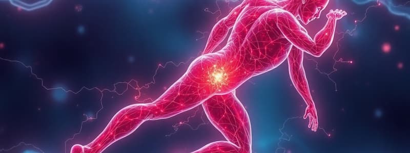
Calcium Homeostasis of Cardiomyocytes
- Calcium ions (Ca²⁺) play a crucial role in various cellular functions, including muscle contraction, fertilization, synaptic transmission, cell division, haemostasis, and apoptosis.
- Changes in intracellular Ca²⁺ concentration can occur very rapidly, for example, during muscle contraction (milliseconds).
- Directly measuring Ca²⁺ in living cells isn't possible.
Introduction - Ca²⁺
- Ca²⁺ acts as a vital secondary messenger.
- Its impact on various cellular processes is significant.
- Ca²⁺-related changes occur quickly, impacting cellular functions rapidly.
- Direct measurement of Ca²⁺ within living cells is not currently possible.
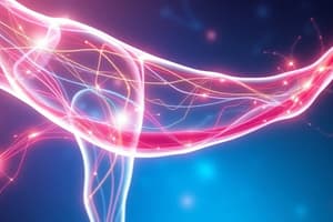
Calcium Cycling in Cardiomyocytes
- The diagram illustrates the complex interplay of various proteins and ion channels involved in regulating calcium levels within cardiomyocytes.
- Specific ion channels and pumps maintain delicate balance of calcium inside and outside the cells.
- Ca²⁺ cycling is a complex process involving intracellular and extracellular mechanisms.
- Intracellular calcium concentrations are tightly regulated.
- A significant amount of Ca²⁺ buffering occurs in the cytosol.
- The resting [Ca²⁺] in the cytosol is approximately 150 nM.
- A 70 µM change in total [Ca²⁺] is needed to elicit a 50% force response.
Origin of Calcium Ions
- Extracellular Ca²⁺ is regulated by several Ca²⁺ channels (e.g., IcaL, IcaT, and NCX).
- Intracellular Ca²⁺ primarily comes from the sarcoplasmic reticulum (SR).
- The release of Ca²⁺ from the SR is influenced by various mechanisms including calcium-induced calcium release (CICR) through ryanodine receptors (RyR2) and inositol-1,4,5-trisphosphate (IP3)-mediated release pathways.
Stop of Ca²⁺ Release
- RyR inactivation and adaptation are essential to regulate calcium release.
- local calcium depletion in the SR regulates the overall process.
Functional CICR Unit ("Couplon")
- A functional unit involves approximately 10-25 DHPRs per 100 RyRs.
Species Dependence
- Short action potentials (APs) are observed in some species (e.g., mice).
- Longer APs are observed in other species like rabbits or humans.
- Mitochondrial Ca²⁺ uptake has a part in regulating intracellular Ca²⁺ regulation.
Calcium Concentration Measurement Techniques
- Non-optical methods include ion-selective electrodes and electrophysiological techniques.
- Optical methods, like fluorescence microscopy (epifluorescence and confocal microscopy) and luminescence, are also used to measure calcium concentrations.
Jablonski Diagram
- The diagram describes the energy levels and transitions of a molecule, in this case, a fluorescent dye, involved in fluorescence measurements during calcium measurements.
- Excitation involves absorption and emission involves fluorescence.
- A Stokes shift appears between excitation and emission wavelengths.
Fluorescence Spectrum
- Excitation and emission spectra are related.
- The shift between excitation and emission wavelengths is vital for precise analysis of calcium levels.
Theory of Fluorescent Measurements
- The process and components of fluorescent measurement are shown.
- A dicroic mirror separates excitation and emission.
- Filters are used to select specific wavelengths.
Confocal Microscopy
- Confocal microscopy involves focusing excitation light and collecting emitted light from a single point within the sample.
- The technique limits out-of-focus light, enhancing resolution and specificity in images.
Selection Criteria for Ca²⁺ Indicators
- Appropriate Ca²⁺ indicators are chosen based on factors like the concentration of Ca²⁺ to be measured, the method of delivery, measuring mode (quantitative or qualitative), the intensity and type of emitted light.
- Simultaneous recording of physiological parameters is important for comprehensive understanding.
Schematic Diagram of Loading Cells Using AM Ester Derivative (Fura-2/AM)
- Loading cells with fluorescent calcium indicators is necessary to measure calcium levels.
- Problems like compartmentalization, incomplete ester hydrolysis, and possible leakage need to be considered.
- The diagram illustrates the process with its limitations.
Fluorescent Ca²⁺ Indicators
- UV indicators include Fura-2, Indo-1 and their derivatives.
- Visible light indicators include Fluo-3, Fluo-4, Rhod-2 and their derivatives.
- Low affinity calcium indicators are useful for identifying calcium concentrations accurately and avoiding false positives.
Fluorescence Excitation Spectra
- FURA-2 and Fluo-3 have specific excitation and emission spectra.
- The spectra vary based on the free Ca²⁺ concentrations.
- These variations in spectra are used to determine intracellular free Ca²⁺ concentrations.
Fluorescent Ca²⁺ Indicators Excited with Visible Light - Advantages
- Visible light indicators are more suitable for various imaging techniques.
- Interference from sample autofluorescence is reduced.
- Cellular photodamage is less of a concern compared to UV indicators.
- Visible dyes tend to absorb more strongly to visible light compared to UV indicators.
Microscope for Epifluorescent Measurement
- Epifluorescence microscopy setup includes a microscope and a filter cube assembly to precisely align excitation and emission filters.
Electrically Stimulated Skeletal Muscle Fibers Filled with Aequorin
- The experiment demonstrates calcium-dependent light emission in skeletal muscle fibers.
- Various parameters, such as membrane potential and isometric tension, are illustrated to show the impact of calcium release.
Isolated Cardiac Myocytes
- Images of isolated cardiomyocytes are shown.
Elementary Calcium Release Events
- The diagram illustrates calcium sparks and waves using fluorescence microscopy.
Epifluoreszcent Image of Control and Diabetic Cells
- This shows images visually comparing normal and diabetic cardiomyocyte behavior.
Epifluoreszcent Calcium Measurement
- An epifluorescence image of cardiomyocytes is displayed.
Confocal Image
- This shows confocal images of A ankyrin-B, B56 alpha, and merged images showing the localization of the proteins in cardiomyocytes.
Calcium Release Events
- Images show calcium sparks and waves in cardiomyocytes.
Confocal Calcium Measurement (Rat Ventricular Myocyte)
- The figure shows simultaneous measurement of fluorescence and membrane potential.
Ca²⁺ - Calmodulin - CaMKII Mediated Signaling
- The roles of calcium, calmodulin, and calcium/calmodulin activated kinase II (CaMKII) in intracellular signaling pathways are highlighted.
Structure of Calmodulin
- The structure of calmodulin (CaM) in different states (apo, Ca2+-bound, and complexed) is illustrated.
Model of the Interaction of CaM with KCNQ Channels
- The interaction of calmodulin (CaM) with potassium channels (KCNQ) is modeled to demonstrate the role of CaM in regulating ion channel activity.
CaMK Mediated Signalization
- The role of CaMKII in signaling pathways is explained.
Domain and Molecular Structure of CaMKII
- The structural domains of CaMKII (cDNA) and important sites involved in activation and regulation are highlighted. This includes the catalytic domain, autoinhibitory domains, and Ca²⁺/calmodulin binding site.
CaMK Isoforms
- Different isoforms (variations) of CaMKII exist.
- The diagram demonstrates the branching structure, highlighting the diversity of isoforms within the CaMKII family and their involvement in various cellular processes.
Role of CaMKII in β-Adrenergic Receptor Signaling
- This describes how CaMKII is part of the signalling pathways regulated by β-adrenergic receptor.
Parallel Pathways of CaMKII Signalization
- This shows the separate signaling pathways of CaMKII and their separate results, including hypertrophy, apoptosis, and myocardial dysfunction.
Ca²⁺ Homeostasis of a Cardiac Myocyte and Integrated Map
- A comprehensive illustration showing the interconnected pathways involved in Ca²⁺ homeostasis within a cardiac myocyte.
- The map highlights the complex interplay of various proteins and pathways involved in Ca²⁺ regulation.
Regulatory Roles of Calcium on Ion Channels
- Calcium ions play vital roles in regulating the activity of other ion channels.
- Numerous mechanisms of this regulation are outlined.
Calcium Cycling in Cardiomyocytes
- A detailed illustration of calcium cycling pathways.
- Important ion channels involved in maintaining Ca²⁺ homeostasis within cardiomyocytes are highlighted.
Pacemaker Activity
- Pacemaker cells exhibit spontaneous activity, generating regular pulses that drive the heartbeat.
Pacemaker Activity in the SA Node (“Membrane Clock”)
- The graphic demonstrates the various ion currents (e.g., Ica,l, If) responsible for the spontaneous depolarization of the SA node.
Local Calcium Releases (LCRs) in Different [Ca]e
- This shows the localization of calcium sparks (LCRs) in various extracellular calcium concentrations.
LCRS during Hyperpolarization
- The figure demonstrates calcium sparks during various depolarization/repolarization potentials in cardiomyocytes.
Correlation between LCR and Spontaneous Beating Frequencies (“Calcium Clock”)
- The image correlates the frequency of calcium sparks to the heartbeat frequency in cardiomyocytes.
The Coupled-Clock Pacemaker System
- This shows a model diagram of the various ion channels and proteins involved in rhythmical behaviour.
Na⁺ Homeostasis of Cardiac Myocytes
- This depicts the ion transport pathways and their regulatory roles in cardiac myocytes.
Interaction between Na⁺ and Ca²⁺ Homeostases of Cardiac Myocytes
- This figure demonstrates interconnected relationship between sodium and calcium.
Afterdepolarizations
- Afterdepolarization (an abnormal electrical event in the heart) is illustrated.
Changes in Calcium Homeostasis in Heart Failure
- Alterations in Ca²⁺ homeostasis are associated with heart failure.
Depolarizing Currents (INa,late és ICa,L)
- The graphic illustrates the currents contributing to depolarization in typical (healthy) and failing cardiac myocytes (HF).
Calcium Transients
- Calcium transients are measured and compared in healthy (AMC) and heart-failing (HF) cardiomyocytes using specialized microscopy techniques.
Pathology
- Electrical dysfunction (arrhythmogenesis) and mechanical dysfunction (heart failure) are common pathological scenarios related to Ca²⁺ dysregulation in the heart.
Molecular Structure
- The various structures involved in Ca²⁺ signalling are illustrated.
Arrhythmogenesis
- The flow chart outlines potential factors linked to the initiation of irregular heartbeats (arrhythmia) in the heart.
Topics
- The list presents a comprehensive overview of the topics covered by the presentation, such as calcium measurements, calcium cycling, transduction of the calcium signal, electrophysiology, and several other aspects of cardiac function and pathology.
Confocal Microscopy
- Illustration details the components of a confocal microscopy setup.
Calcium as a Universal Intracellular Messenger
- Calcium's diverse role as an intracellular signaling molecule in various physiological processes (e.g., electrophysiology, contraction, energy consumption, cell death, transcriptional regulation) is emphasized.
Calcium Cycling (Ventricular Myocyte)
- Illustrates the calcium cycling pathways in ventricular cardiomyocytes
Mechanisms of Ca²⁺ Signalization
- The three main mechanisms by which calcium ions participate in the signalling pathway are illustrated.
Components of Calcium Homeostasis in Cardiac Myocytes
- Key components involved in maintaining calcium homeostasis, including entry, release, extrusion, and sequestration processes, are summarized for cardiomyocytes.
Mechanisms of Ca²⁺ Entry
- Mechanisms that facilitate calcium entry into cardiac cells are listed.
Structure of L-type Ca²⁺ Channels (LTCC)
- The graphic shows the detailed structure of the L-type calcium channel (LTCC) and its various subunits.
Main Facts
- Summary of key characteristics and properties of the L-type calcium channel.
The LTCC during Action Potential
- The figure shows various voltage measurements and currents specific to the L-type calcium channel.
Electrophysiology of L-type Calcium Channel (LTCC)
- Illustrations and figures measuring the voltage-dependent characteristics of the LTCC.
Differences in the Structures of L- and T-type Ca²⁺ Channels
- Structural differences between the L-type and T-type calcium channels (LTCC and TTCC) are compared and highlighted.
I-V Characteristics of LTCC and TTCC
- The functional properties of L-type and T-type calcium channels are illustrated graphically using an I-V curve.
Main Facts (T-type Calcium Channel)
- Summary of important features of the T-type calcium channel (TTCC).
TTX-sensitive Ca²⁺ Channel
- Key characteristics of the TTX-sensitive Ca²⁺ channel are summarized.
Slip Mode Conductance
- Mechanisms and physiological role of slip mode conductance are explained briefly.
IP₃ Signalization of Cardiac Myocytes
- Description of the IP₃ signaling pathway in cardiac myocytes and its connection to calcium release.
Studying That Suits You
Use AI to generate personalized quizzes and flashcards to suit your learning preferences.