Podcast
Questions and Answers
What is the primary function of the diaphragm and chest wall?
What is the primary function of the diaphragm and chest wall?
- To act as a bellows for the lungs (correct)
- To protect the heart from injury
- To filter the air we breathe
- To aid in digestion
What is the significance of the thoracic inlet and outlet?
What is the significance of the thoracic inlet and outlet?
- They are the boundaries of the thoracic cavity (correct)
- They are responsible for regulating blood pressure
- They are the attachment sites for the ribs
- They are the primary sites of gas exchange
What is the role of the pleural fluid in the respiratory system?
What is the role of the pleural fluid in the respiratory system?
- To absorb oxygen from the air
- To regulate body temperature
- To lubricate the movement of the lungs within the thoracic cavity (correct)
- To remove carbon dioxide from the blood
What is unique about the thoracic volume in patients with emphysema?
What is unique about the thoracic volume in patients with emphysema?
What is the primary mechanism of increasing thoracic volume during inspiration?
What is the primary mechanism of increasing thoracic volume during inspiration?
What is the innervation of the diaphragm?
What is the innervation of the diaphragm?
What is the attachment site of the diaphragm?
What is the attachment site of the diaphragm?
What is the primary function of the pleural membranes?
What is the primary function of the pleural membranes?
What structure forms the superior boundary of the thoracic cavity?
What structure forms the superior boundary of the thoracic cavity?
What is the term for the space between the visceral and parietal pleura?
What is the term for the space between the visceral and parietal pleura?
Which of the following structures is NOT part of the thoracic cavity?
Which of the following structures is NOT part of the thoracic cavity?
What is the inferior boundary of the thoracic cavity?
What is the inferior boundary of the thoracic cavity?
What is the function of the pleural fluid?
What is the function of the pleural fluid?
What is the term for the space between the two lungs?
What is the term for the space between the two lungs?
What structures bound the thoracic cavity laterally?
What structures bound the thoracic cavity laterally?
What structure lines the space surrounding the lungs?
What structure lines the space surrounding the lungs?
Which of the following structures forms the bony-cartilaginous ring that the suprapleural membrane attaches to?
Which of the following structures forms the bony-cartilaginous ring that the suprapleural membrane attaches to?
Where does the suprapleural membrane rise up to and extend?
Where does the suprapleural membrane rise up to and extend?
What is the diaphragm?
What is the diaphragm?
What is the function of the hemi-diaphragm?
What is the function of the hemi-diaphragm?
What structures pass through the thoracic inlet?
What structures pass through the thoracic inlet?
What type of fascia do the vessels of the head and neck take as they pass through the thoracic inlet?
What type of fascia do the vessels of the head and neck take as they pass through the thoracic inlet?
What is the thoracic outlet bounded by?
What is the thoracic outlet bounded by?
What passes through the thoracic outlet?
What passes through the thoracic outlet?
Study Notes
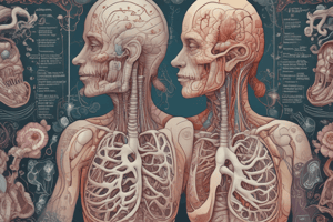
Boundaries of the Thoracic Cavity
- The thoracic cavity is the space occupied by the lungs and their covering of visceral pleura.
- The lungs almost completely fill this space, leaving a tiny gap between the visceral pleura and the parietal pleura.
- The enclosed space between the pleural membranes is called the pleural cavity.
- The thoracic cavity is bounded posteriorly by the thoracic vertebral column, laterally by the ribs, and anteriorly by the sternum.
Thoracic Apertures
- The thoracic inlet, or superior thoracic aperture, is a bony-cartilaginous ring defined by the 1st thoracic vertebra, 1st rib, 1st costal cartilage, and upper margin of the manubrium.
- The suprapleural membrane attaches to this ring and rises upwards, extending 2.5 cm above the medial end of the clavicle.
- The thoracic outlet, or inferior thoracic aperture, is the space bounded by the costal margin in front, the tips of the 11th and 12th ribs, and the inferior margin of the 12th thoracic vertebra.
- The diaphragm closes off the thoracic outlet and is a bi-domed muscular sheet inserting into a central tendon.
Structures Passing Through the Thoracic Inlet
- Structures that pass through the thoracic inlet, such as the trachea, oesophagus, carotid artery, and internal jugular vein, take a sleeve of the fascial membrane as they do so.
- The trachea and oesophagus take pre-tracheal fascia, the vessels of the head and neck take the carotid sheath, and the vessels of the upper limb take the axillary sheath.
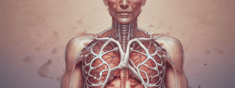
Diaphragm
- The diaphragm is the principal muscle of breathing.
- Each side of the diaphragm is referred to as a hemi-diaphragm.
- The diaphragm descends to enable inspiration and relaxes to allow expiration.
- The thoracic outlet transmits thoracic structures destined for the abdomen and permits passage of venous blood from the lower half of the body back into the heart.
Studying That Suits You
Use AI to generate personalized quizzes and flashcards to suit your learning preferences.
Description
Learn about the boundaries of the chest and the respiratory system in this introductory lecture, covering the thoracic inlet and outlet.