Podcast
Questions and Answers
Which of the following best describes the primary focus of histology?
Which of the following best describes the primary focus of histology?
- The microscopic analysis of cells. (correct)
- The macroscopic study of organ systems.
- The study of ancient artifacts.
- The chemical reactions within cells.
Who is credited as the founder of histology?
Who is credited as the founder of histology?
- Rudolf Virchow
- Anton van Leeuwenhoek
- Robert Hooke
- Marcello Malpighi (correct)
Which of the following is the correct chronological order of these key events in the history of microscopy and histology?
Which of the following is the correct chronological order of these key events in the history of microscopy and histology?
- Development of electron microscope -> Invention of the microscope -> Discovery of cells
- Invention of the microscope -> Discovery of cells -> Development of electron microscope (correct)
- Discovery of cells -> Invention of the microscope -> Development of electron microscope
- Invention of the microscope -> Development of electron microscope -> Discovery of cells
Which of the following statements accurately describes the contribution of Anton van Leeuwenhoek to the field of histology?
Which of the following statements accurately describes the contribution of Anton van Leeuwenhoek to the field of histology?
In the context of preparing a sample for histological examination, what is the primary purpose of fixation?
In the context of preparing a sample for histological examination, what is the primary purpose of fixation?
Which of the following describes the main purpose of the embedding process in histological techniques?
Which of the following describes the main purpose of the embedding process in histological techniques?
What is the most accurate description of the role of xylene in the process of preparing histological samples?
What is the most accurate description of the role of xylene in the process of preparing histological samples?
Which of the following best characterizes the function of hematoxylin and eosin (H&E) staining in histology?
Which of the following best characterizes the function of hematoxylin and eosin (H&E) staining in histology?
Which of the following is the primary reason for using a mounting medium after staining a histological section?
Which of the following is the primary reason for using a mounting medium after staining a histological section?
What is the most significant function of fixatives in the preparation of histological specimens?
What is the most significant function of fixatives in the preparation of histological specimens?
What type of microscopy uses electrons to generate a highly magnified image, allowing for the visualization of cellular ultrastructure:
What type of microscopy uses electrons to generate a highly magnified image, allowing for the visualization of cellular ultrastructure:
Which of the following techniques is best suited for visualizing specific proteins within a tissue sample?
Which of the following techniques is best suited for visualizing specific proteins within a tissue sample?
In the context of tissue processing, what is the purpose of dehydration?
In the context of tissue processing, what is the purpose of dehydration?
Which staining technique allows for the visualization of collagen fibers in tissue sections?
Which staining technique allows for the visualization of collagen fibers in tissue sections?
Which of the following best describes the fundamental difference between light microscopy and electron microscopy?
Which of the following best describes the fundamental difference between light microscopy and electron microscopy?
Which of the following is a primary goal of tissue clearing in histology?
Which of the following is a primary goal of tissue clearing in histology?
In the context of staining techniques, what component of a cell does hematoxylin primarily bind to?
In the context of staining techniques, what component of a cell does hematoxylin primarily bind to?
Why is it crucial to avoid excessive delay between tissue removal (biopsy) and fixation?
Why is it crucial to avoid excessive delay between tissue removal (biopsy) and fixation?
Which statement best describes the role of immersion oil in light microscopy?
Which statement best describes the role of immersion oil in light microscopy?
Which of the following statements correctly differentiates between 'vital' and 'non-vital' staining techniques in histology?
Which of the following statements correctly differentiates between 'vital' and 'non-vital' staining techniques in histology?
What does the term “autolysis” refer to in the context of histology?
What does the term “autolysis” refer to in the context of histology?
What is the purpose of 'decalcification' in histological processing?
What is the purpose of 'decalcification' in histological processing?
Which of the following describes the role of the microtome in histological procedures?
Which of the following describes the role of the microtome in histological procedures?
Which of the following is a common fixative used in histology?
Which of the following is a common fixative used in histology?
Which of the following is a metallic impregnation technique used for staining reticular fibers?
Which of the following is a metallic impregnation technique used for staining reticular fibers?
Which of the following reflects the primary advantage of electron microscopy over light microscopy?
Which of the following reflects the primary advantage of electron microscopy over light microscopy?
What is the main advantage of using immunohistochemistry compared to standard staining techniques like H&E?
What is the main advantage of using immunohistochemistry compared to standard staining techniques like H&E?
The purpose of fixation in preparing tissue samples for histological examination is to:
The purpose of fixation in preparing tissue samples for histological examination is to:
After fixation, tissues are dehydrated using a series of increasing concentrations of:
After fixation, tissues are dehydrated using a series of increasing concentrations of:
Which statement best describes the function of hematoxylin in H&E staining?
Which statement best describes the function of hematoxylin in H&E staining?
What is the term referring to a tissue sample that is taken from a living organism for diagnostic examination?
What is the term referring to a tissue sample that is taken from a living organism for diagnostic examination?
Which of the following best describes the purpose of the clearing step in tissue processing?
Which of the following best describes the purpose of the clearing step in tissue processing?
Which type of microscopy is used for visualizing the three-dimensional surface of a sample?
Which type of microscopy is used for visualizing the three-dimensional surface of a sample?
Which of the staining methods is best suited for visualization of carbohydrate-rich structures, such as glycogen?
Which of the staining methods is best suited for visualization of carbohydrate-rich structures, such as glycogen?
Flashcards
Histology
Histology
Study of tissues, including structure, function, and development.
Zacharias Janssen
Zacharias Janssen
Invented the first compound microscope with two lenses in 1590
Galileo Galilei
Galileo Galilei
Invented a microscope in 1610
Matthias Schleiden
Matthias Schleiden
Signup and view all the flashcards
Theodor Schwann
Theodor Schwann
Signup and view all the flashcards
Rudolf Virchow
Rudolf Virchow
Signup and view all the flashcards
Cell Theory
Cell Theory
Signup and view all the flashcards
Histological study
Histological study
Signup and view all the flashcards
Marcello Malpighi
Marcello Malpighi
Signup and view all the flashcards
Histological Techniques
Histological Techniques
Signup and view all the flashcards
Sample Obtaining
Sample Obtaining
Signup and view all the flashcards
Tissue Fixation
Tissue Fixation
Signup and view all the flashcards
Tissue embedding
Tissue embedding
Signup and view all the flashcards
Tissue Sectioning
Tissue Sectioning
Signup and view all the flashcards
Tissue Staining
Tissue Staining
Signup and view all the flashcards
Mounting
Mounting
Signup and view all the flashcards
Formalin
Formalin
Signup and view all the flashcards
Dehydration
Dehydration
Signup and view all the flashcards
Xylene
Xylene
Signup and view all the flashcards
Optical Microscope
Optical Microscope
Signup and view all the flashcards
Electron microscope
Electron microscope
Signup and view all the flashcards
Eosin
Eosin
Signup and view all the flashcards
Hematoxylin
Hematoxylin
Signup and view all the flashcards
Histoquimica and Citoquimicas
Histoquimica and Citoquimicas
Signup and view all the flashcards
Inmunocitoquimica
Inmunocitoquimica
Signup and view all the flashcards
Inmunofluorescencia
Inmunofluorescencia
Signup and view all the flashcards
Tecnicas de tinción
Tecnicas de tinción
Signup and view all the flashcards
Fixation of Sample
Fixation of Sample
Signup and view all the flashcards
Citology
Citology
Signup and view all the flashcards
Vital staining
Vital staining
Signup and view all the flashcards
Supravital staining
Supravital staining
Signup and view all the flashcards
No vital staining
No vital staining
Signup and view all the flashcards
Hematoxilina-Eosina
Hematoxilina-Eosina
Signup and view all the flashcards
biopsias
biopsias
Signup and view all the flashcards
Citología exfoliativa
Citología exfoliativa
Signup and view all the flashcards
Study Notes
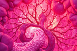
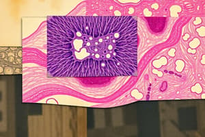
Introducción a la Histología y Técnicas Histológicas
- La histología es el estudio de los tejidos orgánicos, incluyendo su estructura microscópica, desarrollo y funciones
Historia de la Histología
- Culturas antiguas inventaron una lupa rudimentaria alrededor del 3000 a.e.
- Zacharias Janssen creó el primer microscopio compuesto con dos lentes en 1590.
- Galileo Galilei elabora un microscopio compuesto en 1610.
- Robert Hooke examinó laminas delgadas de corcho y llamó células a las celdillas en 1665.
- Anton Van Leeuwenhoek dió a conocer sus observaciones acerca de los glóbulos rojos, espermatozoides y microorganismos en agua en 1673.
- Matthias Schleiden concluyó que los vegetales estaban constituidos por células en 1838.
- Theodor Schwann realizó observaciones animales y concluyó que estaban contituidos por células en 1839.
- Rudolf Virchow estableció que toda célula procede de otra célula en 1850.
- Rudolf Virchow agregó un tercer postulado en 1858.
- Louis Pasteur demuestra la existencia de la vida microbiana en 1878.
- Ernest Ruska y Max Knoli construyeron el microscopio electrónico en 1925.
- Los estudios histológicos fueron posibles a partir del año 1600, cuando se incorporó el microscopio.
- Marcello Malpighi es reconocido como el fundador de la Histología.
- Anton Van Leeuwenhoek nació en Delft, Países Bajos en 1632.
- Las primeras investigaciones histológicas se hicieron en el siglo XVII gracias a la invención del microscopio óptico por Antonio Van Leeuwenhoek.
Microscopios
- El microscopio óptico utiliza luz común para la visualización.
- El microscopio electrónico de transmisión usa un haz de electrones (filamento de tungsteno) y bobinas electromagnéticas.
- El microscopio electrónico de barrido también usa haz de electrones y bobinas electromagnéticas, ofreciendo imágenes tridimensionales
- El poder de resolución del microscopio óptico es de 1 mm (1000 nm).
- El poder de resolución del microscopio electrónico de transmisión es de 0,2 nm (2 Å).
- El poder de resolución del microscopio electrónico de barrido es de 3-5 nm (30-50 Å).
- El microscopio óptico se usa directamente y con fotografía.
- El microscopio electrónico de transmisión usa una pantalla fluorescente y fotografía.
- El microscopio electrónico de barrido usa una pantalla de televisión y fotográfica.
- Microscopio óptico permite ver la estructura de células y tejidos, hongos y bacterias.
- Un microscopio electrónico de transmisión permite ver la ultraestructura de células, organelas, viroides e inclusiones virales.
- Un microscopio electrónico de barrido se usa para el estudio morfológico de la superficie dando imágenes tridimensionales.
Tipos de Microscopios
- Microscopio óptico: Microscopio Óptico Simple y Microscopio Óptico Compuesto.
- Microscopio electrónico: Transmisión, Barrido, Digital y Efecto túnel o cuántico.
Técnica Histológica
- Pasos: Obtención de material, fijación, inclusión, obtención del corte, coloración y montaje final
- Los pasos de la técnica histológica incluyen la fijación, inclusión y tinción
- La fijación conserva la estructura de la célula y componentes extracelulares.
- Se usa formalina, una solución acuosa de formaldehido al 37%.
- Los pasos de la técnica histológica incluyen el lavado y la deshidratación en disoluciones de alcohol.
- Aclaramiento se realiza con disolventes como xileno o tolueno miscibles en la parafina.
- El corte se realiza con un microtomo y un medio de montaje de 5-15 micrones, y permite su observación bajo microscopio
- En las estructuras de membrana se utiliza permanganato y osmio.
- En los lípidos neutros se usa formalina y colorantes que se disuelven en grasa.
Generalidades y Aplicación de la Ténica Hisotógica y de la Microscopía
- La histología se ha desarrollado con el uso de la microscopía electrónica e histoquímica, entre otras técnicas.
Técnicas de Tinción
- Eosina es un colorante ácido (rosado) con carga negativa para grupos catiónicos.
- Hematocilina es un colorante básico (azul) con carga positiva para grupos fosfatos ionizados.
- Ácido peryódico de Schiff (PAS): tiñe hidratos de carbono (púrpura)
- Tricrómica de Gonori: tiñe fibras musculares (rojo).
- Tricrómica de Masson: tiñe cartílago azul/verde, y fibras musculares de rojo.
- Tricrómica de Mallory: tiñe a la queratina (anaranjado, cartílago (azul), matriz ósea (azul intenso), fibras musculares (rojo).
- Weigert para elastina: tiñe fibras elásticas (azul/negro).
Tipos de Coloraciones
- Vitales: Se practican sobre células vivas, introduciendo el colorante en la circulación. Un ejemplo es el verde Jano y el azul de metileno.
- Supravitales: Se realizan en células o tejidos vivos aislados del organismo.
- No vitales: Se realizan en células muertas.
Técnicas de Coloración
- Los colorantes pueden clasificarse en básicos, ácidos, neutros e indiferentes.
- La coloración más común es Hematoxilina-Eosina, que colorea los núcleos celulares y ARN de color índigo, y el citoplasma de color rosado a rojo.
- Los estudios de sangre usan la coloración de Romanowsky.
- El Azul de Metileno fue el primer colorante usado.
- La Fucsina-Resorcina es un colorante usado para fibras elásticas.
- La afinidad por la Plata de las fibras reticulares, han hecho de la coloración Argéntica la más usada para este fin.
- La Tricrómica de Mallory, tiñe los epitelios de rojo y el tejido conectivo de azul.
- La Hematoxilina Férrica resalta las estrías del músculo esquelético.
Clasificación de los Colorantes
- Naturales: Animal y Vegetal (Carmin, Hematoxilina o Arceina).
- Artificiales: Derivados de la anilina. El colorante artificial más usado es la eosina
Fijación de la Muestra
- Este proceso se refiere al tratamiento del tejido con sustancias químicas con varios fines.
- Conservar los tejidos de forma que muestren el mayor parecido posible a su estado in vivo.
- Aumentar la dureza del tejido para facilitar la elaboración de las películas del tejido.
- Destruir bacterias y gérmenes que pudieran encontrarse en ellos.
- Interrumpir los procesos celulares dinámicos que ocurren a la muerte de la célula.
Deshidratación Aclaramiento Inclusión
- Después de la fijación, se elimina el fijador y se deshidrata.
- Se aplica gradualmente una solución de un agente deshidratante como Alcohol Etílico o Acetona (50% hasta 100%).
- El aclaramiento transforma el tejido en transparente en el xileno.
- La inclusión logra infiltrar la parafina líquida al tejido para que disuelva el medio de aclaramiento y penetre el tejido
- El calor evapora el xilol.
Obtención del Material
- Es el primer paso de la técnica histológica, El material puede ser obtenido quirúrgicamente mediante los métodos de incisión o excisión y es importante aplicar el método correcto.
- Mediate citología exfoliativa también se pueden obtener muestras.
- El tejido también puede obtenerse después de la disección de un animal o de una necropsia.
- Los especímenes se pueden analizar "in vitro" o "in vivo".
- Biopsias se realizan por ejemplo con los métodos de sacarocado o Tru-cut.
- Citología se encarga del estudio de las células en cuanto a las propiedades, estructura y funciones.
Studying That Suits You
Use AI to generate personalized quizzes and flashcards to suit your learning preferences.
Related Documents
Description
La histología es el estudio de los tejidos orgánicos, incluyendo su estructura microscópica, desarrollo y funciones. Este tema también cubre la historia de la histología, desde los primeros microscopios hasta los descubrimientos de Rudolf Virchow y Louis Pasteur.