Podcast
Questions and Answers
______ spp. cysts are identified using cultured, Hematoxylin, and Eosin stains.
______ spp. cysts are identified using cultured, Hematoxylin, and Eosin stains.
Acanthamoeba
______ trophozoites are cultured and stained with trichrome and Pap’s stain.
______ trophozoites are cultured and stained with trichrome and Pap’s stain.
Entamoeba gingivalis
The cysts of Giardia intestinalis can be identified using Wet Mount (WM) with Iodine, ______, Trichrome, and Wet Mount under Differential Interference Contrast (DIC).
The cysts of Giardia intestinalis can be identified using Wet Mount (WM) with Iodine, ______, Trichrome, and Wet Mount under Differential Interference Contrast (DIC).
Trichrome
Chilomastix mesnili cysts can be diagnosed using Wet Mount (WM) with Iodine and ______.
Chilomastix mesnili cysts can be diagnosed using Wet Mount (WM) with Iodine and ______.
______ cysts are diagnosable via Iron-hematoxylin and Trichrome staining.
______ cysts are diagnosable via Iron-hematoxylin and Trichrome staining.
Retortamonas intestinalis cysts are exclusively identifiable using ______ staining techniques.
Retortamonas intestinalis cysts are exclusively identifiable using ______ staining techniques.
While most intestinal ameba trophozoites are stained using trichrome, the first trophozoite of ______ is an exception.
While most intestinal ameba trophozoites are stained using trichrome, the first trophozoite of ______ is an exception.
______ trophozoites are identified through culture methods.
______ trophozoites are identified through culture methods.
Flashcards
Concentration Wet Mount (CWM)
Concentration Wet Mount (CWM)
A method for identifying parasites, involving concentration and staining, used for organisms like E.Histolytica and E.Coli.
Trichrome Stain
Trichrome Stain
Staining technique used to differentiate cellular components, aiding in parasite identification for organisms like E.Histolytica and E.Coli.
Wet Mount with Iodine
Wet Mount with Iodine
A staining method used for visualizing parasites in fecal samples, aiding identification. Used for organisms like E.Nana and I.Butschlii.
Culturing Amoebas
Culturing Amoebas
Signup and view all the flashcards
Hematoxylin and Eosin (H&E)
Hematoxylin and Eosin (H&E)
Signup and view all the flashcards
Pap's Stain
Pap's Stain
Signup and view all the flashcards
Iron-Hematoxylin Stain
Iron-Hematoxylin Stain
Signup and view all the flashcards
WM under DIC
WM under DIC
Signup and view all the flashcards
Study Notes
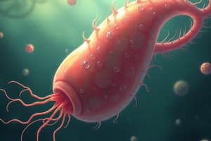
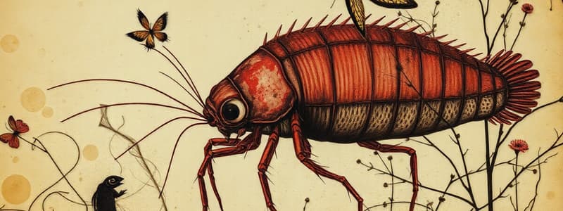
- The images provided show cysts and trophozoites of intestinal and extraintestinal amebas and flagellates, stained with various methods for identification.
Cysts of Intestinal Amebas
- Includes images of E. histolytica, E. hartmanni, E. coli, E. polecki, E. nana, and I. butschlii.
- Staining methods used are concentrated wet mount, wet mount with iodine, trichrome, and CWM with iodine.
Trophozoites of Intestinal Amebas
- Contains images of E. histolytica, E. hartmanni, E. coli, E. polecki, E. nana, and I. butschlii.
- Staining methods are CWM with iodine and trichrome.
- All are stained with trichrome except the first troph of E. histolytica.
Cysts of Extraintestinal Amebas
- Features images of Naegleria fowleri and Acanthamoeba spp.
- Staining methods are culturing and hematoxylin and eosin.
Trophozoites of Extraintestinal Amebas
- Encompasses images of Entamoeba gingivalis, Naegleria fowleri, and Acanthamoeba spp.
- Staining methods are culturing, trichrome, and Pap's stain.
Cysts of Intestinal Flagellates
- Includes images of G. intestinalis, C. mesnili, E. hominis, and R. intestinalis.
- Staining methods are wet mount with iodine, iron-hematoxylin, trichrome, and wet mount under DIC (differential interference contrast).
Trophozoites of Intestinal Flagellates
- Contains images of G. intestinalis, C. mesnili, D. fragilis, T. hominis, E hominis, and R. intestinalis.
- Staining Methods include wet mount with iodine, trichrome, Kohn stain, and Giemsa.
Trophozoites of Extraintestinal Flagellates
- Includes images of T. tenax and T. vaginalis.
- Staining Method is Giemsa.
Studying That Suits You
Use AI to generate personalized quizzes and flashcards to suit your learning preferences.
Related Documents
Description
Images of cysts and trophozoites of intestinal and extraintestinal amebas and flagellates, stained with various methods for identification. Includes E. histolytica, E. hartmanni, E. coli, E. polecki, E. nana, I. butschlii, Naegleria fowleri and Acanthamoeba spp.