Podcast
Questions and Answers
What is the primary form of the organism E. histolytica found in tissues?
What is the primary form of the organism E. histolytica found in tissues?
- Cyst
- Precyst
- Trophozoite (correct)
- Spore
Which structure is characteristic of the mature cyst of E. histolytica?
Which structure is characteristic of the mature cyst of E. histolytica?
- Single nucleus
- Chromatoidal bars with pointed ends
- Glycogen mass present
- 1-4 nuclei (correct)
How does a trophozoite of E. histolytica move?
How does a trophozoite of E. histolytica move?
- By cilia
- By flagella
- By passive flow
- By pseudopodia (correct)
What occurs during the transformation of a trophozoite to a precyst?
What occurs during the transformation of a trophozoite to a precyst?
Which of the following features is NOT present in the immature cyst of E. histolytica?
Which of the following features is NOT present in the immature cyst of E. histolytica?
What is the primary mode of transmission for E.histolytica?
What is the primary mode of transmission for E.histolytica?
What is the infectious form of E.histolytica?
What is the infectious form of E.histolytica?
Where does excystation of E.histolytica occur?
Where does excystation of E.histolytica occur?
In which part of the body does E.histolytica primarily live in its trophozoite form?
In which part of the body does E.histolytica primarily live in its trophozoite form?
Which statement correctly describes the role of carriers of E.histolytica?
Which statement correctly describes the role of carriers of E.histolytica?
Which of the following accurately categorizes Entamoeba histolytica?
Which of the following accurately categorizes Entamoeba histolytica?
What is the primary habitat of Entamoeba histolytica?
What is the primary habitat of Entamoeba histolytica?
Which group has a higher prevalence of Entamoeba histolytica infections?
Which group has a higher prevalence of Entamoeba histolytica infections?
Which of the following is NOT a form of Entamoeba histolytica?
Which of the following is NOT a form of Entamoeba histolytica?
Which statement about Entamoeba histolytica infection is accurate?
Which statement about Entamoeba histolytica infection is accurate?
Flashcards
Intestinal Amoeba
Intestinal Amoeba
A type of amoeba found in the large intestines of humans and animals.
Entamoeba histolytica
Entamoeba histolytica
A pathogenic intestinal amoeba that causes amebiasis.
Entamoeba histolytica Habitat
Entamoeba histolytica Habitat
Lives in the mucous and submucous layers of the large human intestine.
Amebiasis Epidemiology
Amebiasis Epidemiology
Signup and view all the flashcards
Trophozoite
Trophozoite
Signup and view all the flashcards
What is the active form of E. histolytica?
What is the active form of E. histolytica?
Signup and view all the flashcards
How does the trophozoite move?
How does the trophozoite move?
Signup and view all the flashcards
What is contained in the endoplasm?
What is contained in the endoplasm?
Signup and view all the flashcards
What happens before the trophozoite becomes a cyst?
What happens before the trophozoite becomes a cyst?
Signup and view all the flashcards
What are chromatoid bars?
What are chromatoid bars?
Signup and view all the flashcards
E. histolytica Transmission
E. histolytica Transmission
Signup and view all the flashcards
Fecal-oral Transmission
Fecal-oral Transmission
Signup and view all the flashcards
E. histolytica in the Environment
E. histolytica in the Environment
Signup and view all the flashcards
Vector Transmission of E. histolytica
Vector Transmission of E. histolytica
Signup and view all the flashcards
Sexual Contact and E. histolytica
Sexual Contact and E. histolytica
Signup and view all the flashcards
Study Notes
Intestinal Amoeba
- Intestinal amoeba are single-celled protozoa that change shape constantly.
- Locomotion is achieved through an organ called a pseudopodium.
- Classified by habitat: intestinal amoeba (large intestine of humans/animals) and free-living amoeba (small free-living and opportunistic pathogens).
- Pathogenic intestinal amoeba include Entamoeba histolytica.
- Non-pathogenic intestinal amoeba include E. dispar, E. moshkovskii, E. coli, E. polecki, E. hartmanni, E. gingivalis, Endolimax nana, Iodamoeba butschlii.
- Entamoeba histolytica is prevalent in the tropics and subtropics.
- The majority of Entamoeba histolytica cases are asymptomatic.
- It is a common diarrheal pathogen in long-term travelers (>6 months).
- The highest burden of the disease occurs in the tropics of China, Central and South America, and the Indian subcontinents, affecting 10% of the world's population.
- Families of infected people, male homosexuals, those in mental institutions, prisons, and children institutions have a higher prevalence of infection.
- All ages are susceptible, but the rate is lower in infants & children. Females and males have similar likelihood of infection but invasive amebiasis is more common in males.
Classification of Amoeba
- Amoeba are a type of single-celled protozoa.
- They have the ability to constantly change their shape.
- The presence of a pseudopodium (an organ of locomotion) results in their ability to change shape.
Etiological Agent
- Infection is caused by Entamoeba histolytica.
- E. histolytica resides in the human colon, specifically within the mucous and submucous layers of the large intestine.
- Morphology presents in three forms: trophozoite, precyst, and cyst.
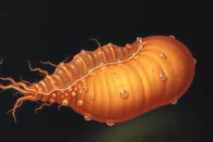
- Trophozoite: the vegetative form found in tissues; irregular shape, 12-60 µm in size; moves unidirectionally using pseudopodia.
- Cytoplasm contains ectoplasm and endoplasm with nuclei, food vacuoles, and phagocytosed red blood cells within the endoplasm.
- Trophozoite is the only form that resides within tissues
- Cysts (10-20 µm, round, thick-walled): highly resistant to harsh digestive environments; survive in external environments. Contains 1-4 nuclei, chromatoid bars (RNA aggregates), and a glycogen mass.
- Cysts are the infectious form.
Life Cycle
- The life cycle occurs within a single host (humans).
- Mature quadrinucleated cysts are the infectious form and are ingested with contaminated food or water.
- In the intestines, cysts transform into trophozoites.
- Trophozoites reside in the lumen of the small intestine, specifically the lower small bowel, and within mucosal crypts in the colon.
- Trophozoites feed on nutrients and bacteria.
- Trophozoites encyst within the colon's lumen, transforming back into cysts, and are discharged in feces.
Pathogenesis
- Infection by E. histolytica is initially asymptomatic in 90% of cases.
- Under particular conditions (host and parasite factors), E. histolytica becomes invasive.
- E. histolytica can cause tissue lysis.
- Factors that increase susceptibility increase invasive potential: poor nutrition, stress, altered colon flora, and low host resistance.
- Trophozoites invading colonic mucosa lead to characteristic ulcerative lesions and profuse bloody diarrhea (amoebic dysentery).
- Males and females are affected equally in terms of infection.
Complications of Intestinal Amebiasis
- Initial lesions of amebic invasion begin as small foci of necrosis within the large bowel mucosa.
- These foci progress into ulcers with a distinctive flask shape (wide base, narrow neck).
- Ulcers can be superficial (heal without scarring) or deep(heal with scarring).
- Ulcers typically locate in the ileocecal region, sigmoidorectal region, or can affect the entire large intestine.
- Confluence of ulcers leads to sloughing of mucosa, leading to secondary bacterial infection.
- Invasive disease can extend to complications such as: fulminant colitis, amoebic appendicitis, intestinal perforation, and amoebic peritonitis and other extraintestinal sites.
Pathogenesis of Extraintestinal Amebiasis
- About 5-10% of patients with intestinal amoebiasis develop extraintestinal amoebiasis 1-3 months after intestinal infection.
- Liver is the most common site of extraintestinal infection, followed by lungs, brain, genitourinary tract, and spleen.
- Characterized by the development of abscesses typically in the upper right lobe of the liver. Abscesses can be multiple.
- Extra-intestinal disease can cause: rupture into lungs or pericardium, skin lesions, right pleura infection, subphrenic abscesses, and peritoneal infection (spreading through blood).
Diagnosis of Intestinal Amebiasis
- Laboratory diagnosis for intestinal disease includes stool microscopy (detecting cysts or trophozoites), stool culture, stool antigen detection (copro-antigen), serology (antibody detection), molecular diagnosis (PCR), and sigmoidoscopy (mucosal scrapings).
Diagnosis of Extraintestinal Amebiasis
- Laboratory tests for extraintestinal amebiasis often include microscopy (detecting trophozoites in pus), molecular diagnosis (PCR of pus), serology, ultrasonography (to visualize the abscess site and extent).
Treatments
- Metronidazole or tinidazole are the drugs of choice for intestinal and hepatic amoebiasis.
- Fluid and electrolyte replacement, symptomatic treatment also recommended for treatment.
- Specific dosage and duration vary depending on the type of amebiasis and/or drug.
Prevention and Control
- Health education, especially for food handlers, children, and maids.
- Good personal hygiene habits (handwashing).
- Good sewage disposal practices.
- Do not use feces as fertilizer.
- Detect and treat cases.
- Boiling of water; thorough cleaning of fruits and vegetables.
- Avoid sexual practices that allow fecal-oral contact.
Prognosis
- The prognosis for amebiasis depends on the location of the infection and the patient's general health prior to infection.
- If infection is limited to the intestines and the patient is healthy, prognosis is generally good.
- Patients with ameboma, peritonitis, and liver infection can have higher mortality rates; fulminant colitis has the highest risk of mortality (>50%).
Studying That Suits You
Use AI to generate personalized quizzes and flashcards to suit your learning preferences.