Podcast
Questions and Answers
What type of intermediate filament is found in epithelial cells?
What type of intermediate filament is found in epithelial cells?
- Nuclear lamins
- Keratin filaments (correct)
- Neurofilaments
- Vimentin filaments
Where are neurofilaments primarily located?
Where are neurofilaments primarily located?
- In the nuclear envelope
- In epithelial cells
- In muscle cells
- In nerve cells (correct)
What process controls the disassembly and reassembly of the nuclear lamina during cell division?
What process controls the disassembly and reassembly of the nuclear lamina during cell division?
- Mitosis and cytokinesis
- Transcription and translation
- Phosphorylation and dephosphorylation (correct)
- Cell differentiation
What happens to lamins during mitosis when they are phosphorylated?
What happens to lamins during mitosis when they are phosphorylated?
Which of the following correctly describes microtubules?
Which of the following correctly describes microtubules?
What are the two proteins that compose the dimer of tubulin?
What are the two proteins that compose the dimer of tubulin?
During cell division, what occurs to cytoplasmic intermediate filaments?
During cell division, what occurs to cytoplasmic intermediate filaments?
What occurs after dephosphorylation of lamins at the end of mitosis?
What occurs after dephosphorylation of lamins at the end of mitosis?
What characterizes the movement cycle of a cilium?
What characterizes the movement cycle of a cilium?
How do flagella differ from cilia in terms of their primary function?
How do flagella differ from cilia in terms of their primary function?
What is the arrangement of microtubules in cilia?
What is the arrangement of microtubules in cilia?
What is the typical duration of a complete cycle of ciliary movement?
What is the typical duration of a complete cycle of ciliary movement?
What propels the movement of cilia and flagella?
What propels the movement of cilia and flagella?
What structural feature characterizes microtubules?
What structural feature characterizes microtubules?
What distinguishes the plus end of a microtubule?
What distinguishes the plus end of a microtubule?
What is the function of γ-tubulin in the centrosome?
What is the function of γ-tubulin in the centrosome?
What describes the dynamic instability of microtubules?
What describes the dynamic instability of microtubules?
Which component of microtubules is involved in structural polarity?
Which component of microtubules is involved in structural polarity?
In animal cells, where is the centrosome typically located?
In animal cells, where is the centrosome typically located?
How many protofilaments constitute a microtubule?
How many protofilaments constitute a microtubule?
Where does microtubule growth primarily occur?
Where does microtubule growth primarily occur?
What type of bonding connects tubulin dimers in microtubules?
What type of bonding connects tubulin dimers in microtubules?
What role do centrioles play in the centrosome?
What role do centrioles play in the centrosome?
What is the term for the behavior of microtubules switching back and forth between polymerization and depolymerization?
What is the term for the behavior of microtubules switching back and forth between polymerization and depolymerization?
What happens to microtubules when their plus end is stabilized?
What happens to microtubules when their plus end is stabilized?
What drives the dynamic instability of microtubules?
What drives the dynamic instability of microtubules?
What is the consequence of losing the GTP cap on a microtubule?
What is the consequence of losing the GTP cap on a microtubule?
How do tubulin dimers carrying GTP behave compared to those carrying GDP?
How do tubulin dimers carrying GTP behave compared to those carrying GDP?
What occurs if the growth of a microtubule is slow?
What occurs if the growth of a microtubule is slow?
What is a possible outcome when a microtubule completely disappears?
What is a possible outcome when a microtubule completely disappears?
What effect does GTP hydrolysis have on the microtubule structure?
What effect does GTP hydrolysis have on the microtubule structure?
What role does the γ-tubulin ring complex play in microtubule dynamics?
What role does the γ-tubulin ring complex play in microtubule dynamics?
Which statement best describes the polarization of differentiated animal cells?
Which statement best describes the polarization of differentiated animal cells?
What is the primary role of kinesins in a nerve cell?
What is the primary role of kinesins in a nerve cell?
How do motor proteins interact with microtubules?
How do motor proteins interact with microtubules?
What role does the tail of a motor protein play in intracellular transport?
What role does the tail of a motor protein play in intracellular transport?
Which motor protein family generally moves toward the minus end of a microtubule?
Which motor protein family generally moves toward the minus end of a microtubule?
What is a primary function of cilia in cells?
What is a primary function of cilia in cells?
Which component of a motor protein dictates its cargo transport nature?
Which component of a motor protein dictates its cargo transport nature?
What structure do axons in nerve cells rely on for the transport of cellular components?
What structure do axons in nerve cells rely on for the transport of cellular components?
In a nerve cell, what direction do microtubules typically point?
In a nerve cell, what direction do microtubules typically point?
What is the main effect of motor proteins on intracellular transport?
What is the main effect of motor proteins on intracellular transport?
Flashcards are hidden until you start studying
Study Notes
Intermediate Filaments and Nuclear Lamina
- Intermediate filaments are classified into four groups: keratin (epithelial cells), vimentin (connective tissue, muscle, glial cells), neurofilaments (nerve cells), and nuclear lamins (nuclear envelope).
- Cytoplasmic intermediate filaments disassemble during mitosis, while nuclear lamina reform after cell division, regulated by phosphorylation and dephosphorylation of lamins.
- Phosphorylation by protein kinases weakens the lamin tetramers, leading to filament disassembly. Dephosphorylation allows for reassembly at the end of mitosis.
Microtubule Structure
- Microtubules consist of tubulin dimers (α-tubulin and β-tubulin), which align to form 13 parallel protofilaments, creating a hollow cylindrical structure.
- Microtubules display structural polarity with a plus end (β-tubulin) and a minus end (α-tubulin).
Centrosome and Microtubule Organization
- The centrosome is the primary microtubule-organizing center in animal cells, typically located near the nucleus.
- It contains centrioles and a matrix of proteins, including gamma-tubulin, which serve as nucleation sites for microtubule growth.
Dynamic Instability of Microtubules
- Microtubules experience dynamic instability, alternating between phases of growth (polymerization) and rapid shrinkage (depolymerization).
- Stabilization of microtubules occurs when their plus end is attached to other structures, preventing disassembly.
GTP Hydrolysis and Microtubule Dynamics
- GTP hydrolysis by tubulin dimers contributes to microtubule dynamic instability. GTP-bound dimers promote growth, while GDP-bound dimers lead to disassembly.
- Loss of the GTP cap results in the peeling away of protofilaments and shrinkage of the microtubule.
Polarity and Transport in Nerve Cells
- Differentiated animal cells exhibit polarity, crucial for the orientation of organelles and transport processes.
- In nerve cells, microtubules in axons point toward axon terminals, facilitating bidirectional transport.
Motor Proteins in Intracellular Transport
- Kinesins and dyneins are the two families of motor proteins that transport cargo along microtubules, with kinesins moving toward the plus end and dyneins toward the minus end.
- Motor proteins possess globular ATP-binding heads for interaction with microtubules and a tail for binding to cargo.
Functionality of Cilia and Flagella
- Cilia beat in a whiplike manner to either move fluid across the cell surface or propel entire single cells, characterized by alternating power and recovery strokes.
- Flagella, longer than cilia, generate regular wave-like movements to propel cells, such as sperm, through fluid environments.
Microtubule Arrangement in Cilia and Flagella
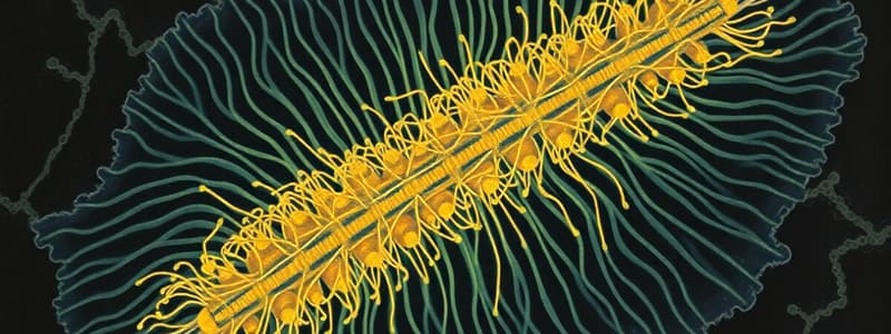
- The internal structure of cilia and flagella features a distinctive arrangement of microtubules, consisting of nine doublets forming a ring with two central microtubules, differing from cytoplasmic microtubules.
Studying That Suits You
Use AI to generate personalized quizzes and flashcards to suit your learning preferences.


