Podcast
Questions and Answers
What are some important functions of the cytoskeleton within the cell?
What are some important functions of the cytoskeleton within the cell?
Maintenance of cell shape, cell adhesion, structural support, cell movement/division, and intracellular movement of proteins and organelles.
What are some important functions of intermediate filaments within a cell?
What are some important functions of intermediate filaments within a cell?
Provides mechanical strength so that cells can withstand stretching; form network surrounding nucleus extending to plasma membrane.
What are some structural characteristics of intermediate filaments?
What are some structural characteristics of intermediate filaments?
Rope-like fibers (flexible), made of different elongated proteins, lots of lateral interactions between subunits.
Describe the subunits and protofilaments in intermediate filaments. How are they used to assemble intermediate filaments?
Describe the subunits and protofilaments in intermediate filaments. How are they used to assemble intermediate filaments?
What are nuclear lamins? What is the function of nuclear lamins within a cell? What happens to nuclear lamins during mitosis?
What are nuclear lamins? What is the function of nuclear lamins within a cell? What happens to nuclear lamins during mitosis?
What are some important functions of microtubules within a cell?
What are some important functions of microtubules within a cell?
What are some structural characteristics of microtubules?
What are some structural characteristics of microtubules?
What is tubulin? Describe the difference between β and α tubulin.
What is tubulin? Describe the difference between β and α tubulin.
Describe the significance of GTP hydrolysis in a microtubule.
Describe the significance of GTP hydrolysis in a microtubule.
What is a GTP cap? Describe how a GTP cap influences polymerization and depolymerization of microtubules.
What is a GTP cap? Describe how a GTP cap influences polymerization and depolymerization of microtubules.
What is dynamic instability? What is the association between dynamic instability and the GTP cap?
What is dynamic instability? What is the association between dynamic instability and the GTP cap?
Describe how a microtubule cycles between growing and shrinking in a test tube.
Describe how a microtubule cycles between growing and shrinking in a test tube.
What is the difference between growing microtubules in a test tube versus in a living cell?
What is the difference between growing microtubules in a test tube versus in a living cell?
Describe the functions of dynein and kinesin. How are they similar?
Describe the functions of dynein and kinesin. How are they similar?
What are some important functions of actin filaments within a cell?
What are some important functions of actin filaments within a cell?
Describe the subunits and protofilaments in actin. How are they used to assemble actin filaments?
Describe the subunits and protofilaments in actin. How are they used to assemble actin filaments?
Describe the three steps of actin polymerization in a test tube.
Describe the three steps of actin polymerization in a test tube.
What happens when preformed actin fragments are added to a test tube with actin monomers and ATP?
What happens when preformed actin fragments are added to a test tube with actin monomers and ATP?
How do cells regulate actin polymerization?
How do cells regulate actin polymerization?
What is myosin II? What is the function of myosin II in human cells?
What is myosin II? What is the function of myosin II in human cells?
Describe the general structure of myosin by explaining the roles of the head, neck, and tail domains.
Describe the general structure of myosin by explaining the roles of the head, neck, and tail domains.
What is unique about myosin II? Why is this unique characteristic important for its function in human cells?
What is unique about myosin II? Why is this unique characteristic important for its function in human cells?
What is the function of the Arp complex?
What is the function of the Arp complex?
Describe how the Arp complex, profilin, and thymosin contribute to the production of membrane protrusions.
Describe how the Arp complex, profilin, and thymosin contribute to the production of membrane protrusions.
What happens to a sarcomere during contraction?
What happens to a sarcomere during contraction?
What is the relationship between a sarcomere, skeletal muscle cell, and myofibril?
What is the relationship between a sarcomere, skeletal muscle cell, and myofibril?
What is the role of cadherins in cell adhesion?
What is the role of cadherins in cell adhesion?
Describe the function of adaptor proteins in cell adhesion.
Describe the function of adaptor proteins in cell adhesion.
What are integrins? How are integrins similar to cadherins?
What are integrins? How are integrins similar to cadherins?
What is signal transduction?
What is signal transduction?
Identify and describe the three steps of cell signaling.
Identify and describe the three steps of cell signaling.
What are effector proteins? Give an example.
What are effector proteins? Give an example.
What is the function of a protein kinase? Why are protein kinases important for enzyme-coupled receptors?
What is the function of a protein kinase? Why are protein kinases important for enzyme-coupled receptors?
What is the epidermal growth factor receptor (EGFR)? What is its ligand?
What is the epidermal growth factor receptor (EGFR)? What is its ligand?
What is Ras? Describe its function in EGFR signaling.
What is Ras? Describe its function in EGFR signaling.
What happens when a signal molecule binds to an EGFR? Describe the signaling process.
What happens when a signal molecule binds to an EGFR? Describe the signaling process.
What is HER2? What is HER2+?
What is HER2? What is HER2+?
What is a tyrosine kinase inhibitor (TKI)?
What is a tyrosine kinase inhibitor (TKI)?
Flashcards
Cytoskeleton functions
Cytoskeleton functions
Maintenance of cell shape, cell adhesion, structural support, cell movement/division, and intracellular movement of proteins and organelles
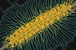
Intermediate filaments
Intermediate filaments
Toughest, most durable filament. Provides mechanical strength so cells can withstand stretching, and forms a network surrounding the nucleus
Structure of intermediate filaments
Structure of intermediate filaments
Rope-like fibers, made of elongated proteins with lateral interactions between subunits
Intermediate filament assembly
Intermediate filament assembly
Signup and view all the flashcards
Nuclear lamins
Nuclear lamins
Signup and view all the flashcards
Microtubule functions
Microtubule functions
Signup and view all the flashcards
Microtubule structure
Microtubule structure
Signup and view all the flashcards
Stability Differences
Stability Differences
Signup and view all the flashcards
What is tubulin?
What is tubulin?
Signup and view all the flashcards
GTP hydrolysis in microtubules
GTP hydrolysis in microtubules
Signup and view all the flashcards
GTP cap influence
GTP cap influence
Signup and view all the flashcards
Dynamic instability definition
Dynamic instability definition
Signup and view all the flashcards
Microtubule cycling
Microtubule cycling
Signup and view all the flashcards
Gamma-tubulin ring complexes
Gamma-tubulin ring complexes
Signup and view all the flashcards
Dynein vs. Kinesin
Dynein vs. Kinesin
Signup and view all the flashcards
Actin filaments functions
Actin filaments functions
Signup and view all the flashcards
Actin filaments
Actin filaments
Signup and view all the flashcards
Significance of ATP hydrolysis
Significance of ATP hydrolysis
Signup and view all the flashcards
3 steps of actin polymerization
3 steps of actin polymerization
Signup and view all the flashcards
Treadmilling
Treadmilling
Signup and view all the flashcards
Study Notes
General Guidelines for Collaborative Study Guide in UT Box
- The study guide on UT Box facilitates class collaboration to answer the questions.
- Course instructors review these answers to ensure accuracy and completeness.
- Answer collaboratively with well founded answers
- Answers should not be generated by AI, use lecture slides and textbook instead
- Student answers use red font for easy feedback from instructor and TAs
- Instructor feedback is provided in blue font, indicating if an answer is correct, incorrect, or incomplete.
Lecture 14: Cytoskeleton, Part I
- Cytoskeleton functions include maintaining cell shape, cell adhesion, structural support, cell movement/division, and intracellular movement of proteins/organelles.
- Intermediate filaments provide mechanical strength to cells, enabling them to withstand stretching.
- Intermediate filaments form a network around the nucleus, extending to the plasma membrane.
- Intermediate filaments are rope-like fibers made of elongated proteins with lateral interactions between subunits.
- Intermediate filament subunits are elongated filaments (homodimers).
- Homodimers bind antiparallel to form a staggered tetramer (protofilament).
- Protofilaments connect with 7 other tetramers.
- Nuclear lamins are a type of intermediate filament subunit forming the nuclear lamina beneath the inner nuclear membrane.
- Nuclear lamins are phosphorylated by kinases during mitosis, causing nuclear membrane disassembly.
- Dephosphorylation of lamins by phosphatases at the end of mitosis allows them to reassemble into the nuclear lamina
- Microtubules provides tracks for transporting vesicles, organelles, and cell components
- Microtubules form the mitotic spindle for chromosome segregation during mitosis
- Microtubules facilitate cell movement via flagella and cilia.
- Microtubules consist of hollow, polarized cylinders of α- and β-tubulin
- Intermediate filaments are flexible, non-polar fibers providing structural support, but not for transport.
- Microtubules and actin filaments are dynamic due to polarity at their ends.
- Intermediate filaments are stable due to identical ends creating no polarity.
- Intermediate filaments are made of subunits having lateral interactions.
- Microtubules are composed of globular subunits with fewer lateral interactions.
- Tubulin is a heterodimer of alpha and beta subunits.
- Beta subunit can bind and hydrolyze GTP to GDP.
- Alpha subunit can bind GTP but cannot hydrolyze it.
- The subunit of microtubules is a heterodimer of alpha and beta tubulin.
- A protofilament consists of tubulin dimers (subunits) bound head-to-toe in a straight line, giving it polarity.
- The protofilament plus end contains the Beta subunit.
- The minus end has the alpha subunit.
- Microtubules are hollow structures composed of 13 parallel protofilaments.
- The plus end grows quicker, indicating a high rate of polymerization,
- The minus end indicates a slower rate.
- GTP hydrolysis in microtubules is crucial for dynamic instability.
- GTP hydrolysis results in rapid polymerization and depolymerization by changing the shape of subunits/protofilaments.
- A GTP cap is region at the growing end of a microtubule where tubulin dimers have GTP.
- The GTP promotes polymerization, but when hydrolyzed to GDP in older subunits, microtubules destabilize
- If tubulin-GTP is added before complete hydrolysis, the microtubule can rescue itself and continue growing.
- Dynamic instability is the rapid switching between microtubule growth and shrinkage.
- This is due to the addition and loss of tubulin dimers at the microtubule plus end.
- A GTP cap regulates this with stability when GTP is present, but rapid depolymerization occurs of GTP is lost.
- Microtubules elongate when tubulin dimers with GTP bind to the plus end.
- Over time, GTP gets hydrolyzed to GDP, weakening the structure.
- A high tubulin-GTP concentration in tests are required for rapid polymerization because the rate of addition is faster than the rate of GTP hydrolysis.
- When concentration decreases, the rate of addition decreases and the GTP cap turns into a GDP cap.
- Microtubules need a high concentration of subunits to grow in a test tube.
- Cells regulate microtubule growth even at low subunit concentrations given regulatory factors to aggregate rapidly.
- Gamma-tubulin ring complexes (γ-TuRCs) serve as nucleation sites for microtubule growth, located on the centrosome surface.
- Gamma-tubulin ring complexes mimic the (+) end and template the 13 protofilaments.
- Cells regulate the number of microtubules using nucleation sites.
- Tubulin-GTP concentration determines the rate of growth.
- Kinesin carries organelles and vesicles toward the plus end of microtubules.
- Dynein also carries organelles and vesicles, however it transports to the minus end of microtubules.
- In neurons, kinesin transports cargo from the cell body to the nerve terminal
- Dynein transports cargo from the nerve terminal back to the cell body.
Lecture 15: Cytoskeleton, Part II
- Functions of actin filaments include: microvilli increase intestinal cell surface area, changing cell shape, cell motility, and contractile ring formation during cell division (cytokinesis).
- Actin is the subunit of actin filaments that are asymmetrical monomers with a plus and minus end binding to ATP.
- Two protofilaments of actin monomers form a helix forming the actin filament.
- Two protofilaments form a helix.
- Actin filaments are helical polymers with a plus end (fast growing) and a minus end (slow growing)
- Actin monomers hydrolyze bound ATP to ADP following incorporation into the actin filament.
- Hydrolysis decreases the stability of the actin subunit.
- Polymerization depends on the amount of actin monomers available.
- The three steps of actin polymerization in a test tube are: nucleation (lag phase), elongation (growth phase), and steady state (equilibrium phase).
- Nucleation is when concentration of actin monomers is high and an initial aggregate of monomers (critical nucleus) will form
- Elongation is when initial aggregates start rapidly growing filaments by subunits attaching to the initial aggregate, decreasing subunit concentration.
- Steady State occurs when concentrations of subunits decline and filaments reach steady state called 'treadmilling' when the rate of addition equals the rate of dissociation.
- Adding preformed actin fragments to a test tube with actin monomers and ATP increases the rate of polymerization.
- There is NO LAG PHASE. This actin polimerization occurs in cells
- Treadmilling involves simultaneous gain of monomers at the plus end and the loss of monomers at minus end.
- Dynamic instability involves rapid switch from growth to shrinkage only at the plus end.
- Actin polymerization is regulated by proteins, such as Arp complex, profilin, and thymosin.
- Thymosin and profilin regulate actin subunits and some regulate actin filaments.
- The Arp complex nucleates actin filament growth by aggregating two actin monomers.
- The Arp complex binds to the minus end of 2 growing actin monomers.
- The aggregates form actin filaments and producing an interconnected network.
- Thymosin allows cells to have a high concentration of actin subunits by preventing assembly.
- Profilin binds to actin and promotes assembly, actively determining when/where polymerization occurs.
- The Arp complex makes branched actin networks driving membrane protrusions.
- Profilin enhances actin polymerization via ATP-actin monomer supply.
- Thymosin regulates monomer availability and coordinates rapid actin assembly for membrane extension.
- Myosin II interacts with actin filaments allowing important muscle contraction.
- Myosin II motor proteins have common features but have a variety in different cell types.
- Myosin II moves towards the plus end of actin filaments.
- The myosin head domains bind to ATP and actin, possessing ATPase activity.
- The neck domains act as lever arms for movement.
- The tail domains allow monomer subunits to dimerize giving aggregate with other dimers to form myosin filaments.
- Myosin II in unique because it is the only myosin that can form bipolar filaments.
- The tail domains of Myosin II bind each other making orientations, producing movement in opposite directions.
- Formin is an actin binding protein, nucleating unbranched actin filaments by binding to the plus end.
- Both Arp and formin promote nucleation.
- Formin allows progressive elongation at the barbed end.
- Arp complexes do nucleation binding to the minus end and attaching to existing filaments.
- Sarcomeres are the basic contractile unit of muscle fibers and contain repeating actin and myosin filaments.
- Actin filaments are anchored at the Z disc by their plus ends, while tropomodulin is at the minus end.
- Myosin II filaments lie in the middle, interacting with actin to shortening distance between Z discs contraction.
- Tropomyosin blocks myosin-binding sites on actin filaments, preventing contraction in a resting muscle.
- Troponin regulates this inhibition by binding to Ca2+ levels.
- Calcium binding causes conformational change causing tropomyosin to shift, exposing myosin-binding sites on actin and allowing muscle contraction.
- Myosin relies on atp to promote sarcomere contraction.
- Myosin lacking ATP or ADP tightly binds as calcium comes back
- ATP then binds to the myosin causing detachment from the subunit.
- ATP hydrolyzes to change myosin position
- Myosin then binds weakly at new filament - Release of ADP causes it to bind tightly again, and repeat.
- Sarcomeres shorten due to actin sliding past myosin during contraction, bringing Z discs closer.
- A sarcomere is a contractile unit with a myofibril, found in the skeletal muscle cell.
- Myofibrils comprise repeating sarcomeres lined end-to-end, with multiple myofibrils spanning muscles.
Lecture 16: Cell Adhesion
- Epithelial tissues (lining of intestinal cells, skin) are tightly bound by cell-cell junctions, have very little EC matrix, and are anchored to the basal lamina.
- The basal lamina is a specialized extracellular matrix providing binding sites for cell-matrix junctions.
- Connective tissues are secreted from sparsely distributed cells form ECM, and reside below the basal lamina and often contains blood vessels.
- Epithelial tissues have both cell-cell and cell-matrix junctions.
- Anchoring junctions use the cytoskeleton in order to connect adjacent cells or the matrix, increasing the mechanical strength of tissue.
- Tight junctions do not act as anchoring junctions.
- Adheren junctions use actin filaments; desmosomes use intermediate filaments and both are anchoring junctions.
- Gap junctions are involved in cell communications instead of anchoring junctions.
- The plasma membrane is spanned by Transmembrane adhesion proteins (TM).
- One end binds the cytoskeleton, the other binds to a structure outside.
- Adhesion can be homophillic or heterophillic.
- Adapter proteins link TM adhesion proteins to the cytoskeleton
- They bind to TM adhesion proteins, as well as actin or intermediate filaments.
- Cadherins mediate cell-cell junctions between proteins.
- Calcium gives shape to catherins.
- Catherins allow cells of the same tissue to adhere to each other preferentially through allowing homophilic binding.
- Catenin is an adapter protein, linking cadherin and actin filaments
- Membrane protrusions from actin polymerization (Arp complex) start the adheren junctions.
- After contact, cadherins and catenins cluster, forming adheren junctions.
- Desmosomes use different cadherin proteins that link to intermediate filaments of actin filaments.
- Adheren junctions are assembled and disassembled in response to cell signals.
- Desmosomes are more stable and are found in tissues that experience mechanical stress.
- Gap junctions allow channels to create between the cells using connexin as a TM protein.
- Glycosylated proteins and polysaccharides are found in the EC matrix and secreted by cells.
- Varying composition for hard or soft structures.
- GAGs are repeating disaccharides with spongy material for compression.
- GAGs when bounded to proteins creates proteoglycans.
- Collagen strengthens and organizes the matrix through aggregates for connective tissues.
- Fibronectin is a scaffolding protein with binding sites for adhesion and matrix proteins.
- ECM has both hard bone and soft cartilage by components.
- Collagen is for support found in ECMs.
- Integrins are TM adhesion proteins like catherins.
- They can attach to both filaments, but instead of catherin homophillic binding, they bind to fibronectin (heterophillic)
- Integrins in call matrix junction through collage bind to ECM fibronectins.
- Hemidesmosomes and actin-linked cell-matrix junctions connect epithelial intgrins to collagen and filaments.
Lecture 17: Cell Signaling, Part I
-
Signal transduction converts one signal form to another.
-
Ach signals elicits different response dependong binding to receptors of different cells
-
The cell is transduced by how the signal is produced.
-
Cell signaling happens in three phases:
- Reception: signals either enter cells or bund receptors in membrane.
- Transduction: signals are converted from the inside, and then amplified signal and recepetors - Response: external signals allow activiation of target proteins and can feedback genes as needed.
-
Primary transduction converts TM receptor to intracellular signal by intracellular domain phosphorylation of RTK or GPCR acrivation,
-
Essential regulation is achieved by a cell by the use of molecular "on" and "off" switches allowing biological and response with certain stimuli.
-
Control systems and processes such as differentiation, growth, etc also help.
-
GTPase and Protein Kinase help during the kinase cascade and cycle.
-
Effector proteins excute cellular signaling event.
-
Some single pass TM receptors are used as stimulus molecules with enzyme or associated in cells.
-
Proteine kinase is important to recptor.
-
Typically phosphoryalte one amino acids by cytoplasmic kinase.
-
Kinases typically phosphorylate one amino acid causing self assembly after binding.
-
Receptor tyrosine kinases (RTKs) binds to tyrosine amino acids with intracellular kinase.
-
the binding cause kinase activation which generates docking sites amplifiying the extracellular signal.
-
EGF receptors are only active as a monomer, where it bind and dimerize leading to down signaling.
-
Grb2 is a scaffolding protein that binds phoshyrated tyrosine on activated RTK allowing GEF to activate Ras protein..
-
Ras is the GTP binding protein which anchors to lipid bilayer transferring TM recepor to plasma.
-
When signal molecule binds the recptor dimerize causing Grb2/Sos recruitment.
-
Ras GEF then allows Ras to activates Raf. It then phosphorylates Erk which activates a transciption fator to gene expression.
-
MAPs (Erk) then go on to active the effector protiend to respond and allows cell dividion.
-
Mutations can lead to tumors by having EGFrs be active even without needed causing constant division.
-
increasing EGFR numbers lead to excess dividing and growth factor.
-
HER2 are human growth division factor.
-
increased division can come from cell division with factor absent, or mutation.
-
TkIs bind intracellular.
Lecture 18: Cell Signaling, Part II
- GPCRs are receptors activating the activation of membrane when linked to proteins or channels of plasms membrane through lipids, neurotransmitters etc.
- They have different units, but some role allow others to activate as the unit binds ot GTP.
- Alpha subunits get attached to gamma subunits allow transduction of the units to swap on TM receptors when activated from the alpha.
- From here, there are multiple different path ways of the proteins getting created.
- Second Messanger is made from being an actvated signal which in the cell and allow increases after binding.
- Cyclic amp (cAMP) is the 2nd messanger when ATP creates and binds to alpha GTP by adenylyl.
- cAMP triggers pKA activitity to allow activity and it allow for protein transfer.
- CREB then binds for alteraion of gene exprression.
- G proteins the activates the cyclades activate the beta and gamma to phosphplipaase activation.
- These messengers then activate protein for expression and increase.
- Signalling can be by neuronic, paracrine or endocrine.
- Endocrine relies on blood.
- Neuronal relies on impulses that stimulate through brain
- Paracrine relies on extracellular matric for a signal to shift for survival.
Paper 2: Ligand-Induced Redistribution of a Human KDEL Receptor from the Golgi Complex to the Endoplasmic Reticulum
- ERD2 is a retrograde protein main for golgi found with kDELS to allow the protein to enter the ER in inactive.
- lysososmes are found in secretions whether from breast or saliva. kDEL keeps proteins inside ER instead.
Studying That Suits You
Use AI to generate personalized quizzes and flashcards to suit your learning preferences.