Podcast
Questions and Answers
What structure acts as the control center for homeostasis related to breathing?
What structure acts as the control center for homeostasis related to breathing?
- Pons
- Thalamus
- Cerebellum
- Medulla Oblongata (correct)
Which term describes the volume of gas left in the lungs after a full exhalation?
Which term describes the volume of gas left in the lungs after a full exhalation?
- Inspiratory Capacity
- Residual Volume (correct)
- Vital Capacity
- Tidal Volume
What component of the respiratory system sends messages to the diaphragm to initiate inhalation?
What component of the respiratory system sends messages to the diaphragm to initiate inhalation?
- Phrenic Nerve (correct)
- Accessory Nerve
- Vagus Nerve
- Intercostal Nerve
Which of the following best describes Tidal Volume?
Which of the following best describes Tidal Volume?
What can contribute to increased resistance in the lungs?
What can contribute to increased resistance in the lungs?
What is the formula for calculating Total Lung Capacity (TLC)?
What is the formula for calculating Total Lung Capacity (TLC)?
Which type of receptor is primarily responsible for detecting carbon dioxide levels in the bloodstream?
Which type of receptor is primarily responsible for detecting carbon dioxide levels in the bloodstream?
What is the term for the volume of gas that can still be inhaled after a normal exhalation?
What is the term for the volume of gas that can still be inhaled after a normal exhalation?
Flashcards
Inspiratory reserve volume (IRV)
Inspiratory reserve volume (IRV)
The amount of air that can be inhaled after a normal breath.
Expiratory reserve volume (ERV)
Expiratory reserve volume (ERV)
The amount of air that can be exhaled after a normal breath.
Vital Capacity (VC)
Vital Capacity (VC)
The total amount of air that can be inhaled and exhaled in one breath.
Functional Residual Capacity (FRC)
Functional Residual Capacity (FRC)
Signup and view all the flashcards
Residual Volume (RV)
Residual Volume (RV)
Signup and view all the flashcards
Tidal volume (TV)
Tidal volume (TV)
Signup and view all the flashcards
Total lung capacity (TLC)
Total lung capacity (TLC)
Signup and view all the flashcards
Homeostasis
Homeostasis
Signup and view all the flashcards
Study Notes
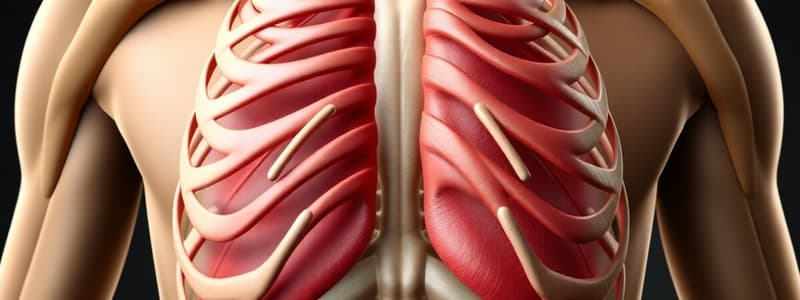
Intercostal Muscles
- The intercostal muscles are a group of muscles located between the ribs.
- There are three layers of intercostal muscles: external, internal, and innermost.
- The external intercostal muscles run diagonally downward and forward, assisting in inhalation.
- The internal intercostal muscles run diagonally downward and backward, helping in exhalation.
- The innermost intercostal muscles are the deepest layer, and their function is similar to the internal intercostal muscles.
- The sternocleidomastoid and pectoral muscles are associated structures, supporting movement of the ribs and thus aiding breathing.
Negative Pressure Breathing
- Breathing is driven by changing pressure in the lungs.
- Air moves from areas of higher pressure to lower pressure.
- Inhalation involves expanding the rib cage and pulling air into the lungs.
- Exhalation involves the rib cage getting smaller, decreasing lung volume and pushing air out.
- The diaphragm, a large muscle under the lungs, moves down during inhalation and up during exhalation.
Homeostasis in Breathing
- Homeostasis (balance) in breathing is maintained through a three-step process.
- Receptors detect changes in CO2 and O2 levels in the blood and cerebrospinal fluid (CSF).
- The control center, mainly the medulla oblongata, processes sensory input and regulates breathing rate.
- Effectors, like the diaphragm and intercostal muscles, are controlled to carry out the process of breathing.
Lung Volumes and Capacities
- Inspiratory Reserve Volume (IRV): The extra air that can be inhaled after a normal breath.
- Expiratory Reserve Volume (ERV): The extra air that can be exhaled after a normal breath.
- Tidal Volume (TV): The amount of air inhaled or exhaled in a normal breath.
- Residual Volume (RV): The amount of air remaining in the lungs after a maximum exhalation.
- Inspiratory Capacity (IC): The total amount of air that can be inhaled after a normal breathing cycle.
- Functional Residual Capacity (FRC): The total amount of air remaining in the lungs after a normal exhalation.
- Vital Capacity (VC): The largest amount of air that can be exhaled after a maximum inhalation.
- Total Lung Capacity (TLC): The total amount of air the lungs can hold. (TLC = VC + RV)
Resistance to Airflow
- Resistance to airflow affects breathing.
- Narrowed airways or other obstacles can hinder the movement of air in the lungs.
- Mucus, blood clots, or other blockage can cause resistance in vessels.
- Conditions like smoking can cause resistance.
Obstructive Lung Diseases
- Emphysema (air sac damage).
- Chronic bronchitis (mucus buildup).
- Bronchiolitis (small airway inflammation).
- Asthma (airway inflammation).
- These conditions can reduce the ability of the lungs to effectively exchange gases.
Compliance
- Compliance is how much the lungs can stretch and expand.
- This is affected by factors like the elasticity of lung tissue, and the presence of blockages during breathing.
Pulmonary Fibrosis
- Scar tissue buildup in the lungs.
- This condition reduces lung compliance, making breathing more difficult for patients.
Pneumothorax
- A collapsed lung.
- This is often treated with a chest tube.
Asbestos
- A known irritant that can lead to the development of pulmonary fibrosis.
Studying That Suits You
Use AI to generate personalized quizzes and flashcards to suit your learning preferences.


