Podcast
Questions and Answers
What is the main indication for using plain film radiography in the urinary tract?
What is the main indication for using plain film radiography in the urinary tract?
- To diagnose urinary tract infections
- To detect urinary tract pathology
- To evaluate renal tract calcifications (correct)
- To stage urological cancer
What has largely replaced intravenous excretion urography (IVU) as an imaging modality?
What has largely replaced intravenous excretion urography (IVU) as an imaging modality?
- Ultrasound (US), CT or MRI or a combination (correct)
- Micturating cystography and cystourethrography
- Plain film radiography
- Radionuclide imaging
What is a contraindication for intravenous excretion urography (IVU)?
What is a contraindication for intravenous excretion urography (IVU)?
- Recurrent urinary tract infection
- Loin pain
- Haematuria
- General contraindications to intravenous watersoluble contrast media (correct)
What is recommended for patients with impaired renal function undergoing IVU?
What is recommended for patients with impaired renal function undergoing IVU?
What is the sensitivity of plain films in detecting renal tract calcifications?
What is the sensitivity of plain films in detecting renal tract calcifications?
What is an alternative imaging modality in patients with contrast medium allergies?
What is an alternative imaging modality in patients with contrast medium allergies?
What is an indication for using IVU?
What is an indication for using IVU?
What is the technique for plain film radiography similar to?
What is the technique for plain film radiography similar to?
What is a contraindication for compression?
What is a contraindication for compression?
What is the purpose of the release film?
What is the purpose of the release film?
What is the purpose of the after micturition film?
What is the purpose of the after micturition film?
Why is tomography performed?
Why is tomography performed?
What is the purpose of a 35° posterior oblique of the kidneys, ureters or bladder?
What is the purpose of a 35° posterior oblique of the kidneys, ureters or bladder?
When is compression released?
When is compression released?
What is the purpose of delayed films?
What is the purpose of delayed films?
Why is a prone abdomen film taken?
Why is a prone abdomen film taken?
What is the typical dosage of low osmolar contrast material for an adult?
What is the typical dosage of low osmolar contrast material for an adult?
Why is dehydration not necessary before the examination?
Why is dehydration not necessary before the examination?
What is the purpose of the preliminary image?
What is the purpose of the preliminary image?
What is the approximate time between the injection of contrast material and the immediate film?
What is the approximate time between the injection of contrast material and the immediate film?
What is the purpose of the 5-min film?
What is the purpose of the 5-min film?
Why is a compression band applied after the 5-min film?
Why is a compression band applied after the 5-min film?
What is the typical location of the x-ray beam for the preliminary image?
What is the typical location of the x-ray beam for the preliminary image?
What is the purpose of the 35° posterior oblique views?
What is the purpose of the 35° posterior oblique views?
Flashcards are hidden until you start studying
Study Notes
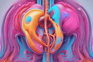
Methods of Imaging the Urinary Tract
- Plain radiography: used to evaluate renal tract calcifications
- Excretion urography (intravenous urogram [IVU]): less frequently used, replaced by US, CT, or MRI
- Ultrasound (US): used for imaging the urinary tract
- Computed tomography (CT): used for urological diagnosis and cancer staging, characterization of renal lesions, and CT urography
- Magnetic resonance imaging (MRI): used for characterization of renal lesions, prostate imaging, bladder imaging, and MR urography
- Micturating cystography and cystourethrography: used for imaging the urinary tract
- Ascending urethrography: used for imaging the urinary tract
- Retrograde pyeloureterography: used for imaging the urinary tract
- Percutaneous renal procedures: include biopsy, cyst puncture, antegrade pyelography, nephrostomy, and percutaneous nephrolithotomy
- Arteriography: used for imaging the urinary tract
- Venography: used for imaging the urinary tract
- Conduitogram: used for imaging the urinary tract
- Radionuclide imaging: used for static renography, dynamic renography, and radionuclide cystography
Plain Film Radiography
- Indications: evaluating renal tract calcifications
- Technique: as for preliminary films for excretion urography
Intravenous Excretion Urography (IVU or IVP)
- Indications: haematuria, renal colic, recurrent urinary tract infection, loins pain, and suspected urinary tract pathology
- Contraindications: general contraindications to intravenous watersoluble contrast media and ionizing radiation
- Contrast medium: low osmolar contrast material (LOCM) 300–370 mg I mL−1
- Patient preparation:
- No food for 5 hours prior to the examination
- Dehydration is not necessary and does not improve image quality
- No routine administration of bowel preparation
- Preliminary image: supine, full-length anterior posterior (AP) of the abdomen, in inspiration
- Technique:
- Venous access is established
- The gauge of the cannula/needle should allow the injection to be given rapidly as a bolus
- Images are taken at 10–14 seconds, 5 minutes, 10 minutes, and after micturition
- Compression is applied midway between the anterior superior iliac spines
- Compression is contraindicated in certain cases, such as after recent abdominal surgery or renal trauma
Studying That Suits You
Use AI to generate personalized quizzes and flashcards to suit your learning preferences.