Podcast
Questions and Answers
What is the primary purpose of the excitation filter in fluorescence microscopy?
What is the primary purpose of the excitation filter in fluorescence microscopy?
- To reflect all wavelengths of light equally
- To block all wavelengths except the emission light
- To allow only the desired excitation wavelength to pass (correct)
- To increase the brightness of the specimen
In fluorescence microscopy, what does the emission wavelength indicate?
In fluorescence microscopy, what does the emission wavelength indicate?
- The specific wavelength that excites the fluorescent molecule
- The longer, lower-energy wavelength emitted by the molecule (correct)
- The initial light source used for imaging
- The higher-energy light absorbed by the fluorescent molecule
Which of the following best describes the role of the dichroic mirror in fluorescence microscopy?
Which of the following best describes the role of the dichroic mirror in fluorescence microscopy?
- To increase the overall resolution of the image
- To absorb all wavelengths of light
- To enhance the contrast of fluorescently labeled structures
- To reflect lower wavelengths while allowing higher wavelengths to pass (correct)
What is a key advantage of using multi-color labeling in fluorescence microscopy?
What is a key advantage of using multi-color labeling in fluorescence microscopy?
What is the primary contribution of the Green Fluorescent Protein (GFP) to biological research?
What is the primary contribution of the Green Fluorescent Protein (GFP) to biological research?
Which color light is typically used to excite a fluorescent molecule that emits green light?
Which color light is typically used to excite a fluorescent molecule that emits green light?
Which variant of Green Fluorescent Protein (GFP) emits red light?
Which variant of Green Fluorescent Protein (GFP) emits red light?
What is the significance of using specific filters in fluorescence microscopy?
What is the significance of using specific filters in fluorescence microscopy?
Which organism is associated with the origin of Green Fluorescent Protein (GFP)?
Which organism is associated with the origin of Green Fluorescent Protein (GFP)?
Which characteristic of fluorescent microscopy enhances the visualization of structures?
Which characteristic of fluorescent microscopy enhances the visualization of structures?
What type of electron microscopy is used to visualize surface structures?
What type of electron microscopy is used to visualize surface structures?
Which radiation type is used in both TEM and SEM?
Which radiation type is used in both TEM and SEM?
What is the primary limitation of a light microscope compared to an electron microscope?
What is the primary limitation of a light microscope compared to an electron microscope?
In which type of microscopy do electrons pass through a thin specimen?
In which type of microscopy do electrons pass through a thin specimen?
What is the resolution of Transmission Electron Microscopy (TEM)?
What is the resolution of Transmission Electron Microscopy (TEM)?
Which application is best suited for Scanning Electron Microscopy (SEM)?
Which application is best suited for Scanning Electron Microscopy (SEM)?
Which of the following is a characteristic feature of Scanning Electron Microscopy (SEM)?
Which of the following is a characteristic feature of Scanning Electron Microscopy (SEM)?
What is a significant advantage of Electron Microscopy over Light Microscopy?
What is a significant advantage of Electron Microscopy over Light Microscopy?
Which microscopy type would you use to visualize viruses?
Which microscopy type would you use to visualize viruses?
What is the approximate magnification capability of Transmission Electron Microscopy (TEM)?
What is the approximate magnification capability of Transmission Electron Microscopy (TEM)?
Flashcards
Visible Spectrum
Visible Spectrum
The range of light visible to the human eye, spanning from violet (around 400 nm) to red (around 700 nm).
Fluorescence Microscopy
Fluorescence Microscopy
A technique that utilizes fluorescent molecules to visualize specific structures or components within a specimen. It involves exciting the molecules with specific wavelengths of light and capturing the emitted light at a longer wavelength.
Excitation Wavelength
Excitation Wavelength
The wavelength of light used to energize (excite) fluorescent molecules.
Emission Wavelength
Emission Wavelength
Signup and view all the flashcards
Fluorescence Microscope Configuration
Fluorescence Microscope Configuration
Signup and view all the flashcards
Dichroic Mirror
Dichroic Mirror
Signup and view all the flashcards
GFP (Green Fluorescent Protein)
GFP (Green Fluorescent Protein)
Signup and view all the flashcards
GFP Variants
GFP Variants
Signup and view all the flashcards
Applications of Fluorescence Microscopy
Applications of Fluorescence Microscopy
Signup and view all the flashcards
Why is fluorescence microscopy useful?
Why is fluorescence microscopy useful?
Signup and view all the flashcards
Electron Microscopy
Electron Microscopy
Signup and view all the flashcards
Transmission Electron Microscopy (TEM)
Transmission Electron Microscopy (TEM)
Signup and view all the flashcards
Scanning Electron Microscopy (SEM)
Scanning Electron Microscopy (SEM)
Signup and view all the flashcards
TEM image
TEM image
Signup and view all the flashcards
SEM image
SEM image
Signup and view all the flashcards
Resolution
Resolution
Signup and view all the flashcards
Magnification
Magnification
Signup and view all the flashcards
Vacuum environment
Vacuum environment
Signup and view all the flashcards
Specimen preparation
Specimen preparation
Signup and view all the flashcards
Study Notes
Imaging Living Organs - Lecture 13
- Overview: The lecture covers advancements in microscopy, focusing on fluorescence and electron microscopy. These techniques offer high sensitivity and resolution for examining biological specimens.
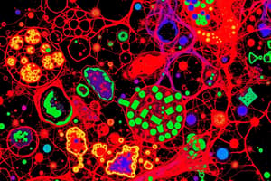
Fluorescence Microscopy
-
Key Concepts - Visible Spectrum:
- The human eye detects light from violet (~400 nm) to red (~700 nm).
- Fluorescence microscopy uses filters to isolate specific wavelengths.
-
How Fluorescence Works:
- Excitation Wavelength: Light of a particular wavelength excites a fluorescent molecule.
- Emission Wavelength: The excited molecule emits light at a longer (lower energy) wavelength. For example, exciting with blue light (~480 nm) results in green emission (~520 nm).
-
Fluorescence Microscope Configuration:
- Pre-treated specimen with fluorescent molecules.
- Excitation Filter: Allows only the desired excitation wavelength to pass (e.g., blue light).
- Dichroic Mirror: Reflects lower wavelengths (e.g., blue light) and allows higher wavelengths to pass. This is to separate light.
- Emission Filter: Blocks unwanted wavelengths to enhance contrast. The result is fluorescently labeled structures visible against a dark background.
-
Applications:
- Identifying specific proteins or structures in cells.
- Multi-color labeling - combining different fluorescent markers for complex imaging.
-
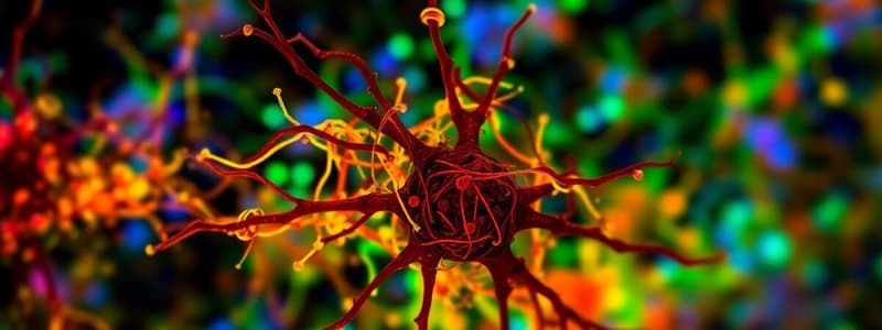
Green Fluorescent Protein (GFP):
- Originates from the bioluminescent jellyfish Aequorea victoria.
- Nobel Prize in Chemistry (2008) for researchers involved.
- Variants include Cyan (CFP), Yellow (YFP), and Red (RFP).
- Uses:
- Studying gene expression and protein localization.
- Labeling cells or structures in live organisms (e.g., zebrafish, mice, pigs).
-
Example Experiments:
- Nerve Regeneration: GFP-tagged nerve cells used to study spinal cord injury.
- Transgenic Animals: GFP used to monitor developmental and disease processes in zebrafish, mice, etc.
Electron Microscopy
-
Key Features:
- Uses electrons instead of light for imaging.
- Provides much higher resolution than light microscopy.
- Requires vacuum environments and extensive specimen preparation.
-
Types of Electron Microscopy:
-
Transmission Electron Microscopy (TEM):
- Electrons pass through a thin specimen.
- Dense areas block electrons (dark spots), while less dense areas allow electrons (light spots).
- Applications: Visualizing organelles (e.g., nucleus, mitochondria), viruses.
-
Scanning Electron Microscopy (SEM):
- Electrons scatter off the specimen's surface.
- Detector captures scattered electrons to create a 3D surface image.
- Applications: Detailed surface structures (e.g., blood cells, insects).
-
-
Comparison of TEM and SEM:
- Aspect | TEM | SEM
- Image Type | 2D | 3D
- Details | Internal structures | Surface structures
- Resolution | ~0.1 nm | ~10 nm
-
Microscope Comparison Table:
- Microscope Type | Magnification | Resolution | Radiation Used | Limitations
- Light | ~400X | ~200 nm | White light | Limited resolution
- SEM | ~20,000X | ~10 nm | Electron beam | Surface details only
Interactive Examples and Key Takeaways
-
Interactive Examples: SEM images (3D): Nerve cells, insects, Velcro hooks; TEM images (2D): Mitochondria, Golgi apparatus, viruses.
-
Key Takeaways:
- Fluorescence microscopy excels at labeling and dynamic cell studies.
- Electron microscopy provides high resolution for ultrastructural and surface details.
- Advancements in microscopy improve understanding of biological processes and disease mechanisms.
Studying That Suits You
Use AI to generate personalized quizzes and flashcards to suit your learning preferences.