Podcast
Questions and Answers
¿Cuál de los siguientes músculos no se inserta en el hueso hioides?
¿Cuál de los siguientes músculos no se inserta en el hueso hioides?
- Estilohioideo
- Trapecio (correct)
- Digástrico
- Hiogloso
El hueso hioides se articula directamente con otros huesos mediante ligamentos.
El hueso hioides se articula directamente con otros huesos mediante ligamentos.
False (B)
¿Qué estructuras anatómicas se relacionan lateralmente con el hueso hioides?
¿Qué estructuras anatómicas se relacionan lateralmente con el hueso hioides?
Arteria carótida, vena yugular interna y nervio hipogloso
El diafragma es inervado principalmente por el nervio ______, que se origina de las vértebras cervicales C3, C4 y C5.
El diafragma es inervado principalmente por el nervio ______, que se origina de las vértebras cervicales C3, C4 y C5.
Empareja las siguientes vértebras cervicales atípicas con sus características distintivas:
Empareja las siguientes vértebras cervicales atípicas con sus características distintivas:
¿Cuál de las siguientes opciones describe mejor la función principal de las articulaciones en el cuerpo humano?
¿Cuál de las siguientes opciones describe mejor la función principal de las articulaciones en el cuerpo humano?
La cifosis es una curvatura de la columna vertebral que presenta una convexidad anterior
La cifosis es una curvatura de la columna vertebral que presenta una convexidad anterior
¿Qué ligamento sostiene la apófisis odontoides en la articulación atlantoaxial?
¿Qué ligamento sostiene la apófisis odontoides en la articulación atlantoaxial?
Las articulaciones ______ también conocidas como articulaciones sinoviales, se caracterizan por permitir una amplia gama de movimientos.
Las articulaciones ______ también conocidas como articulaciones sinoviales, se caracterizan por permitir una amplia gama de movimientos.
Empareje los siguientes orificios del diafragma con las estructuras anatómicas principales que pasan a través de ellos:
Empareje los siguientes orificios del diafragma con las estructuras anatómicas principales que pasan a través de ellos:
¿Cuál de las siguientes opciones describe mejor el proceso facilitado por el movimiento de las costillas durante la respiración?
¿Cuál de las siguientes opciones describe mejor el proceso facilitado por el movimiento de las costillas durante la respiración?
Las articulaciones cigapofisiarias se forman por la unión de los procesos articulares inferiores de una vértebra con los procesos articulares inferiores de la vértebra adyacente.
Las articulaciones cigapofisiarias se forman por la unión de los procesos articulares inferiores de una vértebra con los procesos articulares inferiores de la vértebra adyacente.
¿Qué par craneal inerva el músculo trapecio?
¿Qué par craneal inerva el músculo trapecio?
El músculo serrato anterior se inserta en las costillas 2 a 9, y su función principal es mover la escápula hacia adelante y mantenerla pegada a la ______.
El músculo serrato anterior se inserta en las costillas 2 a 9, y su función principal es mover la escápula hacia adelante y mantenerla pegada a la ______.
Relacione los siguientes músculos principales con sus respectivas inserciones en las costillas:
Relacione los siguientes músculos principales con sus respectivas inserciones en las costillas:
Flashcards
¿Cuáles son los músculos relacionados con el hueso hioides?
¿Cuáles son los músculos relacionados con el hueso hioides?
Digástrico, milohioideo, genihioideo, estilohioideo, omohioideo, esternohioideo, tirohioideo y hiogloso.
¿A qué nivel vertebral se encuentra el hioides?
¿A qué nivel vertebral se encuentra el hioides?
El hueso hioides se ubica a nivel de la tercera vértebra cervical (C3).
¿Cuál es la forma del hueso hioides?
¿Cuál es la forma del hueso hioides?
Semicírculo con un cuerpo mediano arciforme y cuernos laterales (astas mayores y menores).
¿Cuál es la importancia clínica del hueso hioides?
¿Cuál es la importancia clínica del hueso hioides?
Signup and view all the flashcards
¿Cuál es la función del hueso hioides?
¿Cuál es la función del hueso hioides?
Signup and view all the flashcards
¿Dónde se ubica el hueso hioides?
¿Dónde se ubica el hueso hioides?
Signup and view all the flashcards
¿Por qué el hioides es único?
¿Por qué el hioides es único?
Signup and view all the flashcards
¿Con qué se relaciona lateralmente el hioides?
¿Con qué se relaciona lateralmente el hioides?
Signup and view all the flashcards
¿Con qué se relaciona superior e inferiormente el hioides?
¿Con qué se relaciona superior e inferiormente el hioides?
Signup and view all the flashcards
¿Cuáles son las dimensiones del hioides?
¿Cuáles son las dimensiones del hioides?
Signup and view all the flashcards
¿Dónde está ubicada la columna vertebral?
¿Dónde está ubicada la columna vertebral?
Signup and view all the flashcards
¿Qué curvatura tiene convexidad posterior?
¿Qué curvatura tiene convexidad posterior?
Signup and view all the flashcards
¿Qué es un proceso unciforme?
¿Qué es un proceso unciforme?
Signup and view all the flashcards
¿Sinovial =?
¿Sinovial =?
Signup and view all the flashcards
¿Qué vertebras se articulan con las costillas?
¿Qué vertebras se articulan con las costillas?
Signup and view all the flashcards
Study Notes
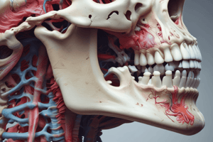
Hioides
- The muscular insertions of the hyoid bone include:
- Suprahioid muscles: digastric, mylohyoid, geniohyoid and stylohyoid.
- Infrahyoid muscles: omohyoid, sternohyoid and thyrohyoid.
- Tongue muscles: hyoglossus.
- The hyoid bone is located at the level of the third cervical vertebra (C3) in standard anatomical position.
- The hyoid bone has a semicircular shape, with an arciform median body and lateral horns( greater and lesser horns).
- Clinically, the hyoid bone is relevant in cases of cervical trauma, and in legal medicine, its fracture may be indicative of strangulation or neck injuries.
- The function of the hyoid bone is to serve as an insertion point for muscles of the neck and larynx, and it aids in swallowing, phonation, and tongue position.
- The hyoid bone is located transversely in the anterior and superior part of the neck, above the larynx, below the tongue, and below and behind the mandible.
- The hyoid bone is considered unique because it does not articulate directly with any other bone, being suspended exclusively by muscles and ligaments.
- Laterally, the hyoid bone is related to the carotid artery, internal jugular vein, and hypoglossal nerve.
- Superiorly, it is related to the base of the tongue and suprahyoid muscles
- Inferiorly, it is related to the thyroid cartilage and infrahyoid muscles.
- The hyoid bone has an average length of approximately 2.5 to 4 cm and a width of approximately 1 to 2 cm, and weighs approximately 3-4 grams, varying slightly among individuals.
Vertebral Column
- The vertebral column is located in the posterior part of the body.
- The vertebrae are divided into 5 sections or regions: cervical, dorsal, lumbar, sacral, and coccygeal.
- The natural anteroposterior curves of the spine are kyphosis and lordosis.
- The joint that unites the atlas and axis (C1 and C2) is called the atlanto-occipital joint.
- The articulation between the ribs and dorsal vertebrae is called costovertebral joints.
- The atlantoaxial articulation is a synovial joint of the pivot type.
- The movements performed by the vertebral column are flexion, extension, rotation, and lateral inclination.
- The bones united in the sacroiliac joint are the sacrum and right and left iliac bones.
- In relation to the cervical region, the occipital bone is found superiorly.
- Kyphosis is the curvature that corresponds to a posterior convexity.
Vertebrae in general
- Uncinate processes are characteristic processes of the cervical vertebrae.
- The apophyseal mass is the name given to the union of the lamina with the pedicle of the vertebra.
- The zygapophyseal joint is formed by the union of the superior articular processes of one vertebra with the inferior articular processes of another.
- The posterior aspect of the vertebral body defines the anterior face of the vertebral foramen.
- There are 23 intervertebral discs in the vertebral column: 6 cervical, 12 thoracic, and 5 lumbar.
- The transverse ligament holds the odontoid process in the atlantoaxial articulation.
- A characteristic shape of the body of a lumbar vertebra that differentiates it from the others is that it is kidney-shaped.
- The promontory is the superoanterior border of the pelvic face of the sacrum.
- Not all cervical vertebrae have bifid spinous processes; only C2 to C6 do.
- The two cylindrical prolongations that emerge from the coccyx articulate with the horns of the sacrum.
Cervical Vertebrae
- The three main functions of the cervical vertebrae are protection, support, and movement.
- A cervical vertebra weighs approximately 20 to 25 grams.
- The alar ligaments runs from the posterior aspect of the dens of the axis to the lateral margins of the foramen magnum.
- The vertebral foramen of the cervical vertebrae is triangular in shape.
- Eight cervical segments of the spinal cord are found in the cervical vertebrae.
- The joint between the facets of the C1 "atlas" vertebra and the occipital condyles is called the atlantooccipital joint.
- The three atypical cervical vertebrae are C1 "ATLAS," C2 "Axis," and C7, the prominent vertebra..
- Cervical vertebrae completely ossify at 25 years of age.
- The atlas (C1) is the cervical vertebra that does not have a body.
- The atlantooccipital joint allows extension and flexion movements.
Articulations
- Synovial joints are also known as diarthrosis.
- The function of joints in the human body is to allow movement of the skeleton, connecting bones to each other and providing stability to the body.
- Joints are classified according to their structure as synovial, cartilaginous, and fibrous.
- Joints can perform at least three types of movements: flexion, extension, abduction, and rotation.
- The components of synovial joints are the articular capsule, synovial membrane, synovial fluid, articular cartilage, and ligaments.
- A flat joint allows displacement movements.
- An example of a hinge joint is the humeroulnar joint.
- The flat joint is also known as arthrodia.
- Cartilaginous joints are synchondrosis and symphysis.
- Two examples of injuries due to overuse of joints are bursitis and tendinitis.
Costovertebral Joint
- Thoracic vertebrae are related in the costovertebral joints.
- The tubercle of the rib and the transverse process of the vertebra are connected by the costotransverse joint
- In the costovertebral joints, the ribs connect to a single vertebra at the level of thoracic vertebrae T1, T2, and T3.
- The radiate ligament corresponds to the costovertebral joint.
- The ligaments related to the costotransverse joint are the middle costotransverse ligament, the superior costotransverse ligament, and the lateral costovertebral ligament.
- Both types of costovertebral joints are innervated by the lateral branches of the posterior branches of the spinal nerves C8-T11.
- Respiration facilitates the movement of the ribs during inspiration and expiration.
- Costovertebral joints allow pump-handle movement and bucket-handle movement.
- The lower ribs, the last five ribs, perform the bucket-handle movement.
- During inhalation, the superior ribs perform an upward and outward movement.
Pectoral Muscles
- The origin of the pectoralis minor is located on the lateral surface and superior border of the 3rd, 4th, and 5th ribs.
- The insertion of the pectoralis minor is on the anterior half of the medial border of the coracoid process.
- The pectoralis minor is located in the anterior portion of the thorax or chest wall, positioned just inferior to the pectoralis major muscle.
- When its fixed point is on the ribs, the pectoralis minor produces the protraction of the coracoid process & depression of the scapula.
- When its point of support is the coracoid process, the pectoralis minor elevates the ribs & serves as an inspiratory muscle.
- The portions that constitute the pectoralis major are clavicular, sternocostal, and abdominal.
- The tendon of termination is inserted on the lateral lip of the intertubercular groove of the humerus.
- The lateral pectoral nerve, which originates from the lateral cord of the brachial plexus (C5, C6 & C7), innervates the pectoralis major.
- The pectoral major adducts the arm & simultaneously brings the shoulder forward. Also, it contributes to shoulder medial rotation & it elevates the trunk in climbing action.
- In Poland's syndrome, congenital anomalies of the pectoral muscle can be observed, which are characterized by the unilateral absence of the pectoralis major.
Diaphragm
- "Diaphragm" comes from the Greek word meaning "divider."
- The diaphragm has three portions:
- Sternal portion: It is inserted into the posterior aspect of the sternum.
- Costal portion: It joins the last six ribs and their costal cartilages.
- Lumbar portion: It anchors to the lumbar vertebrae through the right and left crura.
- The diaphragm is primarily innervated by the phrenic nerve (C3, C4, and C5), which controls its contraction, and also receives fibers from the intercostal and vagus nerves.
- The diaphragm has three main openings:
- Aortic hiatus (T12): Allows passage of the aorta, thoracic duct, and azygos vein.
- Esophageal hiatus (T10): Passage for the esophagus and vagus nerves.
- Vena cava foramen (T8): Passage for the inferior vena cava and branches of the right phrenic nerve.
- The diaphragm is supplied by:
- Main arteries: Superior phrenic (from the thoracic aorta) and inferior phrenic (from the abdominal aorta, main ones).
- Secondary arteries: Musculophrenic and pericardiophrenic (from the internal thoracic) and branches of the intercostals (for the periphery).
- The right hemidiaphragm is higher due to the presence of the liver beneath it, while the left hemidiaphragm is lower because the stomach and spleen are beneath it, and the esophageal hiatus and aorta pass more through the left side.
- The diaphram's primary role is to enable breathing. It contracts to increase the volume of the thoracic cavity, facilitating inspiration, involving coughing, vomiting, and defecation.
- During inspiration, the diaphragm contracts and descends, expanding the lungs; during expiration, it relaxes and ascends, expelling the air.
- The diaphragm makes contact with the lungs and heart on the upper part and with the liver, stomach, spleen, and kidneys on the lower part.
- Common pathologies affecting the diaphragm include diaphragmatic hernia, diaphragmatic paralysis, diaphragmatic spasm (persistent hiccups), and diaphragmatic rupture due to trauma.
Trapezius, latissimus dorsi & Rhomboids
- These muscles connect the axial skeleton with the appendicular skeleton.
- The clavicular insertion of the trapezius is on the lateral third of the posterior border of the clavicle.
- The trapezius covers vertebrae from C1 to T12.
- The trapezius performs elevation, flexion, extension, and rotation.
- The trapezius is innervated from the XI cranial nerve (accessory nerve).
- The trapezius is supplied by the dorsal scapular artery.
- The rhomboids cover vertebrae from C7 to T5.
- The rhomboids insert on the medial border of the scapula.
- Two insertions of the latissimus dorsi are the floor of the intertubercular sulcus of the humerus and the spinous processes of the last 6 or 7 thoracic vertebrae and the 5 lumbar vertebrae, median crest of the sacrum or posterior third of the external lip of the iliac crest or posterolateral face of the last 4 ribs.
- The latissimus dorsi receives irrigation from the thoracodorsal artery in its axillary region.
Serratus Anterior and Posterior
- The serratus anterior is located on the side of the chest, and its main function is to move the scapula forward and keep it attached to the thoracic cage.
- The serratus anterior is inserted on ribs 2 to 9, although sometimes also on the first rib.
- The long thoracic nerve, which comes from the roots C5, C6 and C7 of the brachial plexus, innervates the serratus anterior.
- The main arteries that irrigate the serratus anterior are the lateral thoracic artery, the posterior intercostal arteries, and the thoracodorsal artery.
- The serratus posterior superior is located from the vertebrae C7 to T3 to the ribs 2, 3, and 4.
- The main function of the serratus posterior superior is to elevate the upper ribs, facilitating inspiration.
- The posterior branches of the intercostal nerves from the 2nd to the 5th rib innervates serratus posterior superior
- The serratus posterior inferior is located in the lower part of the back, between the vertebrae T11 to L2 and the ribs 9 to 12.
- The main function of the serratus posterior inferior is to lower the lower ribs and stabilize the lumbar zone.
- The arteries that irrigate serratus posterior inferior are the posterior intercostal arteries, the subcostal artery, and branches of the abdominal aorta artery.
Studying That Suits You
Use AI to generate personalized quizzes and flashcards to suit your learning preferences.