Podcast
Questions and Answers
During embryonic development of cardiac muscle, what crucial process ensures the structural and functional integrity of the developing heart tissue?
During embryonic development of cardiac muscle, what crucial process ensures the structural and functional integrity of the developing heart tissue?
- The random alignment of mesenchymal cells without any specific organization.
- The rapid differentiation of cardiac muscle cells without the formation of cell-to-cell connections.
- The isolation of cardiac muscle cells to prevent premature electrical coupling.
- The formation of complex junctions between interdigitating processes of cardiac muscle cells. (correct)
Given the distinct structural and functional demands placed on the atria and ventricles, how do the T-tubule characteristics in these chambers differ to optimize cardiac performance?
Given the distinct structural and functional demands placed on the atria and ventricles, how do the T-tubule characteristics in these chambers differ to optimize cardiac performance?
- Ventricular T-tubules are smaller or entirely absent to minimize the risk of arrhythmias, whereas atrial T-tubules are larger for increased calcium influx.
- Both atrial and ventricular T-tubules are equally developed to ensure uniform distribution of electrical signals.
- Atrial T-tubules are smaller or entirely absent, while ventricular T-tubules are well-developed and penetrate deeply into the sarcoplasm. (correct)
- Atrial T-tubules are well-developed and larger to enhance the force of contraction, while ventricular T-tubules are reduced to maintain a rapid heart rate.
Cardiac muscle cells are unique due to their ability to coordinate contraction through specialized structures. What role do gap junctions play in this process?
Cardiac muscle cells are unique due to their ability to coordinate contraction through specialized structures. What role do gap junctions play in this process?
- They insulate cardiac muscle cells, ensuring that electrical signals are confined to individual cells.
- They facilitate the rapid spread of contractile stimuli by allowing the passage of ions between cells. (correct)
- They provide structural support, preventing cardiac muscle cells from separating during contraction.
- They regulate the entry of calcium ions into the cells, which is essential for muscle contraction.
Which structural feature of cardiac muscle cells is most directly responsible for the heart's ability to function as a syncytium, enabling rapid and coordinated contraction?
Which structural feature of cardiac muscle cells is most directly responsible for the heart's ability to function as a syncytium, enabling rapid and coordinated contraction?
How does the arrangement of myofibrils within cardiac muscle cells contribute to the functional demands of the heart, particularly in comparison to skeletal muscle?
How does the arrangement of myofibrils within cardiac muscle cells contribute to the functional demands of the heart, particularly in comparison to skeletal muscle?
In cardiac muscle cells, T-tubules and the sarcoplasmic reticulum form dyads, which are critical for excitation-contraction coupling. How does the structure of these dyads enhance cardiac muscle function?
In cardiac muscle cells, T-tubules and the sarcoplasmic reticulum form dyads, which are critical for excitation-contraction coupling. How does the structure of these dyads enhance cardiac muscle function?
Intercalated discs are complex structures that connect adjacent cardiac muscle cells. How do the transverse portions of these discs contribute to the mechanical stability and force transmission in the heart?
Intercalated discs are complex structures that connect adjacent cardiac muscle cells. How do the transverse portions of these discs contribute to the mechanical stability and force transmission in the heart?
Within the intercalated discs of cardiac muscle cells, the lateral portions are crucial for rapid electrical communication. What specific structure within these regions facilitates this function?
Within the intercalated discs of cardiac muscle cells, the lateral portions are crucial for rapid electrical communication. What specific structure within these regions facilitates this function?
Cardiac muscle contraction is intrinsically regulated but also influenced by external factors. How does the autonomic nervous system (ANS) fine-tune cardiac muscle contraction?
Cardiac muscle contraction is intrinsically regulated but also influenced by external factors. How does the autonomic nervous system (ANS) fine-tune cardiac muscle contraction?
In smooth muscle cells, which structural component is responsible for both anchoring thin filaments and transmitting contractile forces to adjacent cells?
In smooth muscle cells, which structural component is responsible for both anchoring thin filaments and transmitting contractile forces to adjacent cells?
How do the unique structural characteristics of smooth muscle cells, such as the lack of T-tubules and the presence of caveolae, influence their contractile properties and response to stimuli?
How do the unique structural characteristics of smooth muscle cells, such as the lack of T-tubules and the presence of caveolae, influence their contractile properties and response to stimuli?
How does the ratio of thin to thick filaments in smooth muscle cells (15:1) affect their contractile mechanism, compared to skeletal muscle (6:1)?
How does the ratio of thin to thick filaments in smooth muscle cells (15:1) affect their contractile mechanism, compared to skeletal muscle (6:1)?
Smooth muscle contraction is regulated by various mechanisms, including calcium-calmodulin interactions and myosin light-chain kinase (MLCK). What critical step is initiated by the calcium-calmodulin complex in this process?
Smooth muscle contraction is regulated by various mechanisms, including calcium-calmodulin interactions and myosin light-chain kinase (MLCK). What critical step is initiated by the calcium-calmodulin complex in this process?
How do the distinct mechanisms of smooth muscle contraction contribute to its ability to maintain prolonged contractions compared to skeletal muscle?
How do the distinct mechanisms of smooth muscle contraction contribute to its ability to maintain prolonged contractions compared to skeletal muscle?
Muscle tissue exhibits varied capacities for regeneration. Which statement accurately compares the regeneration capabilities of skeletal, cardiac, and smooth muscle?
Muscle tissue exhibits varied capacities for regeneration. Which statement accurately compares the regeneration capabilities of skeletal, cardiac, and smooth muscle?
Which of the following best describes the arrangement of smooth muscle layers in the walls of the digestive tract and how this arrangement contributes to its function?
Which of the following best describes the arrangement of smooth muscle layers in the walls of the digestive tract and how this arrangement contributes to its function?
How does the involuntary nature of smooth muscle contraction contribute to the maintenance of homeostasis in the body?
How does the involuntary nature of smooth muscle contraction contribute to the maintenance of homeostasis in the body?
In smooth muscle, what is the functional role of intermediate filaments and how do they interact with dense bodies to facilitate contraction?
In smooth muscle, what is the functional role of intermediate filaments and how do they interact with dense bodies to facilitate contraction?
Smooth muscle lacks the troponin complex found in skeletal and cardiac muscle. How does this absence influence calcium's role in initiating contraction?
Smooth muscle lacks the troponin complex found in skeletal and cardiac muscle. How does this absence influence calcium's role in initiating contraction?
Which of the following statements accurately contrasts the speed and control mechanisms of contraction between smooth and skeletal muscle tissues?
Which of the following statements accurately contrasts the speed and control mechanisms of contraction between smooth and skeletal muscle tissues?
How do the structural differences in T-tubules between atrial and ventricular cardiac muscle cells directly impact the force and speed of contraction in these chambers?
How do the structural differences in T-tubules between atrial and ventricular cardiac muscle cells directly impact the force and speed of contraction in these chambers?
If a researcher is investigating the myogenic properties of cardiac muscle tissue, which specific characteristic would they focus on to understand the intrinsic rhythmicity of heart muscle cells?
If a researcher is investigating the myogenic properties of cardiac muscle tissue, which specific characteristic would they focus on to understand the intrinsic rhythmicity of heart muscle cells?
How does the unique arrangement of myofilaments and dense bodies in smooth muscle cells enable them to sustain prolonged contractions without fatigue, unlike skeletal muscle?
How does the unique arrangement of myofilaments and dense bodies in smooth muscle cells enable them to sustain prolonged contractions without fatigue, unlike skeletal muscle?
Consider a scenario where the intermediate filaments in smooth muscle cells are compromised. What immediate effect would this have on the cell's function?
Consider a scenario where the intermediate filaments in smooth muscle cells are compromised. What immediate effect would this have on the cell's function?
How do the distinct regulatory mechanisms of smooth muscle contraction, involving calcium-calmodulin interactions and myosin light-chain kinase (MLCK), enable smooth muscle to maintain prolonged contractions with minimal energy expenditure, compared to skeletal muscle?
How do the distinct regulatory mechanisms of smooth muscle contraction, involving calcium-calmodulin interactions and myosin light-chain kinase (MLCK), enable smooth muscle to maintain prolonged contractions with minimal energy expenditure, compared to skeletal muscle?
Flashcards
What is cardiac muscle?
What is cardiac muscle?
Found in the heart (myocardium) and small areas in the walls of large blood vessels attached to the heart.
Cardiac Muscle: Embryonic Development
Cardiac Muscle: Embryonic Development
Mesenchymal cells arrange in chainlike arrays around the primitive heart tube, forming complex junctions between interdigitating processes.
Cardiac Muscle in Ventricles
Cardiac Muscle in Ventricles
Thicker muscle, well-developed T-tubules with larger lumens that penetrate the sarcoplasm near the myofibrils' Z discs.
Cardiac Muscle in Atria
Cardiac Muscle in Atria
Signup and view all the flashcards
Cardiac Muscle Cells: Structure
Cardiac Muscle Cells: Structure
Signup and view all the flashcards
Cardiac Muscle Cells: Function
Cardiac Muscle Cells: Function
Signup and view all the flashcards
Cardiac Muscle: Mitochondria
Cardiac Muscle: Mitochondria
Signup and view all the flashcards
Cardiac Muscle: Sarcoplasm
Cardiac Muscle: Sarcoplasm
Signup and view all the flashcards
Cardiac Muscle: Myofibrils
Cardiac Muscle: Myofibrils
Signup and view all the flashcards
Cardiac Muscle: T-tubules
Cardiac Muscle: T-tubules
Signup and view all the flashcards
Intercalated Discs Function
Intercalated Discs Function
Signup and view all the flashcards
Intercalated Discs: Fascia Adherens
Intercalated Discs: Fascia Adherens
Signup and view all the flashcards
Intercalated Discs: Desmosomes
Intercalated Discs: Desmosomes
Signup and view all the flashcards
Intercalated Discs: Gap Junctions
Intercalated Discs: Gap Junctions
Signup and view all the flashcards
Cardiac Muscle Contraction: Control
Cardiac Muscle Contraction: Control
Signup and view all the flashcards
Cardiac Muscle Contraction: Regulation
Cardiac Muscle Contraction: Regulation
Signup and view all the flashcards
What is smooth muscle?
What is smooth muscle?
Signup and view all the flashcards
Smooth Muscle: Structure
Smooth Muscle: Structure
Signup and view all the flashcards
Smooth Muscle Cells: Structure
Smooth Muscle Cells: Structure
Signup and view all the flashcards
Smooth Muscle Cells: Function
Smooth Muscle Cells: Function
Signup and view all the flashcards
Smooth Muscle: Sarcolemma
Smooth Muscle: Sarcolemma
Signup and view all the flashcards
Smooth Muscle: Sarcoplasmic Reticulum
Smooth Muscle: Sarcoplasmic Reticulum
Signup and view all the flashcards
Smooth Muscle: Dense Bodies
Smooth Muscle: Dense Bodies
Signup and view all the flashcards
Smooth Muscle: Intermediate Filaments
Smooth Muscle: Intermediate Filaments
Signup and view all the flashcards
Smooth Muscle: Myofilaments
Smooth Muscle: Myofilaments
Signup and view all the flashcards
Smooth Muscle: Thin Filaments
Smooth Muscle: Thin Filaments
Signup and view all the flashcards
Smooth Muscle: Thick Filaments
Smooth Muscle: Thick Filaments
Signup and view all the flashcards
Smooth Muscle Contraction: Cell Shape
Smooth Muscle Contraction: Cell Shape
Signup and view all the flashcards
Smooth Muscle Contraction: Calcium
Smooth Muscle Contraction: Calcium
Signup and view all the flashcards
Smooth Muscle contraction: control
Smooth Muscle contraction: control
Signup and view all the flashcards
Skeletal Muscle Regeneration
Skeletal Muscle Regeneration
Signup and view all the flashcards
Cardiac Muscle Regeneration
Cardiac Muscle Regeneration
Signup and view all the flashcards
Smooth Muscle Regeneration
Smooth Muscle Regeneration
Signup and view all the flashcards
Study Notes
- Human Histology Lecture is MT120225
- The lecture covers Unit 3: Muscular Tissue
Course Content
- Overview and Classification of Muscle
- Skeletal Muscle
- Cardiac Muscle
- Smooth Muscle
- Muscle Regeneration
- Summary
Unit Intended Learning Outcome
- Students will be able to identify the different types of muscular tissues and their functions by the end of this unit
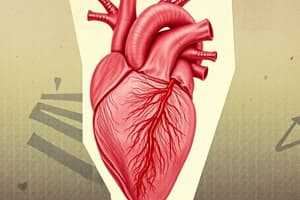
Cardiac Muscle
- Cardiac muscle is found in the heart (myocardium) and small areas in the walls of large blood vessels attached to the heart.
- Mesenchymal cells arrange in chainlike arrays around primitive heart tube.
- Complex junctions form between cardiac muscle cells with inter-digitating processes.
Organization of Cardiac Muscle
- Striations and Intercalated discs are visible.
- Nucleus and cardiac muscle cells are intact.
- Myofibrils are sectioned.
- Also visible are Mitochondria, entrace to T-tubule, Sarcolemma
- Sarcoplasmic reticulum
Cardiac Muscle in Ventricles and Atria
- Ventricle muscle is thicker.
- T-tubules are well developed, with larger lumens to penetrate sarcoplasm near myofibrils' Z discs.
- Atria T-tubules are smaller or absent.
Cardiac Muscle Cells Structure
- Cylindrical and Striated
- Length can be 85-120 um and diameter 15-30 um
- Splits longitudinally and can branch
- Contains one or two ovoid nuclei that are centrally located
Cardiac Muscle Cells Function
- Contraction
- Pacemaker
- Impulse propagation
Mitochondria in Cardiac Muscle Cells
- Occupies up to 40% of cell volume
- Numerous and Larger
Sarcoplasm in Cardiac Muscle Cells
- Is abundant and filled with myofibril
Myofibrils in Cardiac Muscle Cells
- Sparsely and Less organized
- Does consist of sarcomeres
- Does contain thick and thin filaments
- Few differences in protein composition
T-tubules in Cardiac Muscle Cells
- Invagination of the sarcolemma
- Surrounds the Z-lines
- Lumens are bigger
Sarcoplasmic Reticulum in Cardiac Muscle Cells
- Forms Dyads
- One terminal cisternae
- One t-tubule
Intercalated Discs in Cardiac Muscle Cells
- Cell boundaries
- Functions include enhancing molecular and electrical connections and conducting action potentials
- Specialized junctional complexes
- Two regions: transverse and lateral
Intercalated Disc Transverse Portion
- Serves to anchor the myofibrils and keep cells together
- The Fascia Adherens forms broad intercellular junctions
- Thin filaments of sarcomeres attach together.
- Desmosomes have strong intermediate filament coupling between adjacent cells
Intercalated Disc Lateral Portion
- Allows instantaneous spread of contractile stimuli from one cell to another thru Gap junctions
- Provides ionic continuity
Cardiac Muscle Contraction
- Intrinsic and spontaneous
- Supplied with efferent fibers by motor neurons of sympathetic and parasympathetic division of ANS
- Neurotransmitters reach the surface of smooth muscle cells through diffusion and propagate through Gap Junction
- Regulates the rate and strength of muscle contraction
Difference in Cardiac Muscle Contraction
- Calcium ions come from sarcoplasmic reticulum and outside the cell
- Contracts without neural stimulation
- Impulse is generated by Sinoatrial Node which consists of Purkinje fibers
Smooth Muscle
- Smooth muscle comprises the muscular component of walls of visceral organs and blood vessels, and is widely distributed in the body.
Organization of Smooth Muscle
- Caveolae and Dense bodies
- Made of Cytoskeleton, nucleus, filaments, and mitochondria
Smooth Muscle Cells Structure
- Fusiform shape
- Length 20-500 um
- Diameter 2-10 um
- oval nucleus located in the thick part of the cell
Smooth Muscle Cells Function
- Contraction
- Synthesis of proteoglycan, elastin and precursors of collagen fibers
Structure of Sarcolemma in Smooth Muscle Cells
- Does not form T-tubules
- Has Caveolae which are numerous small invaginations containing ion channels
Sarcoplasmic Reticulum Structure in Smooth Muscle Cells
- Poorly developed
Dense Body Structure in Smooth Muscle Cells
- Contains a-actinin
- Corresponds to the Z discs of the sarcomeres of striated muscles
- Attached to sarcolemma or in the intermediate filaments.
Intermediate Filaments Structure
- Made of Desmin
Myofilaments
- Thin and thick filaments (ratio 15:1) is much higher than in skeletal muscle (6:1)
- Are Arranged obliquely
Organization of Smooth Muscle Cell Myofilaments
- Thin filaments surround thick filaments
- Anchored on dense bodies
- Does not have troponin
- Thick filaments scattered all over the sarcoplasm with less Myosin
- Fewer cross bridges
Smooth Muscle Cells - Calmodulin and Myosin light-chain kinase
- Shortening of cells in all directions
- Dense bodies are not arranged in a straight line
- Calcium ions mostly come from extracellular substance
Contractions are stimulated by ANS or hormones
- Contractions are inherently contractile
- Have Interstitial cell of Cajal (small and large intestines)
- Innervated by efferent fibers via motor neurons of the sympathetic and parasympathetic division of the ANS
- Neurotransmitters diffusing to the smooth muscle cell and propagating through gap junctions
Repair and Regeneration of Muscle Tissue
- Skeletal Muscle: Limited, mainly involving satellite cells
- Cardiac Muscle: Very poor, lacks satellite cells
- Smooth Muscle: Rapid, involving mitotic activity of muscle cells
Studying That Suits You
Use AI to generate personalized quizzes and flashcards to suit your learning preferences.