Podcast
Questions and Answers
What initiates the transition from cartilage to bone during skeletal development in Week 7?
What initiates the transition from cartilage to bone during skeletal development in Week 7?
- Differentiation of mesenchymal cells into muscle cells
- Growth of embryonic length
- Development of ossification centers (correct)
- Formation of the cartilaginous skeleton
Which of the following structures first shows signs of ossification in the upper limb during Week 7?
Which of the following structures first shows signs of ossification in the upper limb during Week 7?
- Scapula
- Radius
- Humerus (correct)
- Ulna
What is the purpose of mesenchymal cells during the formation of the cartilaginous skeleton?
What is the purpose of mesenchymal cells during the formation of the cartilaginous skeleton?
- To form muscle tissue that surrounds the skeleton
- To directly ossify into bones
- To condense and create a cartilage model (correct)
- To differentiate into chondrocytes only
During Week 7, what forms from the differentiation of chondrocytes?
During Week 7, what forms from the differentiation of chondrocytes?
What does the Crown-Rump Length (CRL) measure during Week 7?
What does the Crown-Rump Length (CRL) measure during Week 7?
Which event marks the primary phase of skeletal maturation during Week 7?
Which event marks the primary phase of skeletal maturation during Week 7?
What process will the cartilage models undergo in accordance with skeletal development?
What process will the cartilage models undergo in accordance with skeletal development?
By the end of Week 7, the embryo's length progresses to approximately how many millimeters?
By the end of Week 7, the embryo's length progresses to approximately how many millimeters?
What is the primary change occurring to the auricular hillocks by Week 7?
What is the primary change occurring to the auricular hillocks by Week 7?
By Week 7, what is notable about the external auditory canal?
By Week 7, what is notable about the external auditory canal?
Which significant rotation occurs in the midgut loop during Week 6?
Which significant rotation occurs in the midgut loop during Week 6?
What is the main impact of the rotation of the midgut loop?
What is the main impact of the rotation of the midgut loop?
What key process contributes to brain complexity during Week 7?
What key process contributes to brain complexity during Week 7?
During Week 7, which part of the brain is primarily associated with rapid expansion?
During Week 7, which part of the brain is primarily associated with rapid expansion?
What significant cardiovascular development occurs in Week 7?
What significant cardiovascular development occurs in Week 7?
By Week 7, what is the primary focus regarding the development of heart chambers?
By Week 7, what is the primary focus regarding the development of heart chambers?
What significant change occurs in the upper limbs by the end of Week 7?
What significant change occurs in the upper limbs by the end of Week 7?
How do toe rays begin to appear during Week 7 in lower limb development?
How do toe rays begin to appear during Week 7 in lower limb development?
What is the role of apoptosis in toe development during Week 7?
What is the role of apoptosis in toe development during Week 7?
What does the enlargement of the eyes during Week 7 indicate?
What does the enlargement of the eyes during Week 7 indicate?
What signifies the first functional aspect of the retina during Week 7?
What signifies the first functional aspect of the retina during Week 7?
What are auricular hillocks and when do they begin to form?
What are auricular hillocks and when do they begin to form?
What happens to the shape of the eye during Week 7?
What happens to the shape of the eye during Week 7?
Which feature of the face becomes more defined by the end of Week 7?
Which feature of the face becomes more defined by the end of Week 7?
What is the significance of mesenchymal condensation in the formation of the cartilaginous skeleton?
What is the significance of mesenchymal condensation in the formation of the cartilaginous skeleton?
During which week does the mesenchymal condensation occur for limb development?
During which week does the mesenchymal condensation occur for limb development?
How do mesenchymal cells contribute to the skeletal development process?
How do mesenchymal cells contribute to the skeletal development process?
What role does apoptosis play in the development of the fingers during limb differentiation?
What role does apoptosis play in the development of the fingers during limb differentiation?
What is the direct precursor to endochondral ossification?
What is the direct precursor to endochondral ossification?
What physical feature marks the formation of fingers in Week 7?
What physical feature marks the formation of fingers in Week 7?
Which bones begin to take shape through the differentiation of mesenchymal cells into chondrocytes?
Which bones begin to take shape through the differentiation of mesenchymal cells into chondrocytes?
What allows the structured growth of limb bones during endochondral ossification?
What allows the structured growth of limb bones during endochondral ossification?
Flashcards
Mesenchymal Condensation
Mesenchymal Condensation
Mesenchymal cells gather and condense into distinct shapes that will later become bones.
Differentiation into Chondrocytes
Differentiation into Chondrocytes
Mesenchymal cells transform into chondrocytes, which produce the cartilage matrix. These cells are found in the cartilage model of bones such as the humerus, radius, and ulna.
Precursor to Endochondral Ossification
Precursor to Endochondral Ossification
The cartilage formed in the early stages serves as a template for bone development.
Endochondral Ossification
Endochondral Ossification
Signup and view all the flashcards
Digital Rays
Digital Rays
Signup and view all the flashcards
Apoptosis in Interdigital Regions
Apoptosis in Interdigital Regions
Signup and view all the flashcards
Separation of Fingers
Separation of Fingers
Signup and view all the flashcards
Toe Rays in Lower Limbs
Toe Rays in Lower Limbs
Signup and view all the flashcards
Finger Separation
Finger Separation
Signup and view all the flashcards
Finger Formation
Finger Formation
Signup and view all the flashcards
Toe Rays
Toe Rays
Signup and view all the flashcards
Toe Development
Toe Development
Signup and view all the flashcards
Prominent Eyes
Prominent Eyes
Signup and view all the flashcards
Ear Formation
Ear Formation
Signup and view all the flashcards
Eyelid Development
Eyelid Development
Signup and view all the flashcards
Eye Shape
Eye Shape
Signup and view all the flashcards
Ossification
Ossification
Signup and view all the flashcards
Primary Ossification Centers
Primary Ossification Centers
Signup and view all the flashcards
Cartilaginous Skeleton
Cartilaginous Skeleton
Signup and view all the flashcards
Chondrocytes
Chondrocytes
Signup and view all the flashcards
Crown-Rump Length (CRL)
Crown-Rump Length (CRL)
Signup and view all the flashcards
Limb Differentiation
Limb Differentiation
Signup and view all the flashcards
Facial Structure Differentiation
Facial Structure Differentiation
Signup and view all the flashcards
Week 7 of Embryonic Development
Week 7 of Embryonic Development
Signup and view all the flashcards
External Ear Formation
External Ear Formation
Signup and view all the flashcards
Ear Canal Development (Early Stages)
Ear Canal Development (Early Stages)
Signup and view all the flashcards
Midgut Loop Rotation
Midgut Loop Rotation
Signup and view all the flashcards
Impact of Midgut Rotation
Impact of Midgut Rotation
Signup and view all the flashcards
Major Brain Divisions
Major Brain Divisions
Signup and view all the flashcards
Brain Development: Key Processes
Brain Development: Key Processes
Signup and view all the flashcards
Cardiovascular Development in Week 7
Cardiovascular Development in Week 7
Signup and view all the flashcards
Brain Growth and Facial Structure
Brain Growth and Facial Structure
Signup and view all the flashcards
Study Notes
Human Embryology - Week 7
- Learning Outcomes: Students will explain organ refinement and maturation, describe cartilage skeleton formation, elaborate growth milestones (CRL), and analyze limb and facial structure differentiation. CRL at week 7 is approximately 15-20mm.
Skeletal Development: Week 7
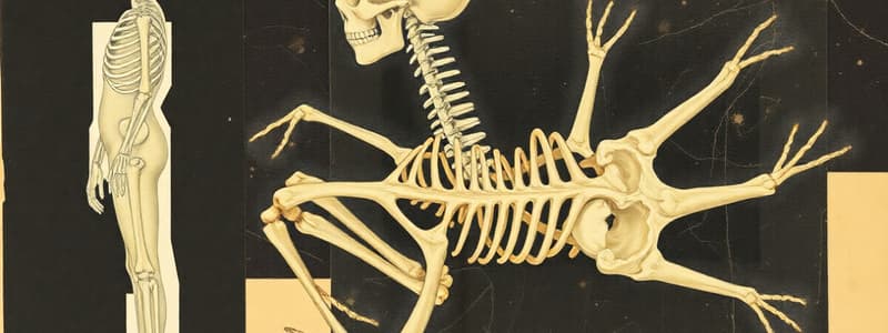
- Overview: Cartilage skeleton formation is the groundwork for future bone development. Ossification begins in specific regions.
- Ossification Centers Appear: Early primary centers develop in the humerus, ulna, and radius. This marks the transition from cartilage to bone, providing skeletal structure.
- Formation of Cartilaginous Skeleton: Mesenchymal cells condense to create cartilage models in developing limbs. These cartilage models are precursors to bones that will undergo endochondral ossification. Chondrocytes (cartilage cells) form the skeletal structure, which will later be replaced by bone.
Ossification Centers in Upper Limb Bones
- Early Primary Ossification Centers: The process starts in the humerus, ulna, and radius (initially cartilaginous), creating primary ossification centers by week 7. The humerus is the first to show ossification, marking a key point in skeletal development.
- From Cartilage to Bone: Mesenchymal cells differentiate into chondrocytes (cartilage-forming cells). These cells multiply and mature, forming the cartilage scaffold that later ossifies into bone. The formation of primary ossification centers kickstarts the gradual replacement of cartilage with bone (endochondral ossification).
Formation of Cartilaginous Skeleton in Week 7
- Mesenchymal Condensation: Mesenchymal cells aggregate and condense into distinct cartilage models. These models form in week 7 and will later undergo endochondral ossification to become limb bones. This process is the initial step in forming the cartilage skeleton.
- Differentiation into Chondrocytes: Mesenchymal cells develop into chondrocytes, which produce the cartilage extracellular matrix. Cartilage models (e.g., humerus, radius, ulna) take shape. This cartilage will eventually ossify into long bones.
- Precursor to Endochondral Ossification: The cartilage created during this stage acts as the template for bone development. Endochondral ossification will replace the cartilage with bone in the coming weeks, allowing limb development.
Limb Development: Weeks 6 & 7
- Digital Rays Appearance: Thickened ridges in the hand plates mark the future fingers, and are sculpted by apoptosis in the interdigital regions.
- Lower Limbs: Slightly lag behind the upper limbs, but show initial signs of lengthening and differentiation. Toe rays appear as thickened ridges on the foot plate.
- Separation of Fingers: Upper limbs exhibit digital rays more prominently, with programmed cell death (apoptosis) separating the finger tissue. This process is crucial in shaping the distinct hand structure.
- Toe Rays in Lower Limbs: Lower limbs lag. Future toes appear as ridges, and apoptosis assists in separation between toes, setting them apart to develop.
Face and Head Development: Week 7
- Overview: Facial features and sensory structures become more distinct.
- Prominent Eyes: Eyes enlarge, with pigmentation beginning to appear in the retina.
- Formation of External Ear: Auricular hillocks form around the first and second pharyngeal arches, and combine to shape the external ear.
Prominent Eyes in Week 7
- Enlargement of Eyes: Increasing size of developing eyes, notably more pronounced and forward-placed. This is essential to craniofacial development.
- Pigmentation of the Retina: Appearance of pigmentation in the retina (light-sensitive layer). This signifies the functional retina and is enabling light detection.
- Development of the Eyelids: Eyelids begin to form, but are not yet fully developed. Growth will continue through succeeding weeks.
- Shape of the Eye: Maintaining a roughly spherical shape, with the cornea and lens at early differentiation stages.
Formation of the External Ear in Week 7
- Auricular Hillocks: Small, tissue bumps (auricular hillocks) form around the first and second pharyngeal arches. These precursors to the external ear are visible by week 7.
- Merging of Hillocks: The auricular hillocks combine to gradually shape the external ear (pinna) over several weeks. Rudimentary form is established, but development continues for a more refined external appearance.
- Development of the Ear Canal: Although the ear canal hasn't fully formed by week 7, the process to establish it begins. The positioning of the pharyngeal arches and the presence of the auricular hillocks support development of the ear canal.
- Functional Aspects of the Ear: Basic development, including the middle ear ossicles, begins in week 7.
Rotation of the Midgut Loop
- Initial 90-Degree Counterclockwise Rotation: The midgut loop rotates 90 degrees counter-clockwise around the superior mesenteric artery (SMA) by week 6. Placement of the small and large intestines within the abdominal cavity is determined by this movement.
- Impact on Organ Positioning: The cranial limb moves to the right side, and the caudal limb to the left, establishing the overall position of digestive organs.
Brain Development in Week 7
- Key Processes: Rapid expansion of the forebrain (cerebral hemisphere growth); increase in neural precursor (neurogenesis) contributing to brain complexity; development of brain cavities for cerebrospinal fluid (CSF) circulation (ventricular system formation).
- Early Functional Insights: Rudimentary neural pathways establish connections; formation of cranial nerves crucial for early sensory functions.
Key Milestones in Brain Development
- Major Divisions Established: Prosencephalon (forebrain), mesencephalon (midbrain), and rhombencephalon (hindbrain).
- Neural Tube Refinement: Advanced folding and segmentation for future brain regions.
- Impact on Facial Structure: Brain growth drives cranial and facial differentiation.
Heart Development in Week 7
- Cardiovascular Highlights: Atrial and ventricular septa begin to form (septation progression). Distinctive left and right atria and ventricles develop (Heart chamber definition).
- Outflow Tract Division: Aorta and pulmonary artery further separate (Outflow tract division).
- Circulatory Function: Increased complexity of blood flow; enhanced pumping efficiency.
Key Features of Heart Development
- Valvular Development: Formation of atrioventricular and semilunar valves.
- Conduction System Establishment: Rudimentary pacemaker activity initiates rhythmic contractions.
- Vascular Network Growth: Expansion of the vascular tree connecting the placenta and developing organs.
Studying That Suits You
Use AI to generate personalized quizzes and flashcards to suit your learning preferences.


