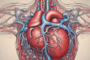
Podcast
Questions and Answers
Which structure carries oxygenated blood away from the heart?
Which structure carries oxygenated blood away from the heart?
- Aorta (correct)
- Inferior vena cava
- Pulmonary veins
- Pulmonary arteries
What is the primary function of the circulatory system?
What is the primary function of the circulatory system?
- To produce red blood cells
- To produce heartbeats
- To filter blood
- To transport gases, hormones, lymph, nutrients, and waste (correct)
What separates the right and left atria in the heart?
What separates the right and left atria in the heart?
- Pulmonary trunk
- Interatrial septum (correct)
- Interventricular septum
- Coronary sinus
How does the right ventricle function in the circulation process?
How does the right ventricle function in the circulation process?
Which of the following chambers of the heart receives deoxygenated blood?
Which of the following chambers of the heart receives deoxygenated blood?
What is the visceral layer of the pericardium referred to as?
What is the visceral layer of the pericardium referred to as?
Which blood vessels return oxygenated blood from the lungs to the heart?
Which blood vessels return oxygenated blood from the lungs to the heart?
Which side of the heart carries deoxygenated blood?
Which side of the heart carries deoxygenated blood?
What primarily distinguishes muscular arteries from elastic arteries?
What primarily distinguishes muscular arteries from elastic arteries?
Which layer is primarily featured in capillaries?
Which layer is primarily featured in capillaries?
What is the primary function of arterioles?
What is the primary function of arterioles?
What characterizes large venules compared to small venules?
What characterizes large venules compared to small venules?
What feature is crucial for the function of veins, especially in the legs?
What feature is crucial for the function of veins, especially in the legs?
What is the primary function of the fibrous pericardium?
What is the primary function of the fibrous pericardium?
Which layer of the pericardium is fused to the surface of the heart?
Which layer of the pericardium is fused to the surface of the heart?
What fills the pericardial cavity?
What fills the pericardial cavity?
In adult circulation, where does deoxygenated blood first enter after the right atrium?
In adult circulation, where does deoxygenated blood first enter after the right atrium?
How does oxygenated blood return to the heart from the lungs?
How does oxygenated blood return to the heart from the lungs?
What is the outermost layer of the pericardium called?
What is the outermost layer of the pericardium called?
Which layer of the serous pericardium is fused to the fibrous pericardium?
Which layer of the serous pericardium is fused to the fibrous pericardium?
In pulmonary circulation, what is the role of the right ventricle?
In pulmonary circulation, what is the role of the right ventricle?
What connects the layers of the serous pericardium?
What connects the layers of the serous pericardium?
Which of the following arrangements accurately describes the order of blood flow through the heart?
Which of the following arrangements accurately describes the order of blood flow through the heart?
What is the function of the ductus venosus in fetal circulation?
What is the function of the ductus venosus in fetal circulation?
How does blood primarily bypass pulmonary circulation in the fetus?
How does blood primarily bypass pulmonary circulation in the fetus?
Which arteries supply the left side of the heart with oxygenated blood?
Which arteries supply the left side of the heart with oxygenated blood?
From which part of the heart does the coronary circulation begin?
From which part of the heart does the coronary circulation begin?
Which layer of a blood vessel is in direct contact with the blood flowing in the lumen?
Which layer of a blood vessel is in direct contact with the blood flowing in the lumen?
What type of muscle is the myocardium?
What type of muscle is the myocardium?
What is the correct pathway of blood flow from the heart to the lungs?
What is the correct pathway of blood flow from the heart to the lungs?
What structure allows blood to flow directly from the right atrium to the left atrium in fetal circulation?
What structure allows blood to flow directly from the right atrium to the left atrium in fetal circulation?
Which feature characterizes cardiac muscle cells and distinguishes them from skeletal muscle cells?
Which feature characterizes cardiac muscle cells and distinguishes them from skeletal muscle cells?
What role does the ductus arteriosus play in fetal circulation?
What role does the ductus arteriosus play in fetal circulation?
What role does the fibrous skeleton of the heart play?
What role does the fibrous skeleton of the heart play?
What type of muscle is primarily found in the tunica media of blood vessels?
What type of muscle is primarily found in the tunica media of blood vessels?
What are intercalated discs primarily composed of?
What are intercalated discs primarily composed of?
Which of the following statements about the conduction system of the heart is correct?
Which of the following statements about the conduction system of the heart is correct?
What function does the endocardium serve?
What function does the endocardium serve?
How do gap junctions in cardiac muscle cells contribute to heart function?
How do gap junctions in cardiac muscle cells contribute to heart function?
What effect does fibrous skeleton have on the heart valves?
What effect does fibrous skeleton have on the heart valves?
Which component helps prevent adjacent cardiac muscle cells from pulling apart during contractions?
Which component helps prevent adjacent cardiac muscle cells from pulling apart during contractions?
Which feature is essential for cardiac muscle to maintain synchronized contractions?
Which feature is essential for cardiac muscle to maintain synchronized contractions?
Flashcards are hidden until you start studying
Study Notes
The Circulatory System
- The circulatory system includes the heart, blood vessels, blood, and the lymphatic system.
- The circulatory system transports gases, hormones, lymph, nutrients and waste.
- The circulatory system protects against disease and fluid loss.
Chambers of the Heart
- The right atrium receives deoxygenated blood from the superior and inferior vena cava, as well as the coronary sinus.
- The left atrium connects to four pulmonary veins and receives oxygenated blood from the lungs.
- The right ventricle connects to the pulmonary trunk which branches into two pulmonary arteries and sends deoxygenated blood to the lungs.
- The left ventricle connects to the aorta and sends oxygenated blood to the rest of the body.
Left vs. Right Side of the Heart
- The left side of the heart carries oxygenated blood.
- The right side of the heart carries deoxygenated blood.
- Veins carry blood to the heart.
- Arteries carry blood away from the heart.
Septa of the heart
- The interatrial septum separates the atria.
- The interventricular septum separates the ventricles.
- The interventricular septum is visible on the outside of the heart as a groove called the interventricular sulcus.
Vessels of the Heart
- The superior and inferior vena cava deliver blood into the right atrium of the heart.
- The pulmonary veins return oxygenated blood from the lungs to the heart, delivering it to the left atrium.
- The pulmonary arteries take deoxygenated blood from the right ventricle to the lungs to be oxygenated.
- The aorta takes oxygenated blood from the left ventricle and delivers it to the body.
The Heart Wall
- The heart wall consists of the epicardium, myocardium, and endocardium.
- The epicardium is the visceral layer of the pericardium and is composed of stratified squamous epithelium and connective tissue.
- The myocardium is cardiac muscle arranged in spiral or circular bundles and is responsible for heart contraction.
- The endocardium is the endothelium that lines the inner surface of the heart and all of the blood vessels and is composed of simple squamous epithelium and connective tissue.
The Fibrous Skeleton of the Heart
- The fibrous skeleton of the heart is connective tissue fibers that surround the muscles of the heart and provide electrical insulation.
- Connective tissue fibers within the heart prevent vessels and valves from stretching due to continuous blood flow.
- Rings of connective tissue between the atria and ventricles keep the heart's openings open.
- Valves are found at the atrioventricular groove between the atria and ventricles and close off certain portions of the heart to regulate blood flow.
Cardiac Muscle
- Cardiac muscle cells are striated, composed of myofibrils, and are arranged into sarcomeres, similar to skeletal muscle cells.
- Cardiac muscle cells are branched and uninucleate unlike skeletal muscle cells.
- Cardiac muscle cells are connected by intercalated discs, containing desmosomes and gap junctions.
- Desmosomes prevent adjacent cells from separating during contraction.
- Gap junctions allow ion exchange between adjacent cells, enabling electrical impulses and coordinated contraction.
The Conduction System
- The conduction system is composed of modified cardiac muscle cells that conduct electrical impulses through gap junctions.
- These modified muscle cells do not contract.
- Components of the conduction system include the SA node, AV node, AV bundle, bundle branches, and Purkinje fibers.
The Pericardium
- The pericardium is a double-walled sac that surrounds the heart and is composed of three layers: the fibrous pericardium, the parietal layer of the serous pericardium, and the visceral layer of the serous pericardium.
- The fibrous pericardium is the outermost layer, composed of dense irregular connective tissue and anchors the heart to surrounding tissues.
- The parietal layer of the serous pericardium is fused to the fibrous pericardium and together they form the pericardial sac.
- The visceral layer of the serous pericardium is called the epicardium and is fused to the surface of the heart, functioning as a key portion of the heart wall.
- The pericardial cavity is the area between the visceral and parietal layers and contains fluid for lubrication.
The Circulatory Route
- Adult circulation is different from fetal circulation.
- Adult circulation:
- Pulmonary circulation: delivers and returns blood to and from the lungs for oxygenation.
- Deoxygenated blood in the right atrium moves into the right ventricle.
- The right ventricle pumps deoxygenated blood to the lungs via the pulmonary arteries.
- Oxygenated blood from the lungs is returned to the left atrium via the pulmonary veins.
- Pulmonary circulation: delivers and returns blood to and from the lungs for oxygenation.
- Fetal circulation:
- Modifications in the fetal circulatory system shunt blood past the lungs and liver.
- The ductus venosus diverts blood from the umbilical vein to the inferior vena cava, bypassing the liver.
- The foramen ovale shunts blood directly from the right atrium to the left atrium, bypassing the lungs.
- The ductus arteriosus diverts blood from the pulmonary trunk to the aorta, bypassing the lungs.
- The umbilical arteries carry partially oxygenated blood from the fetal aorta back to the placenta, entering maternal circulation.
Coronary Circulation
- Coronary circulation supplies the myocardium with oxygen and nutrients.
- Oxygenated blood from the left ventricle enters the aorta, then branches into the right and left coronary arteries.
- Left coronary artery: supplies the left side of the heart with oxygenated blood and branches into the anterior interventricular artery and the circumflex artery.
- Right coronary artery: supplies the right side of the heart with oxygenated blood and branches into the posterior interventricular artery and the right marginal artery.
- Arterioles transition into capillaries, which supply the myocardium.
- Capillaries transition into venules, then into cardiac veins, which drain into the coronary sinus.
- The coronary sinus drains deoxygenated blood into the right atrium, which then pumps it to the lungs for oxygenation.
Blood Vessel Anatomy
- The lumen of a blood vessel is the central area that contains blood.
- The tunica intima is the innermost layer of a blood vessel and is composed of simple squamous endothelium.
- The tunica media is the middle layer and contains circularly arranged smooth muscle and sheets of elastin.
- The tunica externa is the outermost layer and is composed of loosely arranged collagen fibers.
Blood Vessel Arrangement
- Blood flows from the heart into arteries, then arterioles, capillaries, venules, veins, and finally back to the heart.
Arteries
- Arteries transport blood away from the heart.
- Elastic arteries: contain a high proportion of elastin in all three layers, including the tunica media. They are the largest arteries, such as the aorta.
- Muscular arteries: contain more smooth muscle than elastin, with most of the muscle located in the tunica media. These include most arteries in the body, such as the coronary artery.
Arterioles
- Arterioles are the smallest type of arteries and are very muscular.
- Arterioles regulate blood flow and pressure.
Capillaries
- Capillaries only contain a tunica intima - endothelium and a basement membrane.
- Capillaries are the site of nutrient, waste, and gas exchange with cells.
- Capillaries merge into venules.
Venules
- Venules are the smallest veins, analogous to arterioles.
- Small venules: contain tunica intima only.
- Large venules: contain a thin tunica intima and a tunica externa.
Veins
- Veins return blood to the heart.
- Veins contain valves to prevent backflow, especially important in the legs where gravity opposes blood flow.
- Veins have a large lumen and a thin tunica intima, which makes them prone to collapse.
Studying That Suits You
Use AI to generate personalized quizzes and flashcards to suit your learning preferences.


