Podcast
Questions and Answers
What is the primary function of the portal vein?
What is the primary function of the portal vein?
- Drains blood from the abdominal GI tract to the liver (correct)
- Drains blood from the heart to the lungs
- Transports lymphatic fluid to the bloodstream
- Carries oxygen-rich blood from the liver to the body
Which muscle is primarily responsible for maintaining fecal continence?
Which muscle is primarily responsible for maintaining fecal continence?
- Puborectalis (correct)
- Iliococcygeus
- Levator ani
- Pubococcygeus
Which part of the uterus is responsible for the thick muscular wall?
Which part of the uterus is responsible for the thick muscular wall?
- Myometrium (correct)
- Cervix
- Endometrium
- Fundus
Where does fertilization typically occur within the female reproductive system?
Where does fertilization typically occur within the female reproductive system?
What is likely a consequence of injury during childbirth to the pelvic floor muscles?
What is likely a consequence of injury during childbirth to the pelvic floor muscles?
Which of the following describes the position of the uterus when the bladder is full?
Which of the following describes the position of the uterus when the bladder is full?
What does the term 'ectopic pregnancy' refer to?
What does the term 'ectopic pregnancy' refer to?
What is the primary function of the epididymis in the male reproductive system?
What is the primary function of the epididymis in the male reproductive system?
What is the role of the greater vestibular glands?
What is the role of the greater vestibular glands?
What is a common result of cryptorchidism if left uncorrected?
What is a common result of cryptorchidism if left uncorrected?
What is the primary role of the seminal vesicles in male reproductive anatomy?
What is the primary role of the seminal vesicles in male reproductive anatomy?
Which zone of the prostate is most commonly affected by prostate cancer?
Which zone of the prostate is most commonly affected by prostate cancer?
What type of secretion do the bulbourethral glands produce?
What type of secretion do the bulbourethral glands produce?
What is a common symptom of benign hypertrophy of the prostate?
What is a common symptom of benign hypertrophy of the prostate?
Which muscle is primarily responsible for mediating erection in the penis?
Which muscle is primarily responsible for mediating erection in the penis?
Which structure in the kidney is responsible for urine secretion?
Which structure in the kidney is responsible for urine secretion?
What is the main function of the urinary bladder?
What is the main function of the urinary bladder?
How does the left renal vein differ from the right renal vein?
How does the left renal vein differ from the right renal vein?
What area does the urethra naturally traverse as it connects the bladder to the exterior?
What area does the urethra naturally traverse as it connects the bladder to the exterior?
Where does the urine go after it is secreted from the renal pyramids?
Where does the urine go after it is secreted from the renal pyramids?
What is the primary position of the azygos vein in relation to the vertebral column?
What is the primary position of the azygos vein in relation to the vertebral column?
Which of the following structures does not drain into the azygos vein?
Which of the following structures does not drain into the azygos vein?
What is the primary function of the conjoint tendon in relation to the internal and transverse abdominal muscles?
What is the primary function of the conjoint tendon in relation to the internal and transverse abdominal muscles?
Which muscle is primarily responsible for filling the space between the 12th rib and the iliac crest in the posterior abdominal wall?
Which muscle is primarily responsible for filling the space between the 12th rib and the iliac crest in the posterior abdominal wall?
What type of hernia occurs due to increased abdominal pressure causing abdominal contents to protrude?
What type of hernia occurs due to increased abdominal pressure causing abdominal contents to protrude?
Which layer is the deepest in the anterior abdominal wall structure?
Which layer is the deepest in the anterior abdominal wall structure?
What is the primary component of the inguinal canal's posterior wall?
What is the primary component of the inguinal canal's posterior wall?
At what stage of development do the testes begin their descent into the scrotum?
At what stage of development do the testes begin their descent into the scrotum?
Which structure in the abdomen is lined by the visceral peritoneum?
Which structure in the abdomen is lined by the visceral peritoneum?
Which abdominal layer typically provides more stability and compartmentalization?
Which abdominal layer typically provides more stability and compartmentalization?
What is the role of the mesentery in the abdomen?
What is the role of the mesentery in the abdomen?
Which part of the small intestine is primarily responsible for most nutrient absorption?
Which part of the small intestine is primarily responsible for most nutrient absorption?
Which statement correctly describes the pyloric sphincter?
Which statement correctly describes the pyloric sphincter?
What does the term 'retroperitoneal' refer to in abdominal organ positioning?
What does the term 'retroperitoneal' refer to in abdominal organ positioning?
Where does the duodenum primarily receive pancreatic enzymes and bile?
Where does the duodenum primarily receive pancreatic enzymes and bile?
What anatomical feature does the spleen have that distinguishes it from other abdominal organs?
What anatomical feature does the spleen have that distinguishes it from other abdominal organs?
What is the main function of the gallbladder?
What is the main function of the gallbladder?
What distinguishes the liver's visceral surface?
What distinguishes the liver's visceral surface?
Which of the following is true regarding the abdominal aorta's branches?
Which of the following is true regarding the abdominal aorta's branches?
What is unique about the structure of the large intestine's outer longitudinal muscle layer?
What is unique about the structure of the large intestine's outer longitudinal muscle layer?
What type of blood does the azygos vein primarily drain?
What type of blood does the azygos vein primarily drain?
Where does the azygos vein arch before draining into the superior vena cava?
Where does the azygos vein arch before draining into the superior vena cava?
Which of the following is a function of the inguinal canal?
Which of the following is a function of the inguinal canal?
What type of abdominal wall muscle is the external oblique?
What type of abdominal wall muscle is the external oblique?
What does the term 'retroperitoneal' describe?
What does the term 'retroperitoneal' describe?
What fibrous structure connects the testes to the scrotum during descent?
What fibrous structure connects the testes to the scrotum during descent?
How many main layers of muscle does the lateral flat muscle group in the anterior abdominal wall consist of?
How many main layers of muscle does the lateral flat muscle group in the anterior abdominal wall consist of?
Which muscle is primarily responsible for flexing the hip and is located in the posterior abdominal wall?
Which muscle is primarily responsible for flexing the hip and is located in the posterior abdominal wall?
What is the anatomical significance of the cervical constriction of the oesophagus?
What is the anatomical significance of the cervical constriction of the oesophagus?
Which structure forms the floor of the inguinal canal?
Which structure forms the floor of the inguinal canal?
Which function is primarily associated with the mesentery?
Which function is primarily associated with the mesentery?
What anatomical structure supports the anterior abdominal wall and contains the rectus abdominus muscle?
What anatomical structure supports the anterior abdominal wall and contains the rectus abdominus muscle?
What defines the pyloric sphincter?
What defines the pyloric sphincter?
What is the primary role of the testicular gubernaculum during development?
What is the primary role of the testicular gubernaculum during development?
Which part of the small intestine is primarily retroperitoneal?
Which part of the small intestine is primarily retroperitoneal?
What is the primary purpose of the peritoneum in the abdomen?
What is the primary purpose of the peritoneum in the abdomen?
Where does the transverse colon begin?
Where does the transverse colon begin?
What anatomical boundary defines the location of the right lower quadrant in the abdomen?
What anatomical boundary defines the location of the right lower quadrant in the abdomen?
Which structure separates the liver into its two main lobes?
Which structure separates the liver into its two main lobes?
Which muscle is not considered to support the anterior abdominal wall?
Which muscle is not considered to support the anterior abdominal wall?
What anatomical feature does the spleen have that enhances its functions?
What anatomical feature does the spleen have that enhances its functions?
What is the role of the gallbladder?
What is the role of the gallbladder?
What distinguishes the jejunum from the ileum regarding its structure?
What distinguishes the jejunum from the ileum regarding its structure?
Which artery supplies blood to the midgut?
Which artery supplies blood to the midgut?
What is true about the large intestine's wall structure?
What is true about the large intestine's wall structure?
What is the function of the hepatic veins?
What is the function of the hepatic veins?
What is the primary characteristic of the visceral surface of the liver?
What is the primary characteristic of the visceral surface of the liver?
Flashcards are hidden until you start studying
Study Notes
Portal Vein
- Drains blood from the abdominal GI tract, spleen and pancreas
- Carries venous blood to the liver for metabolism
- Metabolised blood drains into the hepatic veins, which drain into the IVC
Pelvic Diaphragm
- Separates pelvis and perineum
- Attachment point for external genitalia
- Diamond shaped with two triangles:
- Urogenital triangle
- Anterior
- Contains urogenital hiatus
- Anal triangle
- Posterior
- Contains anal aperture
- Urogenital triangle
- Formed by levator ani muscle:
- Puborectalis
- Attaches to the pubis bone and goes around the rectum
- Forms a sling around the rectum
- Contributes to faecal continence
- Pubococcygeus
- Lateral to puborectalis
- Attaches to the pubis and coccyx bones
- Iliococcygeus
- Lateral to pubococcygeus
- Attaches to the ilium and coccyx bones
- Puborectalis
Perineal Membrane
- Covers half of the pelvic diaphragm (urogenital triangle)
- Superficial to the pelvic diaphragm
- Opening for the urethra (male and female) and vagina (female)
Deep Perineal Pouch
- Space between perineal membrane and levator ani
- Contains skeletal muscles that form voluntary sphincters around the urethra (external urethral sphincter) and the vagina
Child Birth
- Perineum, levator ani, perineal membrane and deep perineal pouch muscles are often torn or injured
- In females, these muscles support the urethra, vagina, uterus and rectum
- Injury can result in urinary stress incontinence, uterine prolapse and faecal incontinence
Ovary
- Paired
- Intraperitoneal
- Located posterior to the broad ligament and adjacent to the lateral wall of the pelvis
- Ovarian artery supplies the organ
- Ligament of ovary attaches the ovary to the uterus
- Irregular shaped ball
- Two regions:
- Inner medulla
- Contains vessels, nerves and lymphatics
- Outer cortex
- Follicular genesis occurs here
- Inner medulla
- Defined surface epithelium
- Oocytes are surrounded by supporting cells, which prepare the oocyte for ovulation
- Oocyte ovulates into the fallopian tube
Fallopian/Uterine Tubes
- Project laterally from the uterus and open adjacent to ovaries
- Three areas:
- Infundibulum
- Funnel shaped
- Facilitates oocyte collection
- Fimbriae attach to the anterior and lateral part of the ovary and help to catch the oocyte
- Ampulla
- Large
- Labyrinthine lumen
- Fertilization occurs here
- Isthmus
- Narrow
- Connects to uterus
- Infundibulum
Uterus
- Thick walled, muscular
- Facilitates development of the embryo and foetus
- Uterine artery supplies the organ
- Three parts:
- Fundus
- Top part, above fallopian tubes
- Body
- Largest part, below fallopian tubes
- Cervix
- Opening into the vagina
- Fundus
- Two linings:
- Myometrium
- Middle layer made of smooth muscle
- Undergoes hypertrophy and hyperplasia during pregnancy
- Endometrium
- Inner lining
- Supports pregnancy
- Sheds during menstruation
- Myometrium
- Position of the uterus (when the bladder is empty):
- Anteverted relative to the vagina
- Anteflexed over the bladder
- When the bladder is full, uterus is displaced upwards
- Retroverted and retroflexed uterus is displaced to the back and more likely to prolapse
- Uterine prolapse is the uterus pushed through the vagina due to high pressure in the pelvis
Cervix
- Opens inferiorly into the anterior wall of the vagina
- Two parts:
- Supravaginal (above vagina)
- Vaginal (in vagina)
- Uterus is best supported at the cervix
Vagina
- Muscular passage
- Connects uterus to the exterior
- Between the urethra anteriorly and rectum posteriorly
- Fornix is the space created anterior, posterior and lateral to where the cervix enters the vagina
- Vaginal arteries supply the organ
Ectopic Pregnancy
- Fertilization occurs in the ampulla
- Implantation normally occurs in the uterine body
- Ectopic/extrauterine pregnancy is implantation anywhere but the endometrial lining
- The embryo will grow in areas with a large blood supply
- Causes bleeding into the abdominal cavity
- Burst uterine tube is often misdiagnosed as a burst appendix
Peritoneal Folds
- Peritoneum covers the top visceral layer of the pelvis
- Supports and protects the pelvis
- Peritoneal folds form over the pelvic viscera
- Broad ligament is peritoneal folds coming together over the uterine tube
- Ovaries are surrounded by peritoneum and therefore are intraperitoneal
- Ligament of ovary is folds of peritoneum over the ovarian vessels
Peritoneal Pouches
- Created by peritoneal folds
- Recto-uterine pouch
- Between the rectum and uterus
- Deepest part of the abdominal cavity
- Fluid collects here, especially if the patient lies on their back
- Fluid can be aspirated with a syringe through the fornix
- Vesico-uterine pouch
- Between the bladder and uterus
Development of External Genitalia
- Same in males and females
- Genital tubercle develops into the glans penis (male) or glans clitoris (female)
- Testosterone causes more rapid growth of the genital tubercle resulting in it being larger in males
- Urethral folds develop into the urethra (male and female) and labia minora (female)
- Labioscrotal swellings develop into the scrotum (male) and labia majora (female)
Anatomy
- Vulva/pudendum is the erectile tissue and overlying skin which comprises the external genitalia
- Labia minora are two thin skin folds beside the midline, fat free and hair free with connective tissue inside
- Vestibule is the area between each labia minora (contains the urethra and vagina)
- Labia majora are larger skin folds, lateral to labia minora
- Glans clitoris is erectile tissue, exposed, located where the labia minora meet anteriorly
- Perineal membrane and pubic arch are attachment points for the roots of external genitalia
Erectile Tissue
- Erectile tissue is formed by:
- Two corpora cavernosa (anterior to glans clitoris, attaches to pubic arch)
- Two bulbs of vestibule (deep to labia minora)
- Glans clitoris
- Body of clitoris contains corpora cavernosa and bulbs of vestibule
- Erectile tissues are anterior to the urethra
- Greater vestibular glands are posterior to bulbs, have openings into the vestibule, secrete lubricating mucous during arousal
Muscles
- Ischiocavernosus muscle covers crus of clitoris and maintains erection of the clitoris
- Bulbospongiusis muscle encloses the bulbs and greater vestibular glands, supports the pelvic floor, assists in clitoral erection and compresses greater vestibular glands
Pelvic Diaphragm (Male)
- Same as female pelvic diaphragm
- Only one opening in the perineal membrane for the urethra
- Deep perineal pouch also has sphincters around glands
Perineal Membrane (Male)
- Superficial to the pelvic diaghragm
- Covers the urogenital triangle
- Opening for the urethra (male & female) and vagina (female)
Deep Perineal Pouch (Male)
- Space above the perineal membrane
- Contains skeletal muscle that form sphincters around the urethra, glands, & neurovascular structures that supply the penis
Testes
- Function in sperm production
- Septa divides the testes into ~250 lobules
- Lobules contain 3–10 seminiferous tubules
- Each tubule is a highly convoluted loop in the lobule
- The closer to the lumen, the more the sperm differentiate (still non-motile)
- In the mediastinum, tubules end as tubulus erectus, which connects to the rete testis
- Rete testis is an anastomosing channel system, collects sperm from all the seminiferous tubules
- Outer capsules:
- Tunica albugenia surrounds lobules
- Tunica vaginalis covers testes during descent, extension of abdominal peritoneum, surrounds tunica albugenia
Testicular Decent
- Gonadal development occurs along the posterior abdominal wall
- Testes descend towards the inguinal canal by seven months of gestation
- Through the inguinal canal into the scrotum just before birth
- Drags blood vessels, nerves, duct system (vas deferens) and layers of abdominal wall to the scrotum
Cryptorchidism (undescended testes)
- Histological changes à infertility
- Increased risk of testicular cancer
- Needs to be surgically corrected before 18 months
Epididymis
- Coiled tube
- ~5 cm long
- Sperm in the mediastinum of the testes travel through efferent tubules to the epididymis.
- Three parts:
- Capus (head)
- Corpus (body)
- Caudal (tail)
- Surrounded by smooth muscle to assist movement of sperm
- Function to store and mature sperm
- Where sperm acquire motility
- Sperm are stored for ~1 month
Vas Deferens
- Sperm travels from the epididymis to the vas deferens
- Long duct
- Length and complex course results from testicular descent
- Penetrates abdominal wall through the inguinal canal
- Descends along the lateral wall of the pelvic cavity
- Wolffian ducts develop into the vas deferens
- Smooth muscular duct
- Muscular duct which transports spermazoa to ejaculatory duct
- Ejaculation is mediated by the sympathetic nervous system
- Ejaculatory duct is the vas deferens and duct of seminal vesicle
- Function is to transport sperm to ejaculatory duct
- Part of the spermatic cord
Vasectomy
- Ligating the vas deferens to prevent sperm from reaching the penis
- Vas is superficial at the posterior side of the scrotum
- White blood cells assist the epididymis to break down sperm after a vasectomy
Accessory Glands
- Contribute to the volume of ejaculate
Male Reproductive System
- Seminal vesicles
- Posterior surface of bladder
- Ducts of seminal vesicle combine with vas deferens to form ejaculatory duct
- Contribute to volume of semen
- Secrete alkaline fluid
- Help with sperm motility and viability
- Prostate
- Inferior to bladder, anterior to rectum
- Size of a walnut
- Secretes watery liquid with zinc, citric acid, and proteolytic enzymes
- Assists with sperm viability and maturation
- Benign hypertrophy of prostate:
- Occurs in most elderly men
- Hypertrophy of central prostate
- Compresses/blocks urethra causing intermittent urinary stream
- Prostate cancer:
- Common malignancy in men
- Effects the entire prostate
- Treated with radiotherapy, surgery, or androgen deprivation therapy
- Bulbourethral glands
- Pea-shaped, paired
- Located in the deep perineal pouch
- Ducts open into the spongy urethra
- Produce mucous secretion that lubricates urethra
- External genitalia
- Ejaculatory tissues covered by skin
- Scrotum:
- Homologue of labia majora
- Skin fuses in the midline
- Penis
- Covered by skin
- Composed of:
- 2 corpora cavernosa
- 1 corpus spongiosum (expands distally to form glans penis)
- Erection: mediated by parasympathetic nervous system
Kidneys and Urinary System
- Kidneys
- Function to produce urine
- Retroperitoneal, bean shaped
- Extend longitudinally from T12-L3
- Renal capsule lines kidney surface
- Renal sinuses are spaces within the kidney, filled with perinephric fat
- Inner medulla:
- Contains renal pyramids
- Secretory cells secrete urine
- Outer cortex:
- Continuous layer surrounding medulla
- Renal collecting system:
- Urine secreted from the medulla into the hilum
- Urine is collected into the minor calyx, then the major calyx, then the renal pelvis
- Renal hilum:
- Faces anteromedially
- Holds the renal vein, renal artery, and renal pelvis
- Renal artery:
- Supplies blood to each kidney
- Divides into 5 segmental arteries within the hilum
- Accessory renal artery:
- Additional blood supply
- Can enter the kidney via the hilum or the surface
- Renal vein:
- Drains venous blood of the kidneys
- Left renal vein crosses midline and is longer than the right renal vein
- Ureters
- Paired hollow viscus
- Extend from the kidneys into the pelvis
- Constrictions:
- Uretropelvic Junction
- Where ureter crosses the pelvic brim
- Where ureter enters the bladder
- Urinary bladder
- Hollow viscus
- Strong muscular walls
- Function to store urine
- Apex anteriorly, against pubis bone
- Base posteriorly, against uterus in females or rectum in males
- Bladder neck:
- Inferiormost part
- Anchors the bladder
- As the bladder fills, the top expands and is free but the neck stays put
- Base:
- Forms an inverted triangle (trigone)
- Contains 2 uretric orifices and 1 urethral orifice
- Urethra:
- Passes urine from the bladder to the exterior
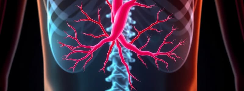
Azygos Vein
- Drains venous blood from the thorax into the superior vena cava (SVC)
- Drains both the parietal and visceral compartments of the thorax, excluding the heart
- Drains venous blood from organs supplied by the descending aorta including the thoracic walls, pericardium, lungs, trachea, bronchi, esophagus, and diaphragm
- The position of the azygos vein against the vertebral column, and the heart's more anterior position, force the azygos vein to arch anteriorly over the right lung root before draining into the SVC.
- An impression of the azygos vein can be seen on the medial surface of the right lung.
GI Tract
- The abdomen is the cavity located between the thorax (above) and pelvis (below).
- The abdominal wall is primarily composed of muscle and associated structures, not bone.
Anterior Abdominal Wall
Bony Framework
- The bony framework consists of the bones of the thorax (superiorly) and pelvis (inferiorly).
- The anterior abdominal wall structures attach to:
- Inferior border of the thorax
- Iliac crest
- Anterior superior iliac spine (ASIS)
- Pubic tubercle
- Pubic crest
Covering Structures
- 8 layers, listed from superficial to deep:
- Skin
- Superficial fascia
- 3 paired muscles (External Oblique, Internal Oblique, Transversus Abdominus)
- 2 vertical muscles (Rectus Abdominus, Pyramidalis)
- Transversalis fascia
- Extra-peritoneal fascia
- Peritoneum
Lateral Flat Muscles (3 Paired, 3 layers)
- 3 paired layers, fleshy laterally, replaced with aponeuroses medially.
- Aponeuroses allow the abdomen to expand and create the linea alba.
- Aponeuroses from both sides interdigitate to form the linea alba, a raphe (seam) that runs along the midline.
External Oblique (EO) Muscles
- Superficial layer
- Fibre direction: inferomedial
- Medial attachment: linea alba
- Superior attachment: ribs (EO fibres overlap the costal margin)
- Posterior attachment: thoracolumbar fascia with the posterior abdominal wall
- Inferior attachment:
- Lateral part: iliac crest and ASIS
- Central part: free
- Medial part: pubic tubercle and pubic crest
- Inguinal Ligament:
- Formed by the free, central part of the inferior border of the EO.
- Thickened as it curves under and extends from ASIS to the pubic tubercle.
Internal Oblique (IO) Muscles
- Intermediate layer
- Fibre direction: superomedial (perpendicular to EO)
- Medial attachment: linea alba
- Superior attachment: costal margin
- Posterior attachment: thoracolumbar fascia
- Inferior attachment: lowermost IO fibres arise from the lateral 2/3 of the inguinal ligament, arch up, then go down to insert on the pubic crest via a conjoint tendon.
Transversus Abdominus (TA) Muscles
- Deep layer
- Fibre direction: transverse
- Medial attachment: linea alba
- Superior attachment: costal margin
- Posterior attachment: thoracolumbar fascia
- Inferior attachment: lowermost TA fibres arise from the lateral 1/3 of the inguinal ligament, arch up, then go down to insert on the pubic crest via a conjoint tendon.
- Conjoint Tendon: a common tendon where the IO and TA muscles insert.
- Transversalis fascia is deep to the TA muscles.
Medial Vertical Muscles
Rectus Abdominus
- Located on either side of the linea alba
- Primary vertical muscle supporting the anterior abdominal wall
- Origin: pubis
- Insertion: ribcage (overlaps costal margin)
- 3 tendinous intersections strengthen the muscle (4 short muscles, not 1 long)
- Rectus Sheath:
- Created by the aponeuroses of the EO, IO, and TA, which pass in front or behind the rectus abdominus.
- Creates a fibrous compartment for the rectus abdominus.
- EO aponeurosis: in front
- IO aponeurosis: splits, passes in front and behind
- TA aponeurosis: behind
Pyramidalis Muscles
- Pyramid-shaped, small, and do not support the anterior abdominal wall.
Testicular Descent
- Testes develop high in the posterior abdominal wall, within extra-peritoneal fat.
- Descent into the scrotum is necessary for spermatogenesis as normal core body temperatures inhibit sperm production.
- A 2-3°C lower temperature is ideal for spermatogenesis.
- Testes connect to the scrotum through the gubernaculum, a fibrous cord present in both males and females.
- Testicular descent occurs around 7 months during gestation.
Inguinal Rings
- To leave the abdomen from extra-peritoneal fat, a hole in the transversalis fascia and EO aponeurosis must be created.
- Deep Inguinal Ring: circular hole in the transversalis fascia.
- Superficial Inguinal Ring: triangular split in the EO aponeurosis (fibrous split at inferior attachment).
- The path between the deep and superficial inguinal rings is oblique.
Inguinal Canal
- The path the testes take during descent into the scrotum.
- It transverses the transversalis fascia (deep inguinal ring), TA, IO, and EO (superficial inguinal ring) layers.
- The inguinal canal is superior to the inguinal ligament, and its boundaries are:
- Floor: inguinal ligament
- Roof: arching IO and TA fibres
- Anterior wall: EO aponeurosis and IO fibres (laterally)
- Posterior wall: transversalis fascia and conjoint tendon (medially)
- Upon emerging from the superficial inguinal ring, it becomes the spermatic cord.
- Women also have inguinal canals, although they are less prominent.
Inguinal Hernia
- The inguinal canal is a communicating structure between the abdomen and the perineum.
- Increased abdominal pressure can lead to abdominal contents protruding through the abdominal wall at weak spots, resulting in a hernia.
- Inguinal hernia: protrusion of abdominal contents into the inguinal canal.
Cryptorchidism
- Undescended testes
- Can be located in the abdomen, inguinal canal, or pre-scrotal region.
- Requires surgical correction before 18 months to 2 years of age.
- Can cause infertility and increases the risk of testicular cancer.
Posterior Abdominal Wall
- Consists of muscle and lumbar vertebrae.
- Attaches to the bony thorax, pelvis, and vertebrae.
- Superiorly: 12th rib
- Medially: lumbar vertebrae
- Laterally: thoracolumbar fascia
- Inferiorly: iliac crest
Psoas Major
- Paired muscle (on either side of the lumbar vertebrae).
- Longitudinally oriented.
- Arises from the lumbar vertebrae bodies.
- Passes inferiorly and under the inguinal ligament.
- Inserts on the lesser trochanter of the femur.
- Psoas Minor: - Lines the surface of the psoas major anteriorly. - Smaller than the psoas major. - Not present in all people, or on both sides.
Quadratus Lumborum
- Lateral to psoas major.
- Paired muscle on either side of psoas major
- Arises from the 12th rib and the tips of the lumbar vertebrae transverse processes.
- Inserts onto the iliac crest.
- Fills the space between the 12th rib and the iliac crest.
Iliacus
- Paired muscle
- Fills the space in the iliacus fossa of the hipbone.
- Situated inferior to the quadratus lumborum and lateral to the psoas major.
Thoracolumbar Fascia
- Muscles of the anterior and posterior abdominal walls, and back muscles are enclosed within the thoracolumbar fascia.
- The fascia of the anterior and posterior abdominal walls meet lateral to the quadratus lumborum.
- The posterior abdominal wall muscles end where the anterior abdominal wall muscles begin.
- It creates a complete muscular wall.
Abdominal Quadrants
- Median Plane: vertically oriented from the xiphoid process to the pubic symphysis.
- Transumbilical Plane: horizontally oriented and crosses the umbilicus.
- Creates 4 quadrants: left upper, right upper, left lower, right lower.
- Used to describe the positioning of abdominal viscera.
Peritoneum
- The innermost layer of the abdominal walls.
- Parietal peritoneum: lines the abdominal wall.
- Visceral peritoneum: lines the viscera.
- Peritoneal cavity: potential space between parietal and visceral peritoneum.
- Intraperitoneal viscus: suspended within the abdominal cavity, completely lined with visceral peritoneum.
- Retroperitoneal viscus:
- Sits against the posterior abdominal wall.
- Outside the abdominal cavity.
- Inner surface lined with parietal peritoneum.
- Example: kidneys.### Mesentery
- A double fold of peritoneum that contains nerves, vessels and provides motility.
- Not all viscera in the abdomen have a mesentery.
- Intraperitoneal viscera have mesentery, retroperitoneal viscera do not.
Oesophagus
- Begins at C6, descends down the midline and passes through the diaphragm at T10.
- Enters the stomach from the right side.
- Muscular tube with inner circular and outer longitudinal muscles.
Oesophageal Constrictions
- Cervical constriction: anatomical sphincter at C6 where the oesophagus begins
- Thoracic constriction: at T4/5, where the oesophagus is compressed from the left side by the bifurcation of the trachea and aortic arch
- Diaphragmatic constriction: occurs at T10, where the oesophagus passes through the diaphragm, creating a functional sphincter.
Oesophagogastric Junction
- Where oesophageal mucosa changes to stomach mucosa.
- Sudden change, indicated by the Z-line.
Stomach
- Located in the left upper quadrant, it's a dilated, hollow, J-Shaped viscus.
- Intraperitoneal, with two openings:
- Cardiac Orifice: proximal, where oesophagus enters the stomach.
- Pyloric Orifice: distal, where the duodenum starts.
- Two curvatures:
- Lesser Curvature: right side.
- Greater Curvature: left side.
- Two surfaces:
- Anterior Surface
- Posterior Surface
- Four parts:
- Cardia: region near cardiac orifice.
- Fundus: part above cardiac orifice, holding gas.
- Body: majority of the stomach.
- Pyloric Part: distal narrowing.
- Pyloric Antrum (wider)
- Pyloric Canal (narrower, more distal)
- Pyloric sphincter/Pylorus: anatomical sphincter at the end of the pyloric canal, controlling chyme entry into the duodenum and aiding absorption.
- Rugae: gastric folds/mucosa on the internal surface.
Small Intestine
- Hollow viscus, divided into three parts:
- Duodenum
- Jejunum
- Ileum
- Function in digestion and absorption.
- Two muscle coats:
- Inner Circular Muscles
- Outer Longitudinal muscles
Duodenum
- Short, retroperitoneal, C-shaped section.
- Located with the right kidney lateral and the head of the pancreas medial to it.
- Four parts:
- 1st part (duodenal cap): immediately after the pylorus.
- 2nd part: descending component, between the right kidney and pancreas.
- Papilla on internal medial wall: secretes pancreatic enzymes and bile into the duodenum.
- Major duodenal papilla: secretes products from the bile duct and main pancreatic duct.
- Minor duodenal papilla: superior to the major, secretes products from the accessory pancreatic duct.
- 3rd part: horizontal, crosses the midline.
- 4th part: hooks around and joins with the jejunum.
Jejunum
- Large, proximal 40% of small intestine.
- Intraperitoneal.
- Resides in the left upper quadrant.
- Larger caliber, holding more gastric contents due to the majority of absorption occurring here.
- Mucosal folds increase surface area.
- Thick-walled.
- Highly vascularized with less fat in the mesentery (vessels and nerves are more visible).
Ileum
- Large, distal 60% of the small intestine.
- Intraperitoneal.
- Resides in the right lower quadrant (changes during pregnancy).
- Lower caliber, holding less gastric contents due to less absorption occurring here.
- Fewer mucosal folds.
- Less vascularized with less fat in the mesentery (vessels and nerves are less visible).
Large Intestine
- Begins at the ileocecal junction where the ileum enters the large intestine.
- Ascending colon: retroperitoneal, large intestine starts to ascend.
- Transverse colon: intraperitoneal, begins at the hepatic/right colic flexure, traverses the midline, and is the longest part.
- Descending colon: retroperitoneal, begins at the splenic flexure/left colic flexure near the spleen, descends, and becomes the sigmoid colon.
- Sigmoid colon: intraperitoneal, bends inferiorly and medially (S-shaped), and becomes the rectum and anal canal.
Large Intestine Wall
- Inner Circular Muscle: complete layer.
- Outer Longitudinal Muscle: split into three bands (tenia coli), which create a baggy appearance due to the incomplete muscle layer (haustra).
- Omental appendices: fatty tags along the tenia coli.
Caecum
- Intraperitoneal.
- Blind pouch.
- Inferior to the ileocecal junction.
- Located in the right lower quadrant.
Appendix
- Intraperitoneal.
- Located in the right lower quadrant, inferior to the ileocecal junction.
- Base of the appendix is located where the three tenia coli bands meet.
- Position varies, but the base remains constant.
Rectum
- Retroperitoneal.
- Tenia coli bands merge to form a complete outer longitudinal muscle layer.
- No tenia coli, haustra, or omental appendices.
Liver
- Located in the right upper quadrant.
- Situated under the right dome of the diaphragm.
- Solid viscus.
- Partially protected by the thoracic cage.
- Intraperitoneal.
- Function in bile production and nutrient metabolism.
Diaphragmatic Surface
- Anterior and superior.
- Where the liver sits against the diaphragm.
- Smooth.
- Partially protected by the ribcage.
- Covered by peritoneum.
Visceral Surface
- Posterior and inferior.
- Interface between the liver and other viscera.
- Concaved due to impressions from adjacent structures.
- Contains the inferior vena cava (IVC) and gallbladder.
- Covered by peritoneum, except for the IVC and gallbladder.
Portal Hepatis
- Hilum, holds vessels, nerves, lymphatics, and ducts.
- Portal Triad:
- Portal Vein: carries venous blood from the GI tract, containing nutrients for metabolism in the liver.
- Hepatic Artery: principle arterial supply to the liver.
- Bile Duct: carries bile from the liver to the duodenum.
Hepatic Veins
- Drain the venous blood from the liver.
- Not located in the hilum.
- Run through grooves.
- Drain directly into the IVC.
Liver Lobes
- Diaphragmatic Surface: falciform ligament separates the liver into two anatomical lobes (larger right and smaller left lobes).
- Visceral Surface:
- Right and Left Anatomical Lobes: fissures and grooves contain structures.
- Left Sagittal Fissure: ligaments run through.
- Right Sagittal Fissure: IVC and gallbladder are located here.
- Between these fissures is the hilum, separating the liver into two more accessory lobes:
- Caudate Lobe: above the hilum.
- Quadrate Lobe: below the hilum.
- Accessory lobes are actually part of the right lobe.
- Right and Left Anatomical Lobes: fissures and grooves contain structures.
Gallbladder
- Located on the visceral surface of the liver.
- Not covered in peritoneum.
- Hollow viscus.
- Resides in the right upper quadrant.
- Four parts:
- Fundus: blind end.
- Body:
- Neck: narrowing.
- Cystic Duct:
- Stores and concentrates bile produced by the liver.
- Overconcentration of bile can lead to gallstones.
Bile Pathway
- Bile travels from the liver through the hepatic ducts to the common hepatic duct.
- The common hepatic duct merges with the cystic duct to form the common bile duct.
- The common bile duct enters the duodenum through the major duodenal papilla.
- The major duodenal papilla has a functional sphincter.
- Contraction of the sphincter (regulated by nerves) closes the sphincter, causing bile to fill and collect in the gallbladder.
- Contraction of the gallbladder pushes bile through the cystic and bile ducts into the duodenum.
Pancreas
- Resides in the left upper quadrant.
- Retroperitoneal.
- Solid viscus.
- Four parts:
- Head: surrounded by the C-shaped duodenum.
- Neck:
- Body:
- Tail: located against the spleen.
- Exocrine and endocrine functions.
Pancreatic Secretions
- Exocrine secretion into the duodenum:
- Main pancreatic duct: collects pancreatic enzymes from the tail, body, neck, and head, and secretes them into the duodenum via the major duodenal papilla.
- Accessory pancreatic duct: collects pancreatic enzymes from the head, and secretes them into the duodenum via the minor duodenal papilla.
Spleen
- Resides in the left upper quadrant.
- Solid viscus.
- Intraperitoneal.
- Associated with GI tract organs.
- Largest lymphatic organ.
- Rich blood supply.
- Two surfaces:
- Diaphragmatic Surface
- Visceral Surface: contains the hilum where the splenic arteries/veins enter/exit.
- Located against the diaphragm and ribs 9, 10, and 11 on the left side.
- Fracture of these ribs can cause damage to the spleen.
- Soft and highly vascularized with a delicate capsule.
- Sharp edges of fractured ribs can lacerate the capsule causing bleeding into the abdominal cavity.
Abdominal Aorta
- Supplies abdominal organs.
- Enters the abdomen at T12.
- Bifurcates into the iliac arteries at L4.
Anterior Surface
- Three Unpaired Branches:
- Supply unpaired viscera (GI tube).
- Celiac Trunk: foregut (abdominal esophagus to the major duodenal papilla).
- Superior Mesenteric Artery: midgut (major duodenal papilla to the distal 1/3 of the transverse colon).
- Inferior Mesenteric Artery: hindgut (distal 1/3 of the transverse colon to ½ way through the anal canal).
- Three Paired Branches:
- Supply paired viscera.
- Suprarenal Arteries: adrenal glands.
- Renal Arteries: kidneys.
- Testicular/Ovarian Arteries: gonads.
Posterior Surface
- Paired arteries supply abdominal wall structures.
- Phrenic Arteries: diaphragm.
- Parietal Arteries: anterior and posterior abdominal walls.
Venous Drainage
- All abdominal venous blood drains into the IVC.
- Venous blood from paired viscera and abdominal walls drains directly into the IVC.
- Venous blood from unpaired viscera (except the liver) drains into the IVC via the portal vein.
Azygos Vein
- Drains venous blood from the thorax into the superior vena cava (SVC)
- Drains both parietal and visceral components of the thorax, excluding the heart
- Receives venous blood from structures supplied by the descending aorta: thoracic walls, pericardium, lungs, trachea, bronchi, esophagus, and diaphragm
- Its drainage pattern varies significantly among individuals
- Located posteriorly against the vertebral column
- Arches anteriorly over the right lung root to drain into the SVC
- Creates an impression on the medial surface of the right lung
Abdominal Cavity
- Located between the thorax (superior) and pelvis (inferior)
- Abdominal wall primarily composed of muscle and coverings, not bone
- Structures attach to the thorax and pelvis
Anterior Abdominal Wall
- Composed of bony framework and lining/covering structures
Bony Framework
- Formed by bones of the thorax (superior) and pelvis (inferior)
- Anterior abdominal wall structures attach to:
- Inferior border of the thorax
- Iliac crest
- Anterior superior iliac spine (ASIS)
- Pubic tubercle (lateral)
- Pubic crest (medial)
Lining/Covering Structures
- Consists of eight layers, arranged from superficial to deep:
- Skin
- Superficial fascia (fat)
- Paired muscles (3 lateral, 2 medial)
- Transversalis fascia
- Extra-peritoneal fascia
- Peritoneum
Lateral Flat Muscles
- Three paired layers located laterally:
- External Oblique (EO)
- Internal Oblique (IO)
- Transversus Abdominus (TA)
- Muscles are fleshy laterally, transitioning to aponeuroses medially, allowing the abdomen to expand
- Aponeuroses from each side meet and interdigitate, forming the linea alba (raphe)
External Oblique (EO) Muscles
- Superficial layer
- Fibers run inferomedially
- Medial attachment: linea alba
- Superior attachment: ribs (EO fibers overlap the costal margin)
- Posterior attachment: thoracolumbar fascia with the posterior abdominal wall
- Inferior attachment:
- Lateral part: iliac crest and ASIS
- Central part: free
- Medial part: pubic tubercle and pubic crest
- Inguinal ligament:
- Formed by the free, central part of the inferior EO border
- Thickened band as it curves inferiorly
- Extends from ASIS to the pubic tubercle
Internal Oblique (IO) Muscles
- Intermediate layer
- Fibers run superomedially (perpendicular to EO)
- Medial attachment: linea alba
- Superior attachment: costal margin
- Posterior attachment: thoracolumbar fascia
- Inferior attachment:
- Lowermost IO fibers originate from the lateral two-thirds of the inguinal ligament, arch superiorly and then inferiorly to insert onto the pubic crest via a conjoint tendon
Transversus Abdominus (TA) Muscles
- Deepest layer
- Fibers run transversely
- Medial attachment: linea alba
- Superior attachment: costal margin
- Posterior attachment: thoracolumbar fascia
- Inferior attachment:
- Lowermost TA fibers originate from the lateral one-third of the inguinal ligament, arch superiorly and then inferiorly to insert onto the pubic crest via a conjoint tendon
- Conjoint tendon: Combined insertion point for the IO and TA muscles
Medial Vertical Muscles
- Located on either side of the linea alba
- Provide vertical support for the anterior abdominal wall
- Origin: pubis
- Insertion: ribcage (overlaps costal margin)
- Three tendinous intersections strengthen the muscle (forming four short muscles, not one long muscle)
- Enclosed within the rectus sheath:
- Formed by aponeuroses of the EO, IO, and TA
- EO aponeurosis: anterior
- IO aponeurosis: splits, passing anterior and posterior
- TA aponeurosis: posterior
- Creates a fibrous compartment for the rectus abdominus
Pyramidalis Muscles
- Pyramid-shaped, small muscle
- Does not support the anterior abdominal wall
Testicular Descent
- Testes develop high in the posterior abdominal wall (extra-peritoneal fat)
- Must descend into the scrotum to be functional, as spermatogenesis is inhibited at core body temperature
- Descent involves moving to the anterior abdominal wall and exiting the abdomen
- Testes in the posterior abdominal wall connect to the scrotum via the gubernaculum (fibrous cord, also present in females)
- Occurs around 7 months of gestation
Inguinal Rings
- For the testes to exit the abdomen from the extra-peritoneal fat, holes need to be created in the transversalis fascia and EO aponeurosis
- Deep inguinal ring: circular hole in the transversalis fascia
- Superficial inguinal ring: triangular split in the EO aponeurosis
- Path between the deep and superficial inguinal rings is oblique
Inguinal Canal
- The path the testes take to descend into the scrotum
- Passes through the following layers:
- Transversalis fascia (deep inguinal ring)
- TA
- IO
- EO (superficial inguinal ring)
- Located superior to the inguinal ligament
- Boundaries:
- Floor: inguinal ligament
- Roof: arching IO and TA fibers
- Anterior wall: EO aponeurosis and IO fibers (laterally)
- Posterior wall: transversalis fascia and conjoint tendon (medially)
- After exiting the superficial inguinal ring, it becomes the spermatic cord
- Females also have inguinal canals (less prominent)
Inguinal Hernia
- The inguinal canal is a pathway from the abdomen to the perineum
- Increased abdominal pressure can cause abdominal contents to protrude through weak spots in the abdominal wall, creating an inguinal hernia
- Inguinal hernia: protrusion of abdominal contents into the inguinal canal
Cryptorchidism
- Undescended testes
- Can be abdominal, inguinal, or pre-scrotal
- Requires surgical correction before 18 months to 2 years
- Can cause infertility
- Increases the risk of testicular cancer
Posterior Abdominal Wall
- Composed of muscle (and lumbar vertebra)
- Attaches to the bony thorax, pelvis, and vertebrae:
- Superiorly: 12th rib
- Medially: lumbar vertebrae
- Laterally: thoracolumbar fascia
- Inferiorly: iliac crest
Psoas Major
- Paired muscle (on either side of the lumbar vertebrae)
- Longitudinally oriented
- Origin: lumbar vertebra body
- Passes inferiorly and under the inguinal ligament
- Insertion: lesser trochanter of the femur
- Psoas minor:
- Lines the anterior surface of the psoas major
- Smaller than the psoas major
- May not be present in all individuals, or on both sides
Quadratus Lumborum
- Lateral to the psoas major
- Paired muscle on either side of the psoas major
- Origin: 12th rib and tips of the lumbar vertebrae transverse processes
- Insertion: iliac crest
- Fills the space between the 12th rib and the iliac crest
Iliacus
- Paired muscle
- Fills the iliacus fossa space of the hipbone
- Inferior to the quadratus lumborum and lateral to the psoas major
Thoracolumbar Fascia
- Encloses the muscles of the anterior and posterior abdominal walls and back muscles
- Fascia of the anterior and posterior abdominal walls meets lateral to the quadratus lumborum
- Posterior abdominal wall muscles terminate where anterior abdominal wall muscles begin
- Creates a complete muscular wall
Abdominal Quadrants
- Median plane: vertical line from the xiphoid process to the pubic symphysis
- Transumbilical plane: horizontal line crossing the umbilicus
- Together, these planes divide the abdomen into four quadrants:
- Left upper quadrant
- Right upper quadrant
- Left lower quadrant
- Right lower quadrant
- Used to describe the positioning of abdominal viscera
Peritoneum
- Innermost layer of the abdominal walls
- Parietal peritoneum: lines the abdominal wall
- Visceral peritoneum: lines the viscera
- Peritoneal cavity: potential space between the parietal and visceral peritoneum
- Intraperitoneal viscus: suspended within the abdominal cavity, completely lined with visceral peritoneum
- Retroperitoneal viscus:
- Adjacent to the posterior abdominal wall
- Located outside of the abdominal cavity
- Inner surface lined with parietal peritoneum
- Examples: ### The Peritoneum
- Parietal peritoneum lines the abdominal wall
- Mesentery is a double fold of peritoneum that allows movement of the intestines
- Viscera with a mesentery are intraperitoneal
- Viscera without a mesentery are retroperitoneal
The Oesophagus
- Begins at C6 and descends down the midline behind the trachea
- Passes through the diaphragm at T10
- Three constrictions: cervical at C6, thoracic at T4/5, diaphragmatic at T10
- Enters the stomach from the right side
- Oesophagogastric junction is where oesophageal mucosa transitions to stomach mucosa
The Stomach
- Intraperitoneal, J-shaped viscus
- Situated in the left upper quadrant
- Two openings: cardiac orifice (proximal) and pyloric orifice (distal)
- Two curvatures: lesser curvature (right) and greater curvature (left)
- Four parts: cardia, fundus, body, and pyloric part.
- Pyloric sphincter at the distal end of the pyloric canal, which controls the flow of chyme into the duodenum.
- Has rugae (gastric folds of mucosa)
Small Intestine
- Intraperitoneal and retroperitoneal organs
- Consists of three parts: duodenum, jejunum, and ileum
- Two complete muscle coats: inner circular and outer longitudinal muscles
- Functions in digestion and absorption
Duodenum
- The shortest segment of the small intestine
- Retroperitoneal and C-shaped
- Four parts: first (duodenal cap), second with major and minor duodenal papillae, third, and fourth
- Papillae secrete pancreatic enzymes and bile
Jejunum
- Intraperitoneal with larger calibre
- The proximal 40% of the small intestine
- Mucosal folds increase surface area
- Left upper quadrant
- Has a thick wall that is highly vascularized with less fat in the mesentery
Ileum
- Intraperitoneal with smaller calibre
- Distal 60% of the small intestine
- Right lower quadrant (position changes in pregnancy)
- Has less mucosal folds, less vascularized, and less fat in the mesentery
Large Intestine
- Starts at the ileocecal junction and includes the ascending, transverse, descending and sigmoid colon
- Tenia coli (incomplete bands of longitudinal muscle) give the large intestine a "baggy" appearance (haustra)
- Has omental appendices (fatty tags)
Caecum
- Intraperitoneal
- Blind pouch inferior to the ileocecal junction
- Right lower quadrant
Appendix
- Intraperitoneal
- Right lower quadrant
- Inferior to the ileocecal junction
- Position varies but the base is always where the three tenia coli bands meet
Rectum
- Retroperitoneal
- Tenia coli bands merge to form a complete outer layer of longitudinal muscles
- No tenia coli, haustra, or omental appendices
Liver
- Solid viscus situated in the right upper quadrant, under the diaphragm
- Intraperitoneal and partially protected by the thoracic cage
- Functions in bile production and nutrient metabolism
Diaphragmatic Surface of the Liver
- Anterior and superior surface
- Smooth and partially protected by the rib cage
- Covered by peritoneum
Visceral Surface of the Liver
- Posterior and inferior surface
- Concaved due to impression of adjacent structures
- Contains the IVC and gallbladder
- Covered by peritoneum (except the IVC and gallbladder)
Portal Heptatis
- Hilum of the liver
- Contains the portal triad (portal vein, hepatic artery, bile duct)
Hepatic Veins
- Drains venous blood from the liver directly into the IVC
- Located in grooves
Liver Lobes
- Diaphragmatic surface: separated into right and left anatomical lobes by the falciform ligament
- Visceral surface: right, left anatomical lobes, caudate and quadrate lobes
- Caudate and quadrate lobes are in the right lobe and separated by the hilum
Gallbladder
- Located on the visceral surface of the liver but not covered by peritoneum
- Hollow viscus situated in the right upper quadrant
- Four parts: fundus, body, neck, and cystic duct
- Stores and concentrates bile produced by the liver
- Gallstones arise from overconcentration of bile
Bile Pathway
- Bile flows from the liver to the duodenum
- Liver to hepatic ducts to common hepatic duct
- Common hepatic duct joins with cystic duct to form the common bile duct
- The common bile duct enters the duodenum via the major duodenal papilla
- Sphincter at the major duodenal papilla closes, causing bile to accumulate in the gallbladder
- Gallbladder contracts, pushing bile to the duodenum
Pancreas
- Solid viscus situated in the left upper quadrant
- Retroperitoneal
- Four parts: head, neck, body and tail
- Exocrine and endocrine functions
Exocrine Function of the Pancreas
- Secretes pancreatic enzymes into the duodenum
- Main pancreatic duct collects enzymes from the tail, body and head and secretes into the duodenum via the major duodenal papilla
- Accessory pancreatic duct secretes enzymes from the head and secretes via the minor duodenal papilla
Spleen
- Situated in the left upper quadrant
- Solid viscus and largest lymphatic organ
- Intraperitoneal and associated with GI tract organs
- Highly vascularized with a delicate capsule
- Soft and prone to laceration
Hilum of the Spleen
- Located on the visceral surface
- Where splenic arteries and veins enter and exit the spleen
Abdominal Aorta
- Supplies abdominal organs
- Enters the abdomen at T12 and bifurcates into iliac arteries at L4
Anterior Surface of the Abdominal Aorta
- Supplies unpaired viscera:
- Celiac trunk: foregut
- Superior mesenteric artery: midgut
- Inferior mesenteric artery: hindgut
- Supplies paired viscera:
- Suprarenal arteries: adrenal glands
- Renal arteries: kidneys
- Testicular/ovarian arteries: gonads
Posterior Surface of the Abdominal Aorta
- Supplies abdominal wall structures:
- Phrenic arteries: diaphragm
- Parietal arteries: anterior and posterior abdominal walls
Venous Drainage
- Drains into the IVC
- Paired structures and abdominal walls drain directly into the IVC
- Unpaired viscera drain into the IVC via the portal vein (except liver)
Studying That Suits You
Use AI to generate personalized quizzes and flashcards to suit your learning preferences.


