Podcast
Questions and Answers
What structure is the basic functional unit of the kidney responsible for urine production?
What structure is the basic functional unit of the kidney responsible for urine production?
- Glomerulus
- Nephron (correct)
- Renal corpuscle
- Collecting duct
Which part of the nephron is responsible for reabsorbing water and sodium?
Which part of the nephron is responsible for reabsorbing water and sodium?
- Proximal convoluted tubule (correct)
- Loop of Henle
- Collecting duct
- Distal convoluted tubule
Which layer of the Bowman's capsule is made up of podocytes?
Which layer of the Bowman's capsule is made up of podocytes?
- Inner visceral layer (correct)
- Capsular space
- Glomerular layer
- Outer parietal layer
What is the purpose of the collecting duct in the kidney?
What is the purpose of the collecting duct in the kidney?
Which of the following regions of the kidney is primarily responsible for containing renal corpuscles?
Which of the following regions of the kidney is primarily responsible for containing renal corpuscles?
Which type of cartilage contains abundant elastic fibers?
Which type of cartilage contains abundant elastic fibers?
What type of collagen can be found in fibro-cartilage?
What type of collagen can be found in fibro-cartilage?
Which of the following sites is typical for hyaline cartilage?
Which of the following sites is typical for hyaline cartilage?
What is a characteristic feature of compact bone?
What is a characteristic feature of compact bone?
Which cells are primarily responsible for bone formation?
Which cells are primarily responsible for bone formation?
Which type of cartilage is NOT covered by a perichondrium?
Which type of cartilage is NOT covered by a perichondrium?
What is the primary function of bone marrow?
What is the primary function of bone marrow?
What type of cartilage is primarily found in the trachea?
What type of cartilage is primarily found in the trachea?
What is the primary function of chondroblasts in cartilage?
What is the primary function of chondroblasts in cartilage?
Which component primarily makes up the extracellular matrix (ECM) of cartilage?
Which component primarily makes up the extracellular matrix (ECM) of cartilage?
What role does the perichondrium play in cartilage?
What role does the perichondrium play in cartilage?
What is a distinguishing feature of articular cartilage compared to other types of cartilage?
What is a distinguishing feature of articular cartilage compared to other types of cartilage?
How are chondrocytes characterized in terms of their cellular structure?
How are chondrocytes characterized in terms of their cellular structure?
Which of the following statements about cartilage healing is correct?
Which of the following statements about cartilage healing is correct?
What percentage of tissue fluid is found in the ground substance of cartilage?
What percentage of tissue fluid is found in the ground substance of cartilage?
Where are chondroblasts primarily located?
Where are chondroblasts primarily located?
What is a distinguishing feature of spongy bone compared to compact bone?
What is a distinguishing feature of spongy bone compared to compact bone?
Which of the following accurately describes the structure of dendrites?
Which of the following accurately describes the structure of dendrites?
What primarily makes up the cytoplasm of the neuron cell body?
What primarily makes up the cytoplasm of the neuron cell body?
What is the role of the axon in a neuron?
What is the role of the axon in a neuron?
Which structure lacks Nissl granules?
Which structure lacks Nissl granules?
What is the primary characteristic of the tunica intima in a blood vessel?
What is the primary characteristic of the tunica intima in a blood vessel?
What happens to Nissl's granules in a neuron during injury?
What happens to Nissl's granules in a neuron during injury?
Which of the following components is abundant in the axon terminals?
Which of the following components is abundant in the axon terminals?
Which layer of an artery is characterized by a thick wall and the presence of an external elastic lamina?
Which layer of an artery is characterized by a thick wall and the presence of an external elastic lamina?
What is the primary function of the alveoli in the respiratory system?
What is the primary function of the alveoli in the respiratory system?
Which type of cells make up the majority of the alveolar epithelium?
Which type of cells make up the majority of the alveolar epithelium?
In veins, which structural characteristic differs from arteries?
In veins, which structural characteristic differs from arteries?
What type of connective tissue is found in the tunica adventitia of an artery?
What type of connective tissue is found in the tunica adventitia of an artery?
What is the role of Type II pneumocytes in the alveoli?
What is the role of Type II pneumocytes in the alveoli?
Which structure is a major component of the conducting portion of the respiratory system?
Which structure is a major component of the conducting portion of the respiratory system?
Which of the following is NOT a feature of capillaries?
Which of the following is NOT a feature of capillaries?
What is the main function of the cuboidal cells in the lungs?
What is the main function of the cuboidal cells in the lungs?
Which type of epithelium is found in the epidermis of the skin?
Which type of epithelium is found in the epidermis of the skin?
What distinguishes thick skin from thin skin?
What distinguishes thick skin from thin skin?
What are the two major layers of the skin?
What are the two major layers of the skin?
Which of the following cell types in the epidermis has an immunological function?
Which of the following cell types in the epidermis has an immunological function?
Which of the following correctly describes the liver's function?
Which of the following correctly describes the liver's function?
What is the structural unit of the liver called?
What is the structural unit of the liver called?
What type of endothelium lines the blood sinusoids in the liver?
What type of endothelium lines the blood sinusoids in the liver?
Flashcards
What is cartilage?
What is cartilage?
A specialized connective tissue that provides support and flexibility. It's firm and rubbery, but lacks blood vessels and cannot heal well.
What is the perichondrium?
What is the perichondrium?
The outer layer of cartilage, responsible for its growth and repair. It's made of cells and fibers.
How does cartilage form?
How does cartilage form?
Cartilage formation is a process where chondroblasts, immature cartilage cells, produce the matrix. They eventually get trapped in lacunae (small spaces) and mature into chondrocytes.
What are chondroblasts?
What are chondroblasts?
Signup and view all the flashcards
What are chondrocytes?
What are chondrocytes?
Signup and view all the flashcards
Why does cartilage heal slowly?
Why does cartilage heal slowly?
Signup and view all the flashcards
How does articular cartilage get nutrients?
How does articular cartilage get nutrients?
Signup and view all the flashcards
Why is the perichondrium important for cartilage health?
Why is the perichondrium important for cartilage health?
Signup and view all the flashcards
Spongy bone
Spongy bone
Signup and view all the flashcards
Bone trabeculae
Bone trabeculae
Signup and view all the flashcards
Bone marrow cavities
Bone marrow cavities
Signup and view all the flashcards
Cell Body (Perikaryon)
Cell Body (Perikaryon)
Signup and view all the flashcards
Nissl’s Bodies
Nissl’s Bodies
Signup and view all the flashcards
Hyaline Cartilage
Hyaline Cartilage
Signup and view all the flashcards
Elastic Cartilage
Elastic Cartilage
Signup and view all the flashcards
Axon
Axon
Signup and view all the flashcards
Tunica Externa
Tunica Externa
Signup and view all the flashcards
Fibrocartilage
Fibrocartilage
Signup and view all the flashcards
Tunica Intima
Tunica Intima
Signup and view all the flashcards
Perichondrium
Perichondrium
Signup and view all the flashcards
Bone Tissue
Bone Tissue
Signup and view all the flashcards
Osteogenic Cells
Osteogenic Cells
Signup and view all the flashcards
Osteoblasts
Osteoblasts
Signup and view all the flashcards
Osteocytes
Osteocytes
Signup and view all the flashcards
Osteoclasts
Osteoclasts
Signup and view all the flashcards
Compact Bone
Compact Bone
Signup and view all the flashcards
Haversian Systems (Osteons)
Haversian Systems (Osteons)
Signup and view all the flashcards
Nephron: What is it?
Nephron: What is it?
Signup and view all the flashcards
Bowman's Capsule: What is it?
Bowman's Capsule: What is it?
Signup and view all the flashcards
Kidney Cortex: What's it?
Kidney Cortex: What's it?
Signup and view all the flashcards
Kidney Medulla: What's it?
Kidney Medulla: What's it?
Signup and view all the flashcards
Kidney Lobule: What is it?
Kidney Lobule: What is it?
Signup and view all the flashcards
Arterial Intima
Arterial Intima
Signup and view all the flashcards
Arterial Media
Arterial Media
Signup and view all the flashcards
Arterial Adventitia
Arterial Adventitia
Signup and view all the flashcards
Venous Intima
Venous Intima
Signup and view all the flashcards
Venous Media
Venous Media
Signup and view all the flashcards
Venous Adventitia
Venous Adventitia
Signup and view all the flashcards
Capillaries
Capillaries
Signup and view all the flashcards
Sinusoids
Sinusoids
Signup and view all the flashcards
Type II pneumocytes
Type II pneumocytes
Signup and view all the flashcards
Skin
Skin
Signup and view all the flashcards
Epidermis
Epidermis
Signup and view all the flashcards
Keratinocytes
Keratinocytes
Signup and view all the flashcards
Melanocytes
Melanocytes
Signup and view all the flashcards
Langerhans cells
Langerhans cells
Signup and view all the flashcards
Liver
Liver
Signup and view all the flashcards
Liver Lobule
Liver Lobule
Signup and view all the flashcards
Study Notes
Cartilage
- Definition: A specialized supportive connective tissue with a firm, non-vascular matrix.
- Histological Structure: Composed of cells and extracellular matrix.
- Extracellular Matrix Components: Fibers (type II collagen fibrils) and ground substance (75% tissue fluid) for nutrient diffusion.
- Cells: Chondroblasts and chondrocytes.
- General Histological Characteristics: Cells are widely separated by abundant extracellular matrix, avascular, poor healing and repairing capacities.
- The cartilage is covered from outside by perichondrium for nutrition and repair.
- The perichondrium is absent in articular cartilage of joints and its nutrition depends from synovial fluid.
- The perichondrium is essential for repair and regeneration, especially in young age.
- Types of Cartilage cells: Chondroblasts and Chondrocytes
- Chondroblasts: Located on inner layer of perichondrium, actively dividing cells, flat, oval, and actively synthesize cartilage matrix, secreting collagen type II. Eventually, chondroblasts change into chondrocytes when trapped inside lacunae.
- Chondrocytes: Old, mature cells, located deeper in the cartilage matrix, rounded cells in lacunae, maintain cartilage matrix.
Types of Cartilage
- Hyaline Cartilage (Most common):
- Covered by perichondrium
- Homogenous and basophilic matrix containing type II collagen fibrils.
- Found in ends of long bones, ends of ribs, trachea, and bronchi.
- Elastic Cartilage:
- Covered by perichondrium.
- Heterogenous and basophilic matrix containing type II collagen fibrils + abundant elastic fibers.
- Flexible
- Found in ear pinna, external ear, and Eustachian tube.
- Fibrocartilage:
- No perichondrium.
- Scanty matrix containing parallel bundles of collagen fibers (type I).
- Chondrocytes within lacunae in between.
- Found in intervertebral discs.
Bone
- Definition: A specialized supportive connective tissue with a hard (solid) matrix.
- Covered externally by periosteum and lined internally by endosteum.
- Formed of Bone cells and solid ECM (calcified calcium & phosphate) with less tissue fluid (25%) and collagen type I.
- Highly vascular.
- Functions: Protects and supports vital organs (brain and spinal cord), storage site for calcium and phosphate, and contains bone marrow which acts as a hematopoietic organ.
- Major Types of Bone Cells: Osteogenic cells, Osteoblasts, Osteocytes, and Osteoclasts.
- Osteogenic Cells: Stem cells within inner layer of periosteum and endosteum.
- Osteoblasts: Present side by side on bone surfaces (periosteum and endosteum), synthesize bone matrix
- Osteocytes: Located within the bone matrix in lacunae, maintain bone tissue.
- Osteoclasts: Present on bone surface (endosteum), in cavities (Howship's lacunae), resorb bone.
Types of Bone Tissue
- Compact Bone: Bone lamellae are regularly arranged, formed of concentric bone lamellae around haversian canals, haversian systems (osteons), contain osteocytes in lacunae in-between lamellae, and has one bone marrow cavity. Found in shafts of long bones.
- Spongy Bone (Cancellous Bone): Branching and anastomosing bone trabeculae with multiple bone marrow cavities, bone lamellae are irregularly arranged, contains osteocytes in-between, and lack haversian systems. Found in ends of long bones, vertebrae, ribs, and skull.
Neuron
- Structure: Cell body (perikaryon), dendrites, and axon.
- Cell Body (perikaryon): Trophic center, receives impulses from other neurons (dendrites), conducts nerve impulse via axon to other neurons.
- Dendrites: Branching extensions on cell bodies, numerous, short, receive impulses from other neurons, become thinner when branching, impulse towards cell body and contains Nissl granules, mitochondria, neurofilaments and microtubules.
- Axon: Single long extension, not branching except at terminal axon terminals, has a constant diameter, impulse away from cell body and contains mitochondria, neurofilaments and microtubules, but no Nissl granules.
- Key Components: Nissl bodies/granules, Golgi complex, mitochondria, neurofilaments, microtubules and cytoplasmic organelles.
Histology of Cell Body
- Nucleus: Single, spherical, large, central, pale vesicular, euchromatin, prominent nucleolus.
- Cytoplasm: Nissl bodies/granules (large basophilic granules scattered in perikaryon and dendrites but not in axon; involved in neurotransmitter transport), free ribosomes & RER; nerve cell Nissl's granules disappear (chromatolysis) after injury.
- Golgi complex: Network around nucleus in cell body, well-developed in EM.
- Mitochondria: Scattered in cytoplasm, abundant in axon terminals.
- Neurofilaments & Microtubules: In cell body, axon and dendrites, supportive and maintain shape, neuro-tubules share in neurotransmitter transport.
Blood Vessel Wall
- Structure: Endothelium, Intima (inner layer), Media (middle layer), Adventitia (outer layer)
- Layers: Endothelium (innermost layer), Subendothelium (layer of connective tissue beneath endothelium), Intima (internal elastic lamina, and smooth muscle layer), Media (smooth muscle cells, connective tissue, and elastic fibers), Adventitia (outermost layer, composed of connective tissue and contains large blood vessels that supply the vessel wall (vasa vasorum)).
Connections Between Arteries & Veins
- Capillaries: Endothelial cell rests on basal lamina, surrounded by pericytes, forms a tube.
- Sinosoids: Specialized capillaries.
- Arteriovenous anastomosis: Direct connections between arterioles and venules.
Respiratory System
- Divided into conducting portion and respiratory portion.
- Conducting portion: Nasal cavity, pharynx, larynx, trachea, bronchi, and bronchioles.
- Respiratory portion: Bronchioles, alveolar ducts, alveolar sacs (alveoli)
- Alveoli: Basic structural and functional unit for gas exchange, thin walls enable CO2 and O2 exchange between inspired air and blood, lined by alveolar epithelium.
- Type I pneumocytes: Majority of alveolar epithelium, simple squamous epithelium, abundant in alveolar space, and involved in gas exchange.
- Type II pneumocytes: Secretory cells, synthesize and secrete pulmonary surfactant, cuboidal-shaped, prevent alveolar collapse, allow for gas exchange between inspired air and blood.
Skin
- Largest organ in the body (about 16% of total body weight).
- Two layers: Epidermis (outer layer, epithelium) and dermis (deeper layer, connective tissue).
- Epidermis cell types: Keratinocytes (85%), Melanocytes, Langerhans cells, Merkel cells.
- Keratinocytes: Stratified squamous epithelium, keratin formation; linked by desmosomes; superficial layers continuously shed off; deep layers dividing, accumulating keratin filaments.
- Thick and Thin skin: Differences in epidermal thickness, presence of sweat glands, and hair follicles.
Liver
- Largest internal organ.
- Mixed exocrine and endocrine gland.
- Endocrine: Synthesis of plasma proteins and coagulation factors.
- Exocrine: Production of bile to aid fat digestion.
- Hepatocytes: Liver cells arranged in anastomosing cords, with spaces (sinusoids) between cords, and bile canaliculi.
- LM & EM of Hepatocytes: Hepatocytes have large nuclei, prominent nucleolus, abundant cytoplasm, extensive SER, RER, Golgi, mitochondria, and glycogen.
- Histological Structure: An anastomosing cords of cells, blood sinusoids (dilated spaces) lined by fenestrated endothelium and permanent macrophages
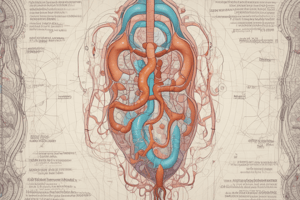
Kidney
- 2 bean-shaped organs with a thin connective tissue capsule and surrounded by adipose tissue.
- Formed of outer cortex and inner medulla.
- Outer Cortex: Reddish due to rich blood supply, granular (renal corpuscles), underneath the capsule.
- Inner Medulla: Gray in color, consists of 6-12 medullary pyramids, each with an apex directed to the minor calyx, opening into major calyx, then to the renal pelvis.
- Urinary passages lined by transitional epithelium.
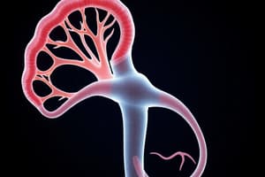
Uriniferous Tubules
- Nephron and collecting ducts form uriniferous tubules.
- Nephrons: Structural and functional units, form urine.
- Renal corpuscle: Double-walled capsule, outer layer (parietal, lined by simple squamous epithelium), inner layer (visceral, modified simple squamous cells called podocytes), and glomerulus.
- Nephrons: Structural and functional units, form urine.
- Collecting ducts: Concentrate and carry urine to ureters.
Studying That Suits You
Use AI to generate personalized quizzes and flashcards to suit your learning preferences.
Related Documents
Description
This quiz covers essential topics in human anatomy, focusing on the structure and function of the kidney and different types of cartilage. Test your knowledge on nephron components, cartilage types, and bone formation. Perfect for students studying anatomy and physiology.