Podcast
Questions and Answers
What is the primary function of the extensor retinaculum?
What is the primary function of the extensor retinaculum?
- To stabilize the wrist during rotation
- To provide muscle attachment
- To flex the wrist
- To hold the long extensor tendons in position (correct)
The extensor retinaculum is located on the front of the wrist.
The extensor retinaculum is located on the front of the wrist.
False (B)
What type of tissue is the extensor retinaculum primarily composed of?
What type of tissue is the extensor retinaculum primarily composed of?
Deep fascia
The extensor retinaculum is responsible for holding the long ______ tendons in position.
The extensor retinaculum is responsible for holding the long ______ tendons in position.
Match the following anatomical structures with their descriptions:
Match the following anatomical structures with their descriptions:
What is the primary function of the extensor in the arm?
What is the primary function of the extensor in the arm?
The extensor is located on the anterior surface of the radius and ulna.
The extensor is located on the anterior surface of the radius and ulna.
What structures do the extensor tendons pass through?
What structures do the extensor tendons pass through?
The extensor converts the grooves on the retinaculum into __________ for the passage of the long extensor tendons.
The extensor converts the grooves on the retinaculum into __________ for the passage of the long extensor tendons.
Match the following anatomical structures with their descriptions:
Match the following anatomical structures with their descriptions:
Study Notes
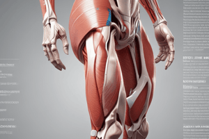
Extensor Retinaculum
- Thickening of deep fascia located at the back of the wrist.
- Holds long extensor tendons in position during movement.
- Converts grooves on the posterior surface of the radius and ulna into six separate tunnels for tendon passage.
- Lateral border is defined by the tendons of abductor pollicis longus and extensor pollicis brevis.
- Medial border is defined by the tendon of extensor pollicis longus.
- The floor of the impression is formed by the scaphoid and trapezium bones.
- The radial artery runs on the floor of the anatomical snuffbox.
- Function: Aids in the adduction of the thumb, index, ring, and little fingers at the metacarpophalangeal joints.
Adductor Pollicis
- Composed of two heads: transverse and oblique.
- Transverse head originates from the third metacarpal bone.
- Oblique head originates from the capitate bone and the bases of the second and third metacarpal bones.
- Plays a crucial role in adduction of the thumb.
Studying That Suits You
Use AI to generate personalized quizzes and flashcards to suit your learning preferences.
Related Documents
Description
Explore the anatomy of the Extensor Retinaculum and the Adductor Pollicis muscle. This quiz covers their definitions, functions, and anatomical relationships. Test your knowledge on these important structures in the human wrist and hand.