Podcast
Questions and Answers
What do the zygomatic bones and temporal bones form?
What do the zygomatic bones and temporal bones form?
The sides of the upper lip.
Which of the following structures are a part of the brachial apparatus?
Which of the following structures are a part of the brachial apparatus?
- Branchial arches (correct)
- Dental lamina
- Pharyngeal pouches (correct)
- Cervical loop
When does the palate begin formation?
When does the palate begin formation?
In the 5th week.
The primary palate contains the maxillary canines and posterior teeth.
The primary palate contains the maxillary canines and posterior teeth.
What is the main function of the cervical loop in root development?
What is the main function of the cervical loop in root development?
Which type of mucosa includes the buccal and alveolar mucosa?
Which type of mucosa includes the buccal and alveolar mucosa?
What happens during the initiation stage of tooth development?
What happens during the initiation stage of tooth development?
Cementum can continue to be produced after tooth eruption.
Cementum can continue to be produced after tooth eruption.
What type of epithelium is found in the lining mucosa?
What type of epithelium is found in the lining mucosa?
What are the primary cells found in the pulp of a tooth?
What are the primary cells found in the pulp of a tooth?
What are the functions of the central cells of the dental papilla?
What are the functions of the central cells of the dental papilla?
What is the largest group of cells found in the dental pulp?
What is the largest group of cells found in the dental pulp?
The two types of cementum are ___ and ___ cementum.
The two types of cementum are ___ and ___ cementum.
Which cell types are found in alveolar bone?
Which cell types are found in alveolar bone?
What mineral composition makes up calcium hydroxyapatite?
What mineral composition makes up calcium hydroxyapatite?
The periodontal ligament (PDL) is innervated and vascular.
The periodontal ligament (PDL) is innervated and vascular.
What are the main types of fiber groups found in the PDL?
What are the main types of fiber groups found in the PDL?
Match the following pulp zones with their descriptions:
Match the following pulp zones with their descriptions:
Which of the following are the four basic histological types of tissues?
Which of the following are the four basic histological types of tissues?
What tissue type covers and lines the external and internal body surfaces?
What tissue type covers and lines the external and internal body surfaces?
Epithelial tissue is vascular.
Epithelial tissue is vascular.
What is the classification of epithelial tissues based on?
What is the classification of epithelial tissues based on?
What are rete ridges?
What are rete ridges?
The _____ layer of the basement membrane is nearer to the epithelium.
The _____ layer of the basement membrane is nearer to the epithelium.
What is the most common cell type in all types of connective tissue?
What is the most common cell type in all types of connective tissue?
Which type of connective tissue serves as a skeletal tissue in the body?
Which type of connective tissue serves as a skeletal tissue in the body?
What type of cartilage is the most common?
What type of cartilage is the most common?
Match the following types of muscle tissue with their characteristics:
Match the following types of muscle tissue with their characteristics:
What is the primary function of nerve tissue?
What is the primary function of nerve tissue?
Mitosis results in daughter cells that are genetically identical to the parent cell.
Mitosis results in daughter cells that are genetically identical to the parent cell.
The _____ period of prenatal development begins at the start of pregnancy.
The _____ period of prenatal development begins at the start of pregnancy.
What happens during the preimplantation period?
What happens during the preimplantation period?
Which layer of the embryo develops into the central nervous system?
Which layer of the embryo develops into the central nervous system?
What does the mandibular arch give rise to?
What does the mandibular arch give rise to?
The frontonasal process gives rise to the upper lip.
The frontonasal process gives rise to the upper lip.
Flashcards are hidden until you start studying
Study Notes
Histology Overview
- Tissues classified into four basic types: Epithelial, Connective, Muscle, Nerve.
Epithelial Tissue
- Covers and lines external and internal body surfaces including vessels and cavities.
- Composed of closely packed polyhedral cells with minimal intercellular substance.
- Cells tightly joined by desmosomes; attach to non-cellular surfaces through hemidesmosomes.
- Avascular, obtaining nutrients by diffusion from adjacent connective tissue; exhibits rapid turnover rates.
Classification of Epithelial Tissues
- Simple epithelium: single layer of cells.
- Stratified epithelium: two or more layers; only the basal layer contacts the basement membrane.
- Stratified squamous epithelium is the most common type.
Rete Ridges
- Extensions of epithelium into connective tissue, known as rete ridges or rete pegs.
Basement Membrane
- Thin acellular layer separating epithelium from connective tissue; consists of two layers:
- Basal lamina (with lamina lucida and lamina densa) and reticular lamina.
- Basal lamina anchors to connective tissue.
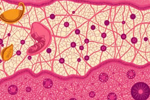
Connective Tissue
- Composed of fewer, more dispersed cells with abundant intercellular substance and fibers.
- Generally vascularized, serving functions like support, transport, insulation, and defense.
- Fibroblast is the predominant cell type, with slower turnover compared to epithelial tissue.
Connective Tissue Layers
- Connective tissue proper has loose and dense layers.
- Papillary layer (loose connective tissue) and reticular layer (dense connective tissue) found under epithelium.
Cartilage
- Firm, non-calcified tissue serving as skeletal support; present in embryonic skeleton and joints.
- Avascular and relies on surrounding tissue for nutrition.
- Chondroblasts produce cartilage matrix; chondrocytes maintain it.
- Three types: Hyaline, Elastic, and Fibrocartilage.
Cartilage Growth
- Interstitial growth: from within the tissue.
- Appositional growth: from the outer layers.
Bone
- Rigid connective tissue that forms the skeleton, with periosteum as the outer covering.
- Compact bone: heavy, dense; Cancellous bone: lighter with spaces.
- Bone matrix formed initially as osteoid by osteoblasts; osteocytes maintain it, while osteoclasts resorb bone.
Blood
- Fluid connective tissue transporting nutrients; composed of plasma and cells.
- Key formed elements: red blood cells (RBCs), white blood cells (WBCs), platelets.
- RBCs transport oxygen and carbon dioxide; WBCs play a role in immune response; platelets assist in clotting.
Muscle Tissue
- Muscular system consists of three types: Smooth (involuntary), Cardiac (involuntary), Skeletal (voluntary).
- Skeletal muscle is striated, made up of myofibrils composed of myofilaments.
Nerve Tissue
- Nerves consist of bundles of neural processes; transmit electrical impulses for muscle contraction, gland stimulation, and sensation.
- Neurons are the functional unit, made up of a cell body, axon, and dendrites.
Developmental Processes
- Mitosis results in two genetically identical daughter cells; meiosis reduces chromosomes for reproductive cells.
Prenatal Development
- Begins at conception; consists of preimplantation, embryonic, and fetal periods.
- Preimplantation: zygote formation and initial cell division.
Embryonic Period
- From implantation to the 8th week; includes formation of bilaminar and trilaminar discs with ectoderm, mesoderm, and endoderm.
- Central nervous system development begins; neural crest contributes to face and neck structures.
Fetal Period
- From week 9 to birth; marked by maturation of the embryo into a fetus.
Facial and Palatal Fusion
- Facial fusion: elimination of grooves between swellings; palatal fusion involves merging of structures from different surfaces.
Brachial Apparatus
- Composed of branchial arches, grooves, and pharyngeal pouches, contributing to head and neck structures.
- First branchial groove forms the external auditory meatus; other grooves disappear by seventh week.
Palatal Development
- Begins in the 5th week; intermaxillary segment forms the primary palate which holds maxillary incisors.### Palate Development
- Primary palate separates nasal and oral cavities by the 5th week.
- Secondary palate forms from the maxillary processes during the 6th week, with palatal shelves fusing medially.
- Secondary palate comprises the posterior two-thirds of the hard palate, containing maxillary canines and posterior teeth, along with the soft palate and uvula.
- Complete fusion of the primary and secondary palates occurs from anterior to posterior.
Nasal Cavity and Septum Development
- Nasal cavity development occurs concurrently with palate formation between the 5th and 12th weeks.
- The vertical nasal septum fuses with the final palate by the 9th week, completing by the 12th week.
- This fusion fully separates the nasal and oral cavities.
Tongue Development
- The tongue develops from the 4th to 8th weeks through structures like the tuberculum impar, lateral lingual swellings, copula, and epiglottic swelling.
- By the end of the 8th week, the tongue has completed the fusion of its swellings and moves into the oral cavity.
Tooth Development
- Initiated in the 7th week with 20 primary teeth, continuing into late teens for 32 permanent teeth.
- Stages of tooth development include: bud stage, cap stage, bell stage, apposition stage, and maturation stage.
- Key structures involved: dental lamina, enamel organ, dental papilla, and dental sac.
Bell Stage
- Inner enamel epithelium (IEE) differentiates into ameloblasts forming enamel matrix.
- Dental papilla consists of outer cells for dentin formation (odontoblasts) and central cells that form primordium.
Apposition Stage
- IEE cells differentiate into preameloblasts, which induce odontoblast differentiation from outer dental papilla cells.
- Odontoblasts begin dentinogenesis from the basement membrane, while preameloblasts differentiate into ameloblasts and start amelogenesis.
Root Development
- Occurs post-crown completion; the cervical loop is crucial for root shape and dentin formation.
- The Hertwig's epithelial root sheath (HERS) is formed from the cervical loop, shaping the root and inducing dentin formation.
Cementum Formation
- After HERS disintegration, undifferentiated dental sac cells become cementoblasts, covering root dentin with cementoid.
Oral Mucosa
- Composed of stratified squamous epithelium and lamina propria, permitting flexibility, moisture, and connective tissue support.
- Categories include lining mucosa, masticatory mucosa, and specialized mucosa.
Lining Mucosa
- Characterized by soft texture, moisture, and stretchability; includes the buccal and labial areas.
- Histologically linked to non-keratinized stratified squamous epithelium.
Masticatory Mucosa
- Features rubbery texture and resiliency; includes attached gingiva and hard palate.
- Comprised of keratinized stratified squamous epithelium with pronounced connective tissue papillae.
Specialized Mucosa
- Present on the tongue within lingual papillae, with varied keratinization.
- Taste buds found in certain papillae types, contributing to taste sensation.
Types of Stratified Squamous Epithelium
- Non-keratinized is the most common and has three layers: basal, intermediate, and superficial.
- Ortho-keratinized is the least common and includes a keratin layer.
- Para-keratinized is found in masticatory mucosa with nucleated cells in the keratin layer.
Lamina Propria
- A support structure carrying nerves and blood vessels, consisting of a papillary layer (loose connective tissue) and a reticular layer (dense connective tissue).
Basement Membrane
- A thin, acellular structure between epithelium and connective tissue composed of a basal lamina and reticular lamina.
Specialized Mucosa of the Tongue
- Covered by lingual papillae, with different types including filiform, fungiform, foliate, and circumvallate, each contributing to taste sensation.
Tooth and Support Tissues
- Enamel: Avascular, non-renewable, produced by ameloblasts, with features like lines of Retzius.
- Dentin: Formed by odontoblasts through dentinogenesis.
- Pulp: Contains fibroblasts and odontoblasts, serving nutritive, formative, sensory, and protective functions.
- Cementum: Avascular, renewable tissue adhering the tooth, produced by cementoblasts.
- Alveolar Bone: Supports teeth, with terms such as cribriform plate and lamina dura.
- Periodontal Ligament (PDL): Connects teeth to the alveolar bone, consists of fiber bundles and cells like fibroblasts and osteoblasts.
Studying That Suits You
Use AI to generate personalized quizzes and flashcards to suit your learning preferences.