Podcast
Questions and Answers
What is the primary purpose of histopathology in the study of tissues?
What is the primary purpose of histopathology in the study of tissues?
- To provide diagnostic information about diseases (correct)
- To classify tissues based on cell types
- To examine cell organization within tissues
- To develop new biomarker therapies
Which of the following best describes the function of desmosomes in epithelial cells?
Which of the following best describes the function of desmosomes in epithelial cells?
- To attach adjacent epithelial cells together (correct)
- To anchor epithelial cells to the basement membrane
- To facilitate nutrient exchange between cells
- To form a barrier against pathogens
What distinguishes stratified epithelial tissue from simple epithelial tissue?
What distinguishes stratified epithelial tissue from simple epithelial tissue?
- Stratified tissue has a single layer of cells
- Stratified tissue has multiple layers of cells (correct)
- Stratified tissue is always glandular
- Stratified tissue is exclusively found in the heart
What is the role of a microtome in tissue study?
What is the role of a microtome in tissue study?
In what context would immunohistochemistry be most effectively applied?
In what context would immunohistochemistry be most effectively applied?
What characteristic is unique to connective tissues, such as blood?
What characteristic is unique to connective tissues, such as blood?
Which type of microscopy is specifically used to visualize the internal structures of a tissue sample?
Which type of microscopy is specifically used to visualize the internal structures of a tissue sample?
In epithelial tissues, where are hemidesmosomes primarily located?
In epithelial tissues, where are hemidesmosomes primarily located?
Which type of epithelium features a single layer that appears to be composed of multiple layers due to the position of the nuclei?
Which type of epithelium features a single layer that appears to be composed of multiple layers due to the position of the nuclei?
What is the primary structural protein produced by keratinocytes that leads to keratinisation?
What is the primary structural protein produced by keratinocytes that leads to keratinisation?
Which junction is primarily responsible for maintaining cell polarity and preventing paracellular transport in epithelial tissues?
Which junction is primarily responsible for maintaining cell polarity and preventing paracellular transport in epithelial tissues?
Which feature differentiates cilia from microvilli?
Which feature differentiates cilia from microvilli?
What type of epithelium is specialized for protection and is typically found in areas such as the skin and the oral cavity?
What type of epithelium is specialized for protection and is typically found in areas such as the skin and the oral cavity?
Which cell type is primarily involved in secretory functions within epithelial tissues?
Which cell type is primarily involved in secretory functions within epithelial tissues?
What unique ability do umbrella cells in transitional epithelium have?
What unique ability do umbrella cells in transitional epithelium have?
Which of the following best describes the apical side of epithelial cells?
Which of the following best describes the apical side of epithelial cells?
Which type of epithelial tissue is characterized by its ability to change shape depending on the state of the organ it lines?
Which type of epithelial tissue is characterized by its ability to change shape depending on the state of the organ it lines?
Which of the following epithelial specializations can move substances along the epithelial surface through their motility?
Which of the following epithelial specializations can move substances along the epithelial surface through their motility?
Flashcards
Cuboidal cell
Cuboidal cell
Cells are round, cube-shaped.
Columnar cell
Columnar cell
Cells are taller than they are wide.
Squamous cell
Squamous cell
Cells are flat and thin.
Ciliated epithelium
Ciliated epithelium
Signup and view all the flashcards
Keratinized epithelium
Keratinized epithelium
Signup and view all the flashcards
Pseudostratified epithelium
Pseudostratified epithelium
Signup and view all the flashcards
Transitional epithelium
Transitional epithelium
Signup and view all the flashcards
Cell polarity
Cell polarity
Signup and view all the flashcards
Tight junction
Tight junction
Signup and view all the flashcards
Gap junction
Gap junction
Signup and view all the flashcards
Histology
Histology
Signup and view all the flashcards
Histopathology
Histopathology
Signup and view all the flashcards
Tissue preparation
Tissue preparation
Signup and view all the flashcards
Microtome
Microtome
Signup and view all the flashcards
Haematoxylin
Haematoxylin
Signup and view all the flashcards
Eosin
Eosin
Signup and view all the flashcards
Four main tissue types
Four main tissue types
Signup and view all the flashcards
Glandular epithelial tissue
Glandular epithelial tissue
Signup and view all the flashcards
Study Notes
Introduction to Histology
- Histology is the study of tissues, focusing on how cells organize into functional tissues.
- It is vital for diagnostics (histopathology) and research into human diseases, drug treatments, and biomarkers.
- Tissue samples need to be fixed (chemically or physically), embedded in resin or wax, and sliced thinly using a microtome for light microscopy.
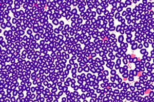
- Staining is crucial for visualizing specific structures. Eosin (red) stains cytoplasm, and haematoxylin (purple) stains the nucleus. H&E staining combines both. Other stains like immunohistochemistry use antibodies to reveal specific proteins.
- Electron microscopy (transmission and scanning) allows for higher resolution imaging.
Four Main Tissue Classifications
- Muscle tissue
- Epithelial tissue
- Connective tissue (e.g., cartilage, blood) - blood fulfills the definition of connective tissue by containing cells suspended in a medium.
- Nervous tissue
Epithelial Tissue
-
Epithelial tissue forms coverings or glands.
-
Covering epithelium: lines cavities and covers surfaces
-
Glandular epithelium: includes secretory epithelial cells with an acinus region for product formation.
-
Epithelial cells adhere to a basement membrane (basal end) via hemidesmosomes and neighbouring cells via desmosomes. This structure helps maintain tissues' integrity and function.
-
Epithelial tissue is classified based on the number of cell layers and cell shape.
-
Number of layers: simple (one layer) / stratified (multiple layers). Simple epithelium lines surfaces where minimal friction exists (e.g., alveoli, pericardium). Stratified epithelium is found in areas experiencing high friction (e.g., mouth, esophagus), offering protection.
-
Cell shape: cuboidal (cube-shaped), squamous (flat), columnar (taller than wide).
-
Specializations:
- Ciliated: have microtubule-based cilia for moving substances (e.g., mucus in the respiratory tract).
- Keratinized: surfaces with protective layers of keratin, derived from dead cells (e.g., skin, hair, nails).
-
Exceptions:
- Pseudostratified: appears stratified but is a single layer with cells positioned at different levels.
- Transitional/urothelium: stratified epithelium with variable cell shape adapting to stretch; umbrella cells in the top layer prevent urine leakage.
Cell Polarity
- Cell polarity refers to the asymmetrical distribution of functions/specializations within a cell, maintaining structure and function.
- The apical (luminal) side faces the exterior/lumen where specializations (microvilli, cilia) reside, while the basal side anchors to the basement membrane. The lateral/intercellular side connects adjacent epithelial cells via cell junctions.
Cell Junctions
- Tight junctions (TJ), adherens junctions (ZA), gap junctions (CJ), desmosomes (D), and hemidesmosomes (HD). These help maintain cell and tissue structure and function.
Cilia vs. Microvilli
- Cilia are motile structures (with microtubules) and are longer, while microvilli (with actin filaments) are smaller and non-motile.
- Microvilli are often highly developed in absorptive tissues, while cilia are found in specific places like respiratory and reproductive tracts.
Keratinization
- A special characteristic of certain squamous epithelial cells.
- Keratin (a protein) is produced and accumulates in cells, which eventually die. This creates a protective layer.
Secretory Epithelial Cells
- Individual secretory cells (like goblet cells) and gland structures (e.g., multicellular glands) are forms of secretory epithelial cells.
Studying That Suits You
Use AI to generate personalized quizzes and flashcards to suit your learning preferences.