Podcast
Questions and Answers
Which of the following is the primary function of hemostasis?
Which of the following is the primary function of hemostasis?
- To decrease oxygen supply to cells
- To accelerate the inflammatory response
- To prevent blood loss when blood vessels are damaged (correct)
- To increase blood flow to injured tissues
Vascular spasm is a short-term response to vessel damage, typically lasting for several hours.
Vascular spasm is a short-term response to vessel damage, typically lasting for several hours.
False (B)
Which mechanism is responsible for vascular spasm?
Which mechanism is responsible for vascular spasm?
- Increased blood pressure
- Release of antithrombin III
- Decreased vessel permeability
- Local myogenic contraction (correct)
Platelet activation is initiated by the contact with ______ fibers within subendothelial layers.
Platelet activation is initiated by the contact with ______ fibers within subendothelial layers.
Which of the following is NOT a characteristic of thrombocytes?
Which of the following is NOT a characteristic of thrombocytes?
Von Willebrand factor (vWF) promotes platelet stickiness.
Von Willebrand factor (vWF) promotes platelet stickiness.
Which of the following substances released by platelets supports thrombocyte aggregation?
Which of the following substances released by platelets supports thrombocyte aggregation?
Platelets are produced in the bone marrow from cells called ______.
Platelets are produced in the bone marrow from cells called ______.
Match the step of platelet plug formation with its description:
Match the step of platelet plug formation with its description:
Which of the following describes the primary function of a platelet plug?
Which of the following describes the primary function of a platelet plug?
Which of the following is a function of endothelial cells?
Which of the following is a function of endothelial cells?
Endothelial cells encourage platelet activation.
Endothelial cells encourage platelet activation.
Nitric oxide produced by endothelium leads to ______.
Nitric oxide produced by endothelium leads to ______.
Which substance produced by endothelium helps platelets stick together and adhere to the walls of blood vessels at the site of a wound?
Which substance produced by endothelium helps platelets stick together and adhere to the walls of blood vessels at the site of a wound?
Match the following substances with their function in relation to the endothelium:
Match the following substances with their function in relation to the endothelium:
What is the critical role of calcium ($Ca^{+2}$) in blood coagulation?
What is the critical role of calcium ($Ca^{+2}$) in blood coagulation?
Clotting factors are primarily synthesized by kidney cells.
Clotting factors are primarily synthesized by kidney cells.
In the coagulation cascade, each clotting factor activates the next in a fixed sequence, ultimately leading to the formation of a ______ net.
In the coagulation cascade, each clotting factor activates the next in a fixed sequence, ultimately leading to the formation of a ______ net.
Which vitamin is required for the synthesis of prothrombin, FVII, FIX, and FX?
Which vitamin is required for the synthesis of prothrombin, FVII, FIX, and FX?
Which of the following is NOT a characteristic of clotting factors?
Which of the following is NOT a characteristic of clotting factors?
Match the clotting factor to its function:
Match the clotting factor to its function:
The extrinsic pathway of coagulation is initiated by damaged endothelium.
The extrinsic pathway of coagulation is initiated by damaged endothelium.
The intrinsic pathway is activated when blood comes in contact with ______.
The intrinsic pathway is activated when blood comes in contact with ______.
In the final common pathway of coagulation, what does prothrombin activator catalyze?
In the final common pathway of coagulation, what does prothrombin activator catalyze?
Fibrinogen is converted to insoluble fibrin threads by thrombin.
Fibrinogen is converted to insoluble fibrin threads by thrombin.
What is the role of Factor XIII (fibrin-stabilizing factor)?
What is the role of Factor XIII (fibrin-stabilizing factor)?
The ______ test is used to evaluate the function of the extrinsic system.
The ______ test is used to evaluate the function of the extrinsic system.
Which of the following indicates that an interaction between both pathways in the coagulation cascade is necessary for hemostasis?
Which of the following indicates that an interaction between both pathways in the coagulation cascade is necessary for hemostasis?
What does a prolonged aPTT indicate?
What does a prolonged aPTT indicate?
The PT and aPTT lab tests accurately predict the bleeding tendency of a patient.
The PT and aPTT lab tests accurately predict the bleeding tendency of a patient.
Match phases from the cell-based model with what they initiate:
Match phases from the cell-based model with what they initiate:
The amplification phase is from?
The amplification phase is from?
During the propagation phase, factors on the thrombocyte surface produce additional FXa, which will convert ______ to fibrin.
During the propagation phase, factors on the thrombocyte surface produce additional FXa, which will convert ______ to fibrin.
Which is true about the intrinsic and extrinsic pathways?
Which is true about the intrinsic and extrinsic pathways?
During clot retraction, serum is absorbed back into the bloodstream.
During clot retraction, serum is absorbed back into the bloodstream.
During vessel repair, which of the following stimulates division of smooth muscle cells and fibroblasts to rebuild the blood vessel wall?
During vessel repair, which of the following stimulates division of smooth muscle cells and fibroblasts to rebuild the blood vessel wall?
Fibrinolysis is the process of clot formation.
Fibrinolysis is the process of clot formation.
Plasminogen becomes ______, which digests fibrin threads.
Plasminogen becomes ______, which digests fibrin threads.
Which of the following naturally prevents clot formation in vessels?
Which of the following naturally prevents clot formation in vessels?
Match the substance to its anticoagulant mechanism:
Match the substance to its anticoagulant mechanism:
Which of the following is an example of an anticoagulant that acts as an antivitamin K?
Which of the following is an example of an anticoagulant that acts as an antivitamin K?
A clot (thrombus) forming in an unbroken blood vessel
A clot (thrombus) forming in an unbroken blood vessel
______ blocks synthesis of thromboxane A2 preventing clot formation.
______ blocks synthesis of thromboxane A2 preventing clot formation.
Flashcards
Function of Hemostasis
Function of Hemostasis
Prevents blood loss when blood vessels are damaged and stops bleeding quickly in a localized manner.
Vascular Spasm
Vascular Spasm
Initiated instantaneously, lasts about 30 minutes, reduces blood loss until other mechanisms take over.
Thrombus
Thrombus
A blood clot that forms inside a blood vessel or the heart.
Embolus
Embolus
Signup and view all the flashcards
Clot prevention
Clot prevention
Signup and view all the flashcards
Anticoagulants
Anticoagulants
Signup and view all the flashcards
von Willebrand factor (vWF)
von Willebrand factor (vWF)
Signup and view all the flashcards
Thrombin
Thrombin
Signup and view all the flashcards
Platelet-derived growth factor (PDGF)
Platelet-derived growth factor (PDGF)
Signup and view all the flashcards
Blood Coagulation
Blood Coagulation
Signup and view all the flashcards
Clot
Clot
Signup and view all the flashcards
Platelet Plug Formation
Platelet Plug Formation
Signup and view all the flashcards
Fibrinolysis
Fibrinolysis
Signup and view all the flashcards
Vitamin K
Vitamin K
Signup and view all the flashcards
t-PA
t-PA
Signup and view all the flashcards
Study Notes
- Hemostasis is the process that prevents blood loss when blood vessels are damaged and stops bleeding in a quick and localized manner
Successful Hemostatic Process
- Speed of reaction is important
- Localization must be at the site of vessel wall injury
- Controllability of the reaction must exist
Hemostatic Processes:
- Vascular spasm
- Platelet plug formation
- Blood coagulation
- Permanent closing of the hole in the vessel
Vascular Spasm
- Occurs instantaneously and lasts for 30 minutes
- Proportional to the vessel wall damage
- Reduces blood loss until other mechanisms can take over
- Mechanisms include nervous reflexes, local myogenic contraction, and vasoconstrictors such as serotonin and thromboxane A₂
Platelet Plug Formation
- Thrombocytes(platelets) are activated by contact with collagen fibers within the subendothelial layers
Thrombocytes
- Round or oval, biconvex disc-shaped cell fragments with a diameter of 2-4 μm
- Thrombocytes lack a nucleus
- Contain granules, mitochondria, rough ER, and ribosomes
- Dense granules contain serotonin, ADP, ATP, and Ca2+
- Serotonin and thromboxane A₂ are vasoconstrictors
- ADP causes stickiness
- α granules contain von Willebrand factor (vWF), platelet factor 4 (PF4), platelet-derived growth factor (PDGF), FV, and FXIII
- vWF acts as a connecting agent
- The average number of thrombocytes is 150 - 400 x 10^9/L
- Thrombocytes have an average lifespan of 8 - 12 days
- Produced in the bone marrow by megakaryocytes through thrombopoiesis, stimulated by thrombopoietin
- Eliminated by the tissue macrophage system in the spleen, liver, and lungs
Substances Released by Platelets
- Platelet-derived growth factor (PDGF) from α-granules is involved in angiogenesis and helps repair damaged vascular walls
- Platelet factor 4 from α-granules is a heparin antagonist
- Von Willebrand factor (vWF) from α-granules helps platelets stick together and adhere to the walls of blood vessels at the site of a wound
- Serotonin from dense granules causes vasoconstriction
- ADP and ATP from dense granules support thrombocyte aggregation
- Ca2+ from dense granules facilitates the activation of many proteins and cytoskeletal rearrangements
- Platelet factor 3 from the cell membrane comprises phospholipids that activate some coagulation factors
- Glycoproteins IIb and IIIa from the cell membrane serve as receptors for fibrinogen and are essential for platelet aggregation
- Thromboxane A2 from the cytoplasm causes vasoconstriction
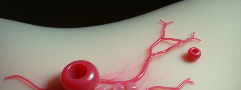
Steps in Platelet Plug Formation
- Adhesion occurs when platelets stick to exposed collagen, leading to activation
- Change of shape is a result of activation
- Release reaction leads to aggregation and is activated by thromboxane A2, serotonin, and ADP
Function of Platelet Plug
- Important for closing minute ruptures in very small blood vessels, which occur hundreds of times daily
- Occludes the hole in the vessel wall without occluding the vessel itself
- Surface of the activated platelet acts as a cofactor for several enzymatic reactions of the coagulation cascade
- Thrombocytopenic purpura is characterized by hundreds of small hemorrhagic areas under the skin and throughout the internal tissues due to a deficiency of platelets
Endothelial Cells
- Play a significant role in some blood functions
- Create a continuous, non-wetting liner of the inner vessel wall
- Attached to each other by a tight junction with varying degrees of tightness
- Functions include regulation of the coagulation cascade, inhibition of platelet activation, regulation of the permeability of the vessels, involvement in the inflammatory process and its regulation, and control of optimal blood flow through the blood vessel
Substances Produced by Endothelium
- Nitric oxide causes Vasodilation
- Prostacyclin causes Vasodilation, antiaggregation, and antiadhesive effects
- Angiotensin II causes Vasoconstriction
- Endothelin 1 causes Vasoconstriction, proadhesive, and proagregative effects
- Platelet-activating factor (PAF) is involved in the aggregation and degranulation of thrombocytes
- von Willebrand factor (vWf) is a prothrombotic factor and helps platelets stick together and adhere to the walls of blood vessels at the site of a wound
- Tissue activator of plasminogen (tPA) facilitates the conversion of plasminogen to plasmin
- Plasminogen activator inhibitor 1 (PAI-1) inhibits tPA
Blood Coagulation
- Blood drawn from the body thickens into a gel through a cascade of reactions
- Each clotting factor activates the next in a fixed sequence, leading to the formation of a fibrin net
- Blood coagulation only occurs at the site of the injury
- Substances required for clotting include Ca+2, enzymes and factors synthesized by liver cells, and substances released by platelets or damaged tissues
Clotting Factors
- Proteins involved in the process of clotting
- Produced mainly in the liver
- Present in the plasma
- Clotting factors are designated by a Roman numeral and a name
- They can function as enzymes or cofactors
Role of Vitamin K in Clotting
- Vitamin K is required for the synthesis of prothrombin, FVII, FIX, and FX
- Sources include food and bacteria in the intestine
- Vitamin K is a fat-soluble vitamin that is absorbed if lipids are present
- It is inactivated by coumarin, which is used in clinical practice
Coagulation Cascade
- Blood coagulation occurs in vitro in three essential steps
- Formation of the prothrombin activator
- Conversion of prothrombin to thrombin
- Conversion of fibrinogen to fibrin threads
- Fibrin threads trap blood cells and proteins
Extrinsic Pathway
- Damaged tissue cells bear tissue factor (TF; thromboplastin)
- FXa combines with FV to form prothrombin activator in the presence of Ca+2, FVIIa, and phospholipids
- Prothrombin activator forms in seconds (15-20 s)
Intrinsic Pathway
- Activated when blood comes in contact with collagen inside damaged endothelium
- Involves the activation of coagulation factors and platelets
- Platelets aggregate, and phospholipids uncover
- Requires several minutes for production of prothrombin activator
Final Common Pathway
- Prothrombin activator (prothrombinase complex) includes FXa and FVa; the presence of Ca+2 and membrane phospholipids is required
- It catalyzes the conversion of prothrombin to thrombin
- Thrombin converts soluble fibrinogen to insoluble fibrin threads and activates FXIII
- Positive feedback cycle
Fibrin
- Produced from fibrinogen (high molecular weight) by thrombin
- Fibrinogen splits into fibrin monomer and low molecular weight peptides (fibrinopeptides A and B)
- Fibrin monomers polymerize to form fibrin threads
- Fibrin-stabilizing factor (FXIII) creates covalent bonds between the fibrin monomer molecules, resulting in multiple cross-linkages between adjacent fibrin threads
- Fibrin threads adhere to damaged surfaces of blood vessels
- A clot consists of a meshwork of fibrin threads running in all directions and entrapping blood cells
- Fibrin stimulates the formation of granulation tissue and initiates mechanisms leading to the repair of the damaged tissue
Testing Functioning of Pathways
- PT (Quicks test) measures prothrombin time, which is normally 12-15 s and evaluates the function of the extrinsic system
- aPTT measures activated partial thromboplastin time, which is normally 30-40 s and evaluates the function of the intrinsic system
- aPTT and/or PT results can detect a deficiency of involved factors but cannot identify which one
- These tests do not predict bleeding tendency accurately
Limitations of Cascade Model
- FVIII or FIX deficiency leads to bleeding even though the extrinsic and common pathways are normal
- FXI deficiency leads to variable hemostatic deficits with bleeding
- FXII deficiency does not lead to bleeding, despite the requirement of this factor for initiating the intrinsic pathway, but aPTT is prolonged
- The intrinsic or extrinsic pathways alone cannot lead to appropriate thrombus production; an interaction between both pathways is necessary for hemostasis "in vivo"
- This interaction can be explained by the cell-based model
Cell-Based Model
- Involves two surfaces: TF-bearing cells and platelets
- Occurs in three phases
- Initiation
- Amplification
- Propagation
Initiation Phase
- Occurs outside of the vascular compartment on many interstitial cells
- TF and FVIIa form the TF/FVIIa complex
- TF/FVIIa activates FIX and FX
- FXa binds to FVa, resulting in the formation of prothrombinase on the surface of TF-bearing cells
- This leads to the generation of a small amount of thrombin
- Amount of thrombin generated is not sufficient to lead to widespread clot formation
Amplification phase
- Small amount of produced thrombin activates more platelets and their flip-flop mechanisms
- Results in the generation of a platelet procoagulant phospholipid surface
- Also activates endothelial cells
- Cleaves FV, FVIII, and FXI on the platelet membrane to their active forms
- FXIa activates FIX to FIXa
- Platelets become coated in activated coagulation factors, leading to an increased amount of thrombin production
Propagation Phase
- Additional FXa produced due to activity of factors on the thrombocyte surface
- FXa and FVa, along with calcium and a phospholipid (surface of activated platelets), form the prothrombinase complex
- A large amount of thrombin is generated, which is enough to catalyze the reaction of converting fibrinogen to fibrin
- Fibrin structure of the clot is stabilized by covalent bonding under the influence of FXIIIa
In vivo Coagulation
- Intrinsic and extrinsic pathways are not redundant
- Operate on different cellular surfaces to fill different roles
- Extrinsic - on the TF-bearing (initiating) cells
- Intrinsic pathway - on platelets
Clot Retraction
- Stabilizes clot
- Actin and myosin in platelets contract within 30-60 minutes
- Platelets pull on fibrin threads, causing clot retraction and expelling serum
- Edges of damaged vessel pulled together
Blood Vessel Repair
- Vessel heals as clot retraction occurs
- Platelet-derived growth factor (PDGF) stimulates the division of smooth muscle cells and fibroblasts to rebuild blood vessel wall
- Vascular endothelial growth factor (VEGF) stimulates endothelial cells to multiply and restore endothelial lining
- Leads to replacement of damaged tissue with a new tissue (other properties than the original)
Fibrinolytic System
- Dissolves small or inappropriate clots and clots at a site of completed repair through fibrinolysis
- Inactive plasminogen incorporated into the clot becomes plasmin, which digests fibrin threads
Clot Prevention
- Smoothness of the endothelium
- Clot formation limited in time and space
- Bloodstream disperses clotting factors
- Antithrombin III with heparin
- Fibrin-antithrombin effect
- Macrophage activity
- Protein C and protein S system (elimination of FVa and FVIIIa)
- Thrombomodulin
Anti-Coagulation Agents
- Suppress or prevent blood clotting
- In vivo: Heparin, released by basophils and coumarin derivatives (antivitamin K)
- In vitro: Stored blood in blood banks treated with citrate phosphate dextrose (CPD) that removes Ca+2
- Thrombolytic agents dissolve clots
- Tissue plasminogen activator (t-PA), Urokinase, and Streptokinase
Inhibition of Coagulation
- Coumarine derivatives act as an antivitamin K, leading to a decrease in production of prothrombin, FVII, FIX, and FX
- Onset of action is gradual, reaching optimal levels in days
- Effect is long-term
- Heparin makes a complex with antithrombin III, enhancing its anticoagulant effect
- Onset of action is very quick
- Effect is short-term
Thrombosis
- Unwanted intravascular clotting causes Clot (thrombus) formation in an unbroken blood vessel
- Forms on rough inner lining of blood vessels
- Can occur if blood flows too slowly (stasis), allowing clotting factors to build up locally and cause coagulation
- Can dissolve spontaneously or dislodge and travel, forming an embolus
- Movable Clot
- Pulmonary embolus
- Aspirin blocks the synthesis of thromboxane A₂ and reduces inappropriate clot formation
Studying That Suits You
Use AI to generate personalized quizzes and flashcards to suit your learning preferences.


