Podcast
Questions and Answers
Which of the following locations is the MOST common site for the abnormal breakdown of red blood cells in hemolytic anemia?
Which of the following locations is the MOST common site for the abnormal breakdown of red blood cells in hemolytic anemia?
- Liver
- Spleen (correct)
- Bone marrow
- Kidneys
What percentage does hemolytic anemia account for of all existing anemias?
What percentage does hemolytic anemia account for of all existing anemias?
- 20%
- 10%
- 5% (correct)
- 1%
Hereditary spherocytosis is classified as which type of hemolytic anemia?
Hereditary spherocytosis is classified as which type of hemolytic anemia?
- Autoimmune
- Acquired
- Extrinsic
- Intrinsic (correct)
Which of the following is an example of an extrinsic cause of hemolytic anemia?
Which of the following is an example of an extrinsic cause of hemolytic anemia?
Which condition is associated with the formation of bilirubin-rich gallstones due to the degradation of hemoglobin?
Which condition is associated with the formation of bilirubin-rich gallstones due to the degradation of hemoglobin?
Which of the following diagnostic findings is specifically associated with intravascular hemolysis, but not extravascular hemolysis?
Which of the following diagnostic findings is specifically associated with intravascular hemolysis, but not extravascular hemolysis?
A low level of haptoglobin is indicative of which condition?
A low level of haptoglobin is indicative of which condition?
What does the osmotic fragility test assess in the context of hemolytic anemia?
What does the osmotic fragility test assess in the context of hemolytic anemia?
In the context of hereditary spherocytosis, what is the role of a splenectomy?
In the context of hereditary spherocytosis, what is the role of a splenectomy?
What specific type of genetic defect leads to sickle cell anemia?
What specific type of genetic defect leads to sickle cell anemia?
Which term describes a disorder caused by inherited mutations leading to structural abnormalities in hemoglobin, resulting in conditions like sickle cell anemia?
Which term describes a disorder caused by inherited mutations leading to structural abnormalities in hemoglobin, resulting in conditions like sickle cell anemia?
What is the implication of a 'left shift' on the oxygen-hemoglobin dissociation curve?
What is the implication of a 'left shift' on the oxygen-hemoglobin dissociation curve?
What is the primary underlying cause of the vaso-occlusive crises seen in sickle cell anemia?
What is the primary underlying cause of the vaso-occlusive crises seen in sickle cell anemia?
A patient with sickle cell anemia is at increased risk for aplastic crisis due to infection with which virus?
A patient with sickle cell anemia is at increased risk for aplastic crisis due to infection with which virus?
In a patient with sickle cell trait, approximately what percentage of hemoglobin is HbA on electrophoresis?
In a patient with sickle cell trait, approximately what percentage of hemoglobin is HbA on electrophoresis?
Which of the following laboratory findings is typical in sickle cell anemia but not in sickle cell trait?
Which of the following laboratory findings is typical in sickle cell anemia but not in sickle cell trait?
In sickle cell anemia, the substitution of valine for glutamic acid in the beta-globin chains results in what change in the hemoglobin molecule?
In sickle cell anemia, the substitution of valine for glutamic acid in the beta-globin chains results in what change in the hemoglobin molecule?
Which of the following is NOT a basic treatment principle for sickle cell anemia?
Which of the following is NOT a basic treatment principle for sickle cell anemia?
Which of the following can contribute to the development of hemolytic anemia?
Which of the following can contribute to the development of hemolytic anemia?
Which inherited defect leads to sickle cell anemia?
Which inherited defect leads to sickle cell anemia?
Thalassemia is classified as what type of disorders?
Thalassemia is classified as what type of disorders?
Which of the following is an acquired mutation in the PIGA gene (phosphatidylinositol glycan)?
Which of the following is an acquired mutation in the PIGA gene (phosphatidylinositol glycan)?
Mechanical trauma to red blood cells and defective cardiac valves are associated with which type of hemolytic anemia?
Mechanical trauma to red blood cells and defective cardiac valves are associated with which type of hemolytic anemia?
Enzyme deficiencies, such as glucose-6-phosphate dehydrogenase deficiency, can lead to what type of hemolytic anemia?
Enzyme deficiencies, such as glucose-6-phosphate dehydrogenase deficiency, can lead to what type of hemolytic anemia?
Deficient hemoglobin synthesis is typically associated with what specific syndrome?
Deficient hemoglobin synthesis is typically associated with what specific syndrome?
Which of the following is an autoimmune cause of hemolytic anemia?
Which of the following is an autoimmune cause of hemolytic anemia?
Which condition involves increased destruction of RBCs by phagocytes in the spleen?
Which condition involves increased destruction of RBCs by phagocytes in the spleen?
A patient presents with hemoglobinemia, hemoglobinuria, and hemosiderinuria. Which type of hemolysis is MOST likely occurring?
A patient presents with hemoglobinemia, hemoglobinuria, and hemosiderinuria. Which type of hemolysis is MOST likely occurring?
Which of the following clinical features is associated with hemolytic anemia?
Which of the following clinical features is associated with hemolytic anemia?
What is the expected finding for haptoglobin levels in a patient experiencing hemolysis?
What is the expected finding for haptoglobin levels in a patient experiencing hemolysis?
Flashcards
Hemolytic Anemia
Hemolytic Anemia
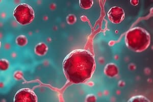
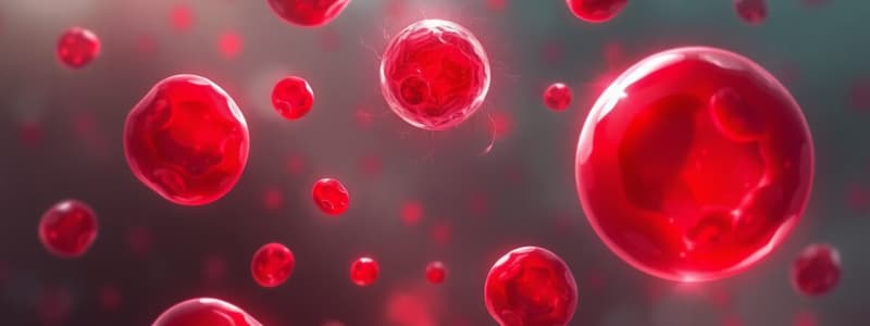
A form of anemia due to the abnormal breakdown of red blood cells, either in the blood vessels or elsewhere in the body.
Hereditary Spherocytosis
Hereditary Spherocytosis
An inherited intrinsic defect in the red blood cell membrane.
Sickle Cell Anemia
Sickle Cell Anemia
An inherited defect in the hemoglobin molecule B globin chain.
Thalassemia
Thalassemia
Signup and view all the flashcards
Glucose-6-Phosphate Dehydrogenase Deficiency (G6PD)
Glucose-6-Phosphate Dehydrogenase Deficiency (G6PD)
Signup and view all the flashcards
Paroxysmal Nocturnal Hemoglobinuria (PNH)
Paroxysmal Nocturnal Hemoglobinuria (PNH)
Signup and view all the flashcards
Immunohemolytic Anemia
Immunohemolytic Anemia
Signup and view all the flashcards
Intravascular Hemolysis
Intravascular Hemolysis
Signup and view all the flashcards
Extravascular Hemolysis
Extravascular Hemolysis
Signup and view all the flashcards
Splenomegaly
Splenomegaly
Signup and view all the flashcards
Hemoglobin/Hematocrit levels in Hemolytic Anemia
Hemoglobin/Hematocrit levels in Hemolytic Anemia
Signup and view all the flashcards
Coombs test
Coombs test
Signup and view all the flashcards
Osmotic fragility test
Osmotic fragility test
Signup and view all the flashcards
Hereditary Spherocytosis cause
Hereditary Spherocytosis cause
Signup and view all the flashcards
Sickle cell Anemia
Sickle cell Anemia
Signup and view all the flashcards
Sickle cell anemia pathogenesis
Sickle cell anemia pathogenesis
Signup and view all the flashcards
Sickle cell anemia - two major consequences
Sickle cell anemia - two major consequences
Signup and view all the flashcards
Sickle cell trait
Sickle cell trait
Signup and view all the flashcards
Sickle cell anemia lab findings
Sickle cell anemia lab findings
Signup and view all the flashcards
Normal Hemoglobin
Normal Hemoglobin
Signup and view all the flashcards
Study Notes
Hemolytic Anemia Overview
- Hemolytic anemia is a form of anemia resulting from hemolysis, or the abnormal breakdown of red blood cells, that occurs in the blood vessels or elsewhere in the body.
- It commonly occurs in the spleen, but can occur in the reticuloendothelial system or mechanically.
- Hemolytic anemia accounts for 5% of all existing anemias.
- Consequences range from general symptoms to life-threatening systemic effects.
- The general classification is either intrinsic or extrinsic, which determines treatment.
Common Hemolytic Anemias
- Hereditary spherocytosis is an inherited intrinsic defect in the red blood cell membrane.
- Sickle cell anemia is an inherited defect in the hemoglobin B globin chain.
- Thalassemia is an inherited disorder in globin genes decreasing the synthesis of alpha or beta globin.
- Glucose-6-phosphate dehydrogenase deficiency (G6PD) is caused by exposure to toxins, drugs, or infectious agents.
- Paroxysmal nocturnal hemoglobinuria (PNH) occurs due to an acquired mutation in the PIGA gene (phosphatidylinositol glycan).
- Immunohemolytic anemia is caused by antibodies binding to determinants on red cell membranes induced by exogenous agents, drugs, and chemicals.
Classifying Hemolytic Anemia
- Extrinsic hemolytic anemia:
- Antibody-mediated which may be a transfusion reaction or disease of the newborn.
- Due to Autoantibodies which may be drug associated
- Mechanical trauma to RBC from defective heart valves.
- Disseminated intravascular coagulation (blood clots).
- Infections such as malaria.
- Intrinsic hemolytic anemia:
- Membrane abnormalities like hereditary spherocytosis.
- Enzyme deficiencies such as glucose 6 phosphate dehydrogenase deficiency.
- Disorders of hemoglobin synthesis like sickle cell disease.
- Deficient hemoglobin synthesis like thalassemia syndromes.
- Acquired intrinsic red cell membrane defect, paroxysmal nocturnal hemoglobinuria (PNH).
Intravascular vs Extravascular Hemolysis
- Intravascular hemolysis causes:
- Membrane defects, enzyme defects and hemoglobin defects in the red cell.
- Auto immune reactions such as warm, cold and drug associated
- Transfusion reactions
- Or non-immune reactions from hypersplenism, fragmentation syndromes, infections and drugs
- Extravascular hemolysis involving phagocytes in the spleen is caused by defects that increase the destruction of RBCs.
- In extravascular hemolysis, hyperbilirubinemia and jaundice occur from the degradation of hemoglobin in macrophages.
- Extravascular hemolysis can lead to splenomegaly and cholelithiasis with bilirubin-rich gallstones.
- Intravascular hemolysis: RBCs explode within the circulation.
- Intravascular hemolysis can be due to:
- Turbulence from defective heart valve defects, bacterial toxins, heat shock or complement fixation.
- Intravascular hemolysis causes hemoglobinemia, hemoglobinuria and hemosiderinuria (3H's) with a loss of iron.
- Turbulence from defective heart valve defects, bacterial toxins, heat shock or complement fixation.
Clinical Manifestations of Hemolytic Anemia
- Common symptoms: Weakness and dizziness
- Other signs: Fever, weight loss, fatigue, pallor and jaundice
- Dark urine and gallstones
- Splenomegaly as well as extramedullary hematopoiesis and thinning of cortical bone
Lab values in Hemolytic Anemia
- Hemoglobin and hematocrit are usually low.
- Haptoglobin usually combines with hemoglobin and a low level is a sign of hemolytic anemia.
- When hemoglobin is broken down to bilirubin, increased levels may signal hemolytic anemia.
- Other laboratory tests for hemolytic anemia:
- Hemoglobin electrophoresis
- Osmotic fragility test is preformed to look for RBCs that are more fragile then others.
- Glucose 6 phosphate dehydrogenase (G6PD) deficiency
- Urine test for hemoglobin
Lab tests
- Findings during hemolysis:
- Haptoglobin decreases because it binds free hemoglobin.
- Lactate dehydrogenase is elevated because it is released from lysis of red blood cells.
- Peripheral blood smear shows abnormal red blood cells based on cause of anemia.
- Reticulocyte count increases due to marrow response to anemia.
- Unconjugated bilirubin increases due to increased hemoglobin breakdown.
- Urinalysis has urobilinogen and tests positive for blood due to free hemoglobin and its metabolites.
Pathogenesis of Hereditary Spherocytosis
- Hereditary Spherocytosis are inherited defects in the RBC cell membrane proteins that stabilize the lipid layer (spectrin, ankyrin, and band 3).
- These mutations result in spherocytosis.
- Splenectomy corrects the anemia but not the spherocytosis.
- Clinical findings:
- Splenomegaly, usually 500-1000 gms (200 gms is normal)
- Anemia and jaundice
- Parvovirus marked tropism increases osmotic fragility.
- Marrow may become depleted of RBC progenitors increasing the anemia and requiring transfusions.
Hemolytic Anemia destruction
- Abnormal HgB qualitative: Sickle Cell Anemia
- Abnormal HgB quantitative: Alpha and Beta Thalassemia
- Enzyme defects: G6PD and PK deficiency
- Membrane defects : Eliptocytosis and Hereditary Spherocytosis
- Acquired hemolytic anemia: PNH
- TTP ,Thrombotic Cytopenia Purpura
Sickle Cell Anemia Overview
- Hemoglobinopathy is an inherited disorder caused by inherited mutations leading to structural abnormalities in hemoglobin which is the most common familial hemolytic anemia.
- Occurs in 1 out of 350 African Americans but can affect people of any race or ethnicity.
- Sickle cell anemia is caused by a mutation in the B-globin chains that leads to sickle deoxygenated hemoglobin (HbS), which is also the most common familial hemolytic anemia
- The pathogenesis involves a single amino acid substitution in the B globin chains resulting in deoxygenated hemoglobin, leading to distortion of red blood cell shape.
- Normal hemoglobin is composed of tetramers with 2 pairs of similar chains, alpha and beta (96% HbA (alpha2beta2), 1% fetal HbF, and HgA2.
- Inpatients with HbS completely replaces HbA with HbS.
- Heterozygous carriers (trait) only have half replaced by HbS because HbC, another mutant Beta-globin, has a lysine residue instead of the normal glutamic acid residue at position 6.
- The mutation produces the sickle cell trait.
Oxygen Hemoglobin Dissociation Curve
- Left shift: Decreased unloading of O2 at the tissue level
- Decreased H+ (alkalosis), temperature, 2,3-diphosphoglycerate, stored red blood cells
- Right shift: Increased unloading of O2 at the tissue level
- Increased H+ (acidosis), temperature, 2,3-diphosphoglycerate, pregnancy, chronic anemia, arterial hypoxemia
Sickle Cell Anemia Consequences
- Sickle cell anemia involves 2 major consequences related to the sickling of red blood cells
- Severe hemolytic anemia is produced by red blood cell membrane damage.
- Vascular obstruction results in ischemia and pain.
- Clinical features involve an unremitting course with crisis, hand-foot syndrome due to ischemia, acute chest syndrome due to sluggish blood flow in pulmonary circulation, stroke (a leading cause of death), retinopathy, and aplastic crisis (parvovirus infection).
Sickle Cell Trait
- Sickle cell trait is an inherited blood disorder but (not considered a disease) that affects 1 to 3 million Americans.
- These individuals usually live normal lives without signs or symptoms, can involve many races and ethnicities (>100 million worldwide) and carry only one gene, compared to 2 in patients with sickle cell anemia.
- Electrophoresis shows HbA 60% and HbS %.
sickle cell anemia laboratory findings
- The RED BLOOD CELL's 5-50% sickled
- Low hemoglobin: 5-10 gms/dl(Normal 14-18M,12-15F)
- HgA 0%, HbS 90%
- Increased reticulocytes, platlets, and leukocytes
- Routine DNA screening required
Electrophoretic pattern in Sickle Cell Disease
- BABA is normal
- BABS is sickle cell trait
- BSBS is sickle cell anemia
- With electropheresis direction running from (positive to negative)
- Beta polypeptide (Hb-A) amino acid sequence: H3N+ Val His Leu Thr Pro Glu Glu
- Sickle cell and Beta polypeptide Hb-S amino acid sequence: H3N+ Val His Leu Thr Pro Val Glu
Hemoglobin Electrophoresis
- The substitution of Valine by Glutamic acid in Beta chains results in decreased mobility of HbS in the electric field compared to HbA due to less negative charge on Beta -globin chains
Treatment
- Basic treatment principles include: Hydration, oxygenation, pain management, blood transfusions, bone marrow transplant, hydroxyurea, herbal remedies, nutraceuticals, dietary modifications, and lifestyle changes.
Studying That Suits You
Use AI to generate personalized quizzes and flashcards to suit your learning preferences.