Podcast
Questions and Answers
What is the primary function of the right side of the heart in relation to the lungs?
What is the primary function of the right side of the heart in relation to the lungs?
- Pumps deoxygenated blood to the lungs for oxygenation (correct)
- Pumps oxygenated blood to the body
- Drains waste products from the heart
- Pumps blood through the coronary arteries
Which artery branches directly from the left coronary artery?
Which artery branches directly from the left coronary artery?
- Marginal artery
- Anterior interventricular artery (correct)
- Right coronary artery
- Posterior interventricular artery
What is the role of the cardiac veins in the circulatory system?
What is the role of the cardiac veins in the circulatory system?
- Supply blood to the aortic valve
- Remove waste products from the myocardium (correct)
- Pump deoxygenated blood back to the lungs
- Deliver oxygenated blood to coronary arteries
During which phase of the cardiac cycle is blood delivered to the myocardium?
During which phase of the cardiac cycle is blood delivered to the myocardium?
Which of the following describes systemic circulation?
Which of the following describes systemic circulation?
What is the primary function of the heart valves?
What is the primary function of the heart valves?
How many atrioventricular (AV) valves are present in the heart?
How many atrioventricular (AV) valves are present in the heart?
Which of the following statements best describes the function of the chordae tendinae?
Which of the following statements best describes the function of the chordae tendinae?
Where is the tricuspid valve best heard when listening to heart sounds?
Where is the tricuspid valve best heard when listening to heart sounds?
What triggers the semilunar valves to open?
What triggers the semilunar valves to open?
Which valve prevents backflow of blood into the right ventricle?
Which valve prevents backflow of blood into the right ventricle?
Which valve is also known as the mitral valve?
Which valve is also known as the mitral valve?
How many cusps does the aortic valve have?
How many cusps does the aortic valve have?
Which chamber of the heart receives oxygenated blood from the lungs?
Which chamber of the heart receives oxygenated blood from the lungs?
What separates the left atrium from the right atrium?
What separates the left atrium from the right atrium?
Which part of the heart is primarily responsible for pumping oxygenated blood to the body?
Which part of the heart is primarily responsible for pumping oxygenated blood to the body?
What is the function of the atrioventricular valves?
What is the function of the atrioventricular valves?
The apex of the heart is located in which intercostal space?
The apex of the heart is located in which intercostal space?
Which statement is true regarding the fibrous skeleton of the heart?
Which statement is true regarding the fibrous skeleton of the heart?
What describes the coronary circulation?
What describes the coronary circulation?
What type of blood flows through the right ventricle?
What type of blood flows through the right ventricle?
Which veins drain directly into the right atrium?
Which veins drain directly into the right atrium?
What is the primary purpose of coronary angiography?
What is the primary purpose of coronary angiography?
Which component is part of the conduction system responsible for initiating electrical impulses in the heart?
Which component is part of the conduction system responsible for initiating electrical impulses in the heart?
What effect does sympathetic stimulation have on the heart?
What effect does sympathetic stimulation have on the heart?
Which nerve is primarily responsible for parasympathetic regulation of the heart?
Which nerve is primarily responsible for parasympathetic regulation of the heart?
What role do afferent fibres parallel to the vagus nerve play in cardiac activity?
What role do afferent fibres parallel to the vagus nerve play in cardiac activity?
What is the primary function of the fibrous skeleton of the heart?
What is the primary function of the fibrous skeleton of the heart?
Which of the following statements is true regarding the cardiac plexus?
Which of the following statements is true regarding the cardiac plexus?
What can occur as a result of cardiac ischemia?
What can occur as a result of cardiac ischemia?
What happens to the foramen ovale at birth?
What happens to the foramen ovale at birth?
Which structure constricts to become the ligamentum arteriosum after birth?
Which structure constricts to become the ligamentum arteriosum after birth?
Which condition is associated with the aortic valve?
Which condition is associated with the aortic valve?
What is a common complication of congenital heart defects?
What is a common complication of congenital heart defects?
Which cardiac issue is related to the myocardium?
Which cardiac issue is related to the myocardium?
What is the fate of the ductus venosus after birth?
What is the fate of the ductus venosus after birth?
Which of the following is NOT a component of the clinical correlations listed?
Which of the following is NOT a component of the clinical correlations listed?
Which structure serves as a conduit for blood flow between the pulmonary trunk and the aorta in a fetus?
Which structure serves as a conduit for blood flow between the pulmonary trunk and the aorta in a fetus?
Which condition is associated with the coronary arteries?
Which condition is associated with the coronary arteries?
What occurs to the pulmonary trunk at birth?
What occurs to the pulmonary trunk at birth?
Flashcards are hidden until you start studying
Study Notes
Heart Anatomy
- The heart is a cone-shaped, muscular organ that pumps blood through the body.
- It is approximately the size of a fist, weighing around 300 grams and measuring 11cm long.
- Located within the mediastinum, it is protected by the ribs and sternum.
- The heart lies between ribs 2-5 to the left of the midline, with the apex (bottom) positioned in the 5th intercostal space at the midclavicular line.
Heart Chambers
- The heart is divided into four chambers:
- Two atria (upper chambers)
- Two ventricles (lower chambers)
- The atria and ventricles are separated by the interatrial septum (between atria) and the interventricular septum (between ventricles).
Heart Valves
- Valves are essential for ensuring unidirectional blood flow within the heart.
- There are four valves:
- Two atrioventricular (AV) valves:
- Tricuspid valve (right AV valve): located between the right atrium and right ventricle; three cusps.
- Bicuspid/mitral valve (left AV valve): located between the left atrium and left ventricle; two cusps
- Two semilunar (SL) valves:
- Pulmonary valve: three cusps; located between the right ventricle and pulmonary trunk.
- Aortic valve: three cusps; located between the left ventricle and aorta.
- Two atrioventricular (AV) valves:
Atrioventricular (AV) Valves
- Open when blood flows from the atria to the ventricles.
- Close during ventricular contraction (systole).
- Chordae tendinae, attached to papillary muscles, prevent the valves from everting (turning inside out) and ensure blood does not flow back into the atria.
- The closing of AV valves (tricuspid and mitral) produces S1 ("Lub") heart sound.
Semilunar (SL) Valves
- Open during ventricular contraction (systole) as blood is ejected from the ventricles.
- Close during ventricular relaxation (diastole) to prevent blood from flowing back into the ventricles.
- The closing of SL valves (pulmonic and aortic) produces the S2 ("Dub") heart sound.
Heart Circulation
- Pulmonary Circulation (blue):
- Involves the right side of the heart and the lungs.
- Deoxygenated blood is pumped from the right ventricle to the lungs for oxygenation.
- Systemic Circulation (red):
- Involves the left side of the heart and the rest of the body.
- Oxygenated blood is pumped from the left ventricle to the body's organs and tissues to deliver oxygen and nutrients.
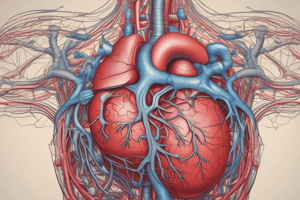
Coronary Circulation
- Supplies the heart muscle with oxygen and nutrients.
- Consists of coronary arteries that deliver blood and cardiac veins that remove waste.
Coronary Arteries
- Main Branches:
- Right Coronary Artery (RCA):
- Marginal artery
- Posterior interventricular artery
- Left Coronary Artery (LCA):
- Anterior interventricular artery (LAD)
- Circumflex artery
- Right Coronary Artery (RCA):
Cardiac Veins
- Drain into the coronary sinus, which empties into the right atrium.
- Anterior cardiac veins drain directly into the right atrium.
Fibrous Skeleton of the Heart
- A meshwork of dense connective tissue.
- Functions as an insulator for electrical impulses, preventing direct conduction from the atria to the ventricles.
- Supports and anchors the heart valves.
Regulation of Cardiac Activity
- Conduction system:
- Sinoatrial (SA) node (pacemaker): initiates electrical impulses.
- Atrioventricular (AV) node: delays impulse conduction.
- AV bundle (bundle of His): conducts impulses to the ventricles.
- Right and left bundle branches: conduct impulses to specific ventricular regions.
- Purkinje fibers: conduct impulses throughout the ventricular myocardium.
- Autonomic nervous system (ANS):
- Sympathetic: increases heart rate, force of contraction, and dilates coronary vessels.
- Parasympathetic: decreases heart rate and force of contraction.
Foetal Circulation
- Before birth, the circulatory system undergoes significant adjustments to adapt to the absence of respiratory function in the womb.
- Key features of foetal circulation:
- Foramen ovale: an opening between the right and left atria, allowing blood to bypass the lungs.
- Ductus arteriosus: a vessel connecting the pulmonary trunk to the aorta, allowing blood to bypass the pulmonary circulation.
- Umbilical cord: supplies the fetus with oxygen and nutrients from the placenta, and removes waste products.
Changes at Birth
- When the newborn takes its first breath, the lungs expand, increasing pulmonary blood flow.
- The foramen ovale closes, preventing blood from bypassing the lungs.
- The ductus arteriosus constricts and eventually becomes the ligamentum arteriosum.
- The umbilical cord is tied and severed, leading to closure of the umbilical vessels.
Clinical Correlations
- Valves: Mitral valve prolapse, Tricuspid valve stenosis, Pulmonary valve stenosis, Aortic valve stenosis
- Myocardium: Heart failure, Myocarditis, Myocardial infarction, Hypertrophic cardiomyopathy, Dilated cardiomyopathy
- Pericardium: Pericarditis, Pericardial effusion, Cardiac tamponade, Constrictive pericarditis
- Conduction System: Arrhythmias, Heart block, Congenital conduction defects, Atrial fibrillation, Ventricular fibrillation
- Coronary Arteries: Coronary artery disease (CAD), Myocardial infarction (MI), Angina, Coronary artery aneurysm
- Aorta and Great Vessels: Aortic aneurysm, Aortic dissection, Coarctation of the aorta, Atherosclerosis
- Pulmonary Arteries: Pulmonary embolism, Pulmonary hypertension
- Pulmonary Veins: Pulmonary venous congestion, Pulmonary venous obstruction, Pulmonary hypertension
- Chambers of the Heart (Atria and Ventricles): Atrial fibrillation, Atrial septal defect, Ventricular septal defect, Congestive heart failure (CHF)
- Ventricular Septum: Ventricular septal defect (VSD), Myocardial infarction involving the septum, Hypertrophic cardiomyopathy
- Veins (e.g., coronary veins): Coronary sinus thrombosis, Venous insufficiency, Varicose veins, Deep vein thrombosis (DVT)
- Pericardial Cavity: Pericardial effusion, Cardiac tamponade
- Coronary Circulation (Arteries and Veins): Coronary artery disease (CAD), Angina, Coronary vein thrombosis, Coronary artery bypass grafting (CABG)
- Fibrous Skeleton of the Heart: Valve regurgitation, Valve stenosis, Heart block (due to electrical disruption)
- Autonomic Nervous System (ANS) Influence on Heart: Tachycardia, Bradycardia, Vasovagal syncope, Autonomic dysfunction
- Foetal Circulation: Patent ductus arteriosus (PDA), Patent foramen ovale (PFO), Congenital heart defects
Studying That Suits You
Use AI to generate personalized quizzes and flashcards to suit your learning preferences.