Podcast
Questions and Answers
What is the primary function of the heart's right ventricle?
What is the primary function of the heart's right ventricle?
- To prevent the backflow of blood
- To receive oxygenated blood from the lungs
- To distribute blood to the rest of the body
- To pump deoxygenated blood to the lungs (correct)
Which chamber of the heart receives blood from the superior vena cava?
Which chamber of the heart receives blood from the superior vena cava?
- Right atrium (correct)
- Right ventricle
- Left ventricle
- Left atrium
What distinguishes systemic circulation from pulmonary circulation?
What distinguishes systemic circulation from pulmonary circulation?
- Systemic circulation is shorter in distance from the heart
- Pulmonary circulation is strictly for oxygenated blood
- Systemic circulation serves the entire body (correct)
- Systemic circulation involves arteries only
What valve prevents backflow of blood from the right ventricle to the right atrium?
What valve prevents backflow of blood from the right ventricle to the right atrium?
Which statement correctly describes the function of the coronary circulation?
Which statement correctly describes the function of the coronary circulation?
Why is the left side of the heart muscle thicker than the right side?
Why is the left side of the heart muscle thicker than the right side?
Which of the following statements about the heart's structure is true?
Which of the following statements about the heart's structure is true?
What is the role of the semilunar valve in the heart?
What is the role of the semilunar valve in the heart?
What is the primary function of the heart?
What is the primary function of the heart?
Which valve is located between the left atrium and the left ventricle?
Which valve is located between the left atrium and the left ventricle?
What component of the heart initiates electrical impulses for heartbeats?
What component of the heart initiates electrical impulses for heartbeats?
What condition is characterized by the blockage of arteries due to fatty plaque buildup?
What condition is characterized by the blockage of arteries due to fatty plaque buildup?
During which phase of the cardiac cycle does the heart fill with blood?
During which phase of the cardiac cycle does the heart fill with blood?
What is the name of the valves that prevent blood from flowing backward into the ventricles?
What is the name of the valves that prevent blood from flowing backward into the ventricles?
Which of the following best describes the myocardium?
Which of the following best describes the myocardium?
Which structures help to connect cardiomyocytes in the heart?
Which structures help to connect cardiomyocytes in the heart?
What is angina a result of?
What is angina a result of?
What is the role of the coronary arteries?
What is the role of the coronary arteries?
What is recorded in an electrocardiogram (ECG)?
What is recorded in an electrocardiogram (ECG)?
What happens to the heart's pacemaker activity after the atrioventricular (AV) node?
What happens to the heart's pacemaker activity after the atrioventricular (AV) node?
How many times does the human heart beat in a day, approximately?
How many times does the human heart beat in a day, approximately?
Which chambers of the heart contract simultaneously during the cardiac cycle?
Which chambers of the heart contract simultaneously during the cardiac cycle?
Flashcards
Pulmonary Circulation
Pulmonary Circulation
The system that circulates blood between the heart and the lungs, carrying deoxygenated blood to the lungs for oxygenation and returning oxygenated blood to the heart.
Systemic Circulation
Systemic Circulation
The system that circulates blood between the heart and the rest of the body, delivering oxygenated blood to tissues and collecting deoxygenated blood.
Coronary Circulation
Coronary Circulation
The part of the circulatory system that delivers blood to the heart muscle itself.
Right Atrium
Right Atrium
Signup and view all the flashcards
Right Ventricle
Right Ventricle
Signup and view all the flashcards
Left Atrium
Left Atrium
Signup and view all the flashcards
Left Ventricle
Left Ventricle
Signup and view all the flashcards
Tricuspid Valve
Tricuspid Valve
Signup and view all the flashcards
Left Atrium to Aorta
Left Atrium to Aorta
Signup and view all the flashcards
Bicuspid (Mitral) Valve
Bicuspid (Mitral) Valve
Signup and view all the flashcards
Aortic Semilunar Valve
Aortic Semilunar Valve
Signup and view all the flashcards
Myocardium
Myocardium
Signup and view all the flashcards
Epicardium
Epicardium
Signup and view all the flashcards
Endocardium
Endocardium
Signup and view all the flashcards
Coronary Arteries
Coronary Arteries
Signup and view all the flashcards
Atherosclerosis
Atherosclerosis
Signup and view all the flashcards
Angina
Angina
Signup and view all the flashcards
Myocardial Infarction
Myocardial Infarction
Signup and view all the flashcards
Cardiac Cycle
Cardiac Cycle
Signup and view all the flashcards
Systole
Systole
Signup and view all the flashcards
Diastole
Diastole
Signup and view all the flashcards
Sinoatrial (SA) Node
Sinoatrial (SA) Node
Signup and view all the flashcards
Atrioventricular (AV) Node
Atrioventricular (AV) Node
Signup and view all the flashcards
Study Notes
Heart Anatomy and Function
-
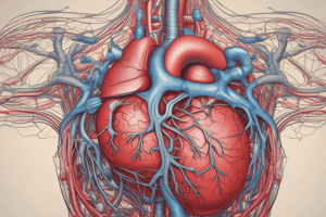
The heart pumps blood through three circulatory systems: coronary (heart itself), pulmonary (heart and lungs), and systemic (body's systems).
-
The right side of the heart, responsible for pulmonary circulation, has a thinner muscle wall than the left, which pumps blood to the entire body.
-
The human heart is roughly the size of a clenched fist and comprises four chambers: two atria and two ventricles.
-
The right atrium receives deoxygenated blood from the superior and inferior vena cava, and the coronary sinus.
-
Deoxygenated blood flows from the right atrium to the right ventricle through the tricuspid valve.
-
The right ventricle pumps blood through the pulmonary arteries to the lungs for oxygenation.
-
The left atrium receives oxygenated blood from the lungs via pulmonary veins.
-
Oxygenated blood flows from the left atrium to the left ventricle through the bicuspid/mitral valve.
-
The left ventricle pumps blood through the aorta to the body's organs and muscles.
-
The heart's pumping action, double circulation, is observed in all mammals.
Heart Structure and Layers
-
The heart has three layers: epicardium (outer), myocardium (middle, muscle), and endocardium (inner lining).
-
The pericardium is a membranous layer surrounding the heart, aiding in protection and reducing friction.
-
Coronary arteries, branching from the aorta, supply oxygenated blood to the heart muscle.
-
Coronary veins collect deoxygenated blood from the heart muscle and return it to the right atrium.
Cardiac Cycle
-
The cardiac cycle involves the coordinated filling and emptying of blood within the heart.
-
The cycle is initiated by electrical signals causing heart muscle contraction (systole) and relaxation (diastole).
-
Atrial contraction forces blood through atrioventricular valves into ventricles.
-
Ventricular contraction forces blood through semilunar valves into arteries.
-
"Lup" sound: closing of atrioventricular valves
-
"Dup" sound: closing of semilunar valves
Cardiomyocytes and Pacemaker
-
Cardiomyocytes (heart muscle cells) are striated (like skeletal muscle) but pump rhythmically and involuntarily (like smooth muscle).
-
Intercalated disks connect cardiomyocytes uniquely in cardiac muscle.
-
Cardiomyocytes are self-stimulated, and isolated cells will beat under proper conditions.
-
The heart's internal pacemaker, the sinoatrial (SA) node, initiates electrical signals.
-
Electrical signals spread to the atria, pause at the atrioventricular (AV) node, and then spread to the ventricles via the bundle of His, bundle branches, and Purkinje fibers.
-
This delay allows atria to completely empty into ventricles before ventricular contraction.
Electrocardiogram (ECG)
-
Electrical impulses in the heart generate electrical currents, measurable as an electrocardiogram (ECG) on the skin using electrodes.
-
ECGs record the electrical activity of cardiac muscle.
Heart Diseases
- Atherosclerosis involves fatty plaque buildup in arteries, potentially leading to angina or myocardial infarction (heart attack) in the coronary arteries.
Studying That Suits You
Use AI to generate personalized quizzes and flashcards to suit your learning preferences.


