Podcast
Questions and Answers
What structure do hair cells use to generate receptor potentials in the auditory system?
What structure do hair cells use to generate receptor potentials in the auditory system?
- Cochlear nucleus
- Medial geniculate body
- Superior olivary nucleus
- Organ of Corti (correct)
Which pathway do auditory impulses follow after leaving the cochlear nuclei?
Which pathway do auditory impulses follow after leaving the cochlear nuclei?
- Medial geniculate body to auditory cortex (correct)
- Inferior colliculi to thalamus
- Basil ganglia to temporal lobe
- Superior olivary nuclei to lateral lemniscus
What is the primary role of the middle ear in sound conduction?
What is the primary role of the middle ear in sound conduction?
- House the cochlear nerve endings
- Convert sound waves into nerve impulses
- Transport fluid to the inner ear
- Amplify sound waves through the ossicles (correct)
Which statement accurately describes the difference between air and bone conduction?
Which statement accurately describes the difference between air and bone conduction?
What is the final destination of auditory impulses in the brain for sound perception?
What is the final destination of auditory impulses in the brain for sound perception?
What initiates the generation of action potentials in the hearing pathway?
What initiates the generation of action potentials in the hearing pathway?
Why does bone conduction transmit weaker sound than air conduction?
Why does bone conduction transmit weaker sound than air conduction?
Which structure does the sound wave reach after traveling through the fluid in the inner ear?
Which structure does the sound wave reach after traveling through the fluid in the inner ear?
What is the primary role of inner hair cells?
What is the primary role of inner hair cells?
What initiates the pressure wave in the cochlea when sound waves are transmitted?
What initiates the pressure wave in the cochlea when sound waves are transmitted?
Which statement about outer hair cells is correct?
Which statement about outer hair cells is correct?
What connects the stereocilia of inner hair cells?
What connects the stereocilia of inner hair cells?
What allows outer hair cell stereocilia to interact with sound waves?
What allows outer hair cell stereocilia to interact with sound waves?
What happens when the stereocilia of inner hair cells bend?
What happens when the stereocilia of inner hair cells bend?
Which of the following statements about the transmission of sound waves in the cochlea is NOT true?
Which of the following statements about the transmission of sound waves in the cochlea is NOT true?
What is the distribution of hair cells within the Organ of Corti?
What is the distribution of hair cells within the Organ of Corti?
What is the primary consequence of defective K+ transporters in the inner ear?
What is the primary consequence of defective K+ transporters in the inner ear?
Which syndrome is associated with a defective Na+/K+/2Cl- symport and results in deafness?
Which syndrome is associated with a defective Na+/K+/2Cl- symport and results in deafness?
What factor contributes to the development of Meniere's disease?
What factor contributes to the development of Meniere's disease?
How do loop diuretics affect hearing when used for an extended period?
How do loop diuretics affect hearing when used for an extended period?
Which treatment strategy is suggested for managing Meniere's disease?
Which treatment strategy is suggested for managing Meniere's disease?
Which of the following is a symptom of Meniere's disease?
Which of the following is a symptom of Meniere's disease?
What physical structure lies below the dura mater and is involved in endolymph absorption?
What physical structure lies below the dura mater and is involved in endolymph absorption?
What effect does the accumulation of endolymph have on hair cells?
What effect does the accumulation of endolymph have on hair cells?
What is the role of the endolymphatic duct in the inner ear?
What is the role of the endolymphatic duct in the inner ear?
What happens to outer hair cells when stereocilia bend toward the shortest one?
What happens to outer hair cells when stereocilia bend toward the shortest one?
Which mechanism initially allows K+ to enter hair cells when stereocilia bend toward the longest one?
Which mechanism initially allows K+ to enter hair cells when stereocilia bend toward the longest one?
Which of the following is NOT a consequence of insufficient endolymph reabsorption?
Which of the following is NOT a consequence of insufficient endolymph reabsorption?
What occurs when K+ channels close due to stereocilia bending toward the shortest one?
What occurs when K+ channels close due to stereocilia bending toward the shortest one?
What triggers the opening of voltage-gated Ca2+ channels in hair cells?
What triggers the opening of voltage-gated Ca2+ channels in hair cells?
What is the result of glutamate release in the synapse with cochlear nerve fibers?
What is the result of glutamate release in the synapse with cochlear nerve fibers?
What effect does bending of stereocilia towards the shortest one have on neurotransmitter release?
What effect does bending of stereocilia towards the shortest one have on neurotransmitter release?
Which structure is responsible for head movement in response to sound?
Which structure is responsible for head movement in response to sound?
What condition must be met for an action potential to be generated in hair cells?
What condition must be met for an action potential to be generated in hair cells?
Which part of the cochlea vibrates maximally at high frequency sounds?
Which part of the cochlea vibrates maximally at high frequency sounds?
How is the localization of sound primarily achieved?
How is the localization of sound primarily achieved?
What does a higher amplitude in sound waves trigger?
What does a higher amplitude in sound waves trigger?
Which part of the primary auditory cortex processes low frequency sounds?
Which part of the primary auditory cortex processes low frequency sounds?
Which nucleus determines the time delay for sound localization?
Which nucleus determines the time delay for sound localization?
What sensory aspect does the lateral superior olivary nucleus primarily assess?
What sensory aspect does the lateral superior olivary nucleus primarily assess?
What occurs when a sound source is closer to the ipsilateral side?
What occurs when a sound source is closer to the ipsilateral side?
What happens when sound is perceived louder in one ear?
What happens when sound is perceived louder in one ear?
How does the cochlea contribute to the perception of pitch?
How does the cochlea contribute to the perception of pitch?
Which mechanism does NOT contribute to the localization of sound?
Which mechanism does NOT contribute to the localization of sound?
What is indicated when sound arrives at both ears at the same time?
What is indicated when sound arrives at both ears at the same time?
Which type of sounds only activate hair cells with the highest excitability?
Which type of sounds only activate hair cells with the highest excitability?
What determines the reflected wave's delay in localization?
What determines the reflected wave's delay in localization?
Flashcards are hidden until you start studying
Study Notes
Hearing Pathway
- Receptors: Hair cells located in the organ of Corti within the inner ear
- Receptor Potential Generation: Hair cells generate a receptor potential, triggering neurotransmitter release onto cochlear nerve endings.
- Action Potential Generation: If the threshold is reached, action potentials are generated in the cochlear nerve.
- Brain Stem Transmission: Impulses are conducted to the cochlear nuclei in the brain stem.
- Superior Olivary Nuclei: The impulses are transmitted to the superior olivary nuclei on both sides of the brain.
- Inferior Colliculi and Medial Geniculate Bodies: Impulses travel to the inferior colliculi and then to the medial geniculate bodies.
- Primary Auditory Area: Impulses reach the superior temporal gyrus in the cerebral cortex, housing the primary auditory area responsible for sound perception.
Air and Bone Conduction
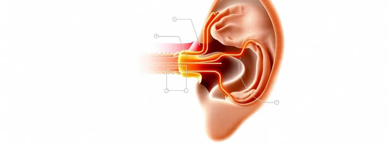
- Air Conduction: Sound waves travel through the external auditory canal, vibrate the tympanic membrane, and transmit to the middle ear via the ossicle chain. The ossicle chain then transmits the sound waves to the oval window, which vibrates the fluid in the inner ear. This fluid vibration reaches the hair cells in the organ of Corti.
- Bone Conduction: Sound waves reach the fluid in the inner ear by directly vibrating the bones of the skull. The vibrating object is in contact with the skin, and the sound waves travel through the temporal bone to the inner ear fluid.
Endolymph Transport
- Bony Labyrinth: The bony outer wall of the inner ear, including the vestibulum, semicircular canals, and cochlea.
- Membranous Labyrinth: Located within the bony labyrinth, filled with endolymph.
- Perilymph: Fills the space between the bony and membranous labyrinths.
- Endolymphatic Duct: Collects endolymph from the membranous labyrinth and transports it to the endolymphatic sac.
- Endolymphatic Sac: Located below the dura mater, responsible for absorbing excess endolymph into its blood vessels.
Meniere's Disease
- Cause: Decreased endolymph removal from the inner ear.
- Contributing Factors: Viral and bacterial infection, autoimmune reactions, genetic causes related to endolymph reabsorption.
- Consequences:
- Recurring Vertigo: Dizziness and a sensation of spinning despite being stationary.
- Sensorineural Hearing Loss: Accumulation of endolymph in the membranous cochlea damages hair cells.
- Tinnitus: Threatening damage to outer hair cells causes their contractions, leading to ringing in the ear.
- Feeling of Fullness in the Ear: Increased pressure in the membranous labyrinth causes this sensation.
Hair Cells
- Location: Organ of Corti, situated on the basilar membrane of the scala media.
- Structure: Four rows of hair cells, including three rows of outer hair cells and one row of inner hair cells.
- Stereocilia: Each hair cell has stereocilia, organized in increasing length to one side of the cell.
- Tip Links: Connect stereocilia, allowing coordinated movement.
Inner Hair Cells
- Sensory Function: Important for sensory function.
- Afferent Nerve Fibers: Most afferent nerve fibers originate from inner hair cells.
- Sound Perception and Signal Transduction: Responsible for both sound perception and signal transduction.
- Stereocilia and Endolymph Flow: Their stereocilia are not connected to the tectorial membrane and are influenced by endolymph flow in the inner corner of the scala media.
- Depolarization and Impulse Transmission: Stereocilia bending causes depolarization and impulse transmission along afferent nerve fibers to the brain.
Outer Hair Cells
- Afferent and Efferent Fibers: Have few afferent fibers but receive most efferent fibers from the superior olivary nuclei.
- Large Receptive Fields: 100-150 hair cells can send impulses to a single cochlear nerve fiber.
- Stereocilia and Tectorial Membrane: Stereocilia are connected to the tectorial membrane, their movement is influenced by its interaction.
- Motor Protein Prestin: Present in the outer hair cell membrane, involved in depolarization.
- Depolarization and Contraction: Depolarization causes outer hair cells to contract, shortening the distance between the basilar and tectorial membranes.
- Hyperpolarization and Relaxation: Hyperpolarization causes outer hair cells to relax, increasing the distance between the basilar and tectorial membranes.
Sound Transmission through the Inner Ear
- Stapes Vibration: When sound waves reach the middle ear, the stapes footplate vibrates the perilymph.
- Perilymph Vibration: Due to perilymph's lower acoustic inertia compared to air, amplification is required to transmit sound from air to fluid.
- Pressure Wave Transmission: Vibrations in the scala vestibuli create a pressure wave, traveling to the helicotrema and then back into the scala tympani.
- Basilar Membrane Vibration: This pressure wave vibrates the basilar membrane, eventually dissipating through the round window back into the middle ear.
Sound Coding
- Frequency/Pitch Coding: Dependent on cochlea properties. The basilar membrane's stiffness and width vary along its length, determining which frequencies stimulate specific regions.
- High Frequency: Base area of the cochlea (narrow, stiff)
- Low Frequency: Apex area of the cochlea (wide, flexible)
- Different Nerve Fibers: Specific locations of the basilar membrane send impulses through different nerve fibers to distinct areas of the auditory cortex — this allows for differentiation of frequencies.
- Amplitude/Loudness Coding: Greater amplitude sound waves cause larger vibrations of the basilar membrane, leading to increased activity of hair cells.
- Increased Impulse Frequency: Hair cells respond with a greater firing rate.
- Different Cell Sensitivity: Hair cells have different sensitivity to stimuli; soft sounds only activate highly excitable cells, while louder sounds activate more cells with higher thresholds.
- Auditory Cortex Interpretation: The auditory cortex interprets impulse frequency as loudness. High-frequency impulses correlate with loud sounds, and low-frequency impulses correlate with soft sounds.
- Localization Coding: Determined through time and loudness delays, allowing us to distinguish sound source direction.
- Time Delay: The ear closest to the sound source receives sound quicker than the opposite ear, creating a delay that is used for localization.
- Loudness Delay: Sounds are perceived as louder in the ear closest to the source and softer in the ear furthest from the source.
- Olivary Nuclei Processing: The superior olivary nuclei process information from both ears, allowing us to determine both time and loudness delays.
Time Delay Localization
- Medial Superior Olivary Nucleus: Determines the time delay between input from both ears, using excitatory input from both.
- Ipsilateral Sound Source: Impulses from the closer side arrive before impulses from the opposite side; the ipsilateral medial superior olivary nucleus will be activated once the threshold is reached. No signals from the ipsilateral side would be sent to the brain, indicating that the sound is closer to the ipsilateral side.
- Contralateral Sound Source: Impulses from both ears arrive simultaneously, the contralateral medial superior olivary nucleus generates an action potential. This signals that the sound source is closer to the contralateral side.
Loudness Delay Localization
- Lateral Superior Olivary Nucleus: Determines loudness difference, using excitatory input from the ipsilateral side and inhibitory input from the contralateral side.
- Ipsilateral Sound Source: Ipsilateral signals excite the lateral superior olivary nucleus, while contralateral signals arrive later and cause inhibition of it. This results in signals from the ipsilateral side being sent to the brain, indicating the sound source is closer to the ipsilateral ear.
- Contralateral Sound Source: Signals from both sides arrive simultaneously, causing excitation and inhibition simultaneously. The lateral superior olivary nucleus does not send impulses to higher brain centers, indicating the sound source is closer to the contralateral ear.
Vertical Localization
- Auricula Shape: The ear's shape reflects sound waves into the external auditory canal, creating a direct wave and a reflected wave.
- Wave Comparison: The comparison between the time delays of the direct and reflected waves helps localize the sound source vertically.
- Long Delay: Sound comes from below.
- Short Delay: Sound comes from front or above.
- Unilateral Localization: The ear can isolate the sound source vertically.
Audiometry, Weber, and Rinne Tests
- Audiometry: A hearing test that assesses hearing thresholds for different frequencies.
- Weber Test: Evaluates bone conduction by placing a vibrating tuning fork on the middle of the head. This test helps determine if hearing loss is conductive or sensorineural.
- Rinne Test: Compares air conduction to bone conduction by placing a vibrating tuning fork first on the mastoid bone and then near the external auditory canal. Result patterns can indicate different hearing loss types.
Studying That Suits You
Use AI to generate personalized quizzes and flashcards to suit your learning preferences.


