Podcast
Questions and Answers
What is the normal range of CVP, representing the filling pressure of the right side of the heart?
What is the normal range of CVP, representing the filling pressure of the right side of the heart?
0 to +8 mmHg
What is a potential complication of invasive intra-arterial pressure monitoring?
What is a potential complication of invasive intra-arterial pressure monitoring?
Arterial thrombosis
What is the site of CVP measurement?
What is the site of CVP measurement?
Right atrium or superior vena cava
What is a potential complication of invasive intra-arterial pressure monitoring?
What is a potential complication of invasive intra-arterial pressure monitoring?
What has been challenged in regards to CVP monitoring?
What has been challenged in regards to CVP monitoring?
What is the reference point for levelling the transducer?
What is the reference point for levelling the transducer?
What is included in invasive haemodynamic monitoring?
What is included in invasive haemodynamic monitoring?
What is the significance of cardiac output monitoring in haemodynamic monitoring?
What is the significance of cardiac output monitoring in haemodynamic monitoring?
What is the primary purpose of haemodynamic accuracy in monitoring?
What is the primary purpose of haemodynamic accuracy in monitoring?
What is the name of the test used to check the accuracy of subsequent haemodynamic pressure values?
What is the name of the test used to check the accuracy of subsequent haemodynamic pressure values?
What are the other possible sites for haemodynamic monitoring?
What are the other possible sites for haemodynamic monitoring?
What is the benefit of haemodynamic monitoring in periods of fluid volume instability?
What is the benefit of haemodynamic monitoring in periods of fluid volume instability?
What is the importance of interpreting CVP in association with other haemodynamic variables?
What is the importance of interpreting CVP in association with other haemodynamic variables?
What does a dominant a wave in CVP waveform indicate?
What does a dominant a wave in CVP waveform indicate?
What is the significance of an absent x descent in CVP waveform?
What is the significance of an absent x descent in CVP waveform?
What does a sharp y descent in CVP waveform indicate?
What does a sharp y descent in CVP waveform indicate?
What is the significance of a prominent x and y descent in CVP waveform?
What is the significance of a prominent x and y descent in CVP waveform?
What does a cannon a wave in CVP waveform indicate?
What does a cannon a wave in CVP waveform indicate?
What is the significance of a dominant v wave in CVP waveform?
What is the significance of a dominant v wave in CVP waveform?
What is the main function of Central Venous Pressure (CVP) in haemodynamic monitoring?
What is the main function of Central Venous Pressure (CVP) in haemodynamic monitoring?
What does an exaggerated x descent in CVP waveform indicate?
What does an exaggerated x descent in CVP waveform indicate?
What does the 'a' wave in CVP waveform analysis represent?
What does the 'a' wave in CVP waveform analysis represent?
What is the primary use of CVP monitoring in diagnosing cardiovascular conditions?
What is the primary use of CVP monitoring in diagnosing cardiovascular conditions?
What is one of the causes of raised CVP?
What is one of the causes of raised CVP?
What is the significance of the 'x' wave in CVP waveform analysis?
What is the significance of the 'x' wave in CVP waveform analysis?
What is one of the conditions that can be diagnosed using CVP monitoring?
What is one of the conditions that can be diagnosed using CVP monitoring?
What is the significance of the 'v' wave in CVP waveform analysis?
What is the significance of the 'v' wave in CVP waveform analysis?
Flashcards are hidden until you start studying
Study Notes
Haemodynamic Monitoring
- Involves the measurement of pressure, flow, and oxygenation within the cardiovascular system
- Can be divided into non-invasive and invasive methods
Non-Invasive Monitoring
- Includes measurement of:
- Heart rate, pulses, ECGs
- Blood pressure, capillary refill
- Respiratory rate, pulse oximetry
- Urine output
- Body temperature, skin temperature
Invasive Monitoring
- Includes measurement of:
- Systemic arterial pressure
- Central venous pressure (CVP)
- Pulmonary artery pressure
- Cardiac output
Principles of Haemodynamic Monitoring
- Zeroing and levelling the transducer system ensures accuracy of readings
- Fast-Flush Square-Wave Testing allows for checking the accuracy of subsequent haemodynamic pressure values
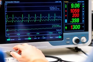
Invasive Intra-Arterial Pressure Monitoring
- Commonly placed in the radial artery, but can also be in the brachial, femoral, dorsalis pedis, and axillary arteries
- Indicated when precise and continuous monitoring is required, especially in periods of fluid volume, cardiac output, and blood pressure instability
- Complications include:
- Infection
- Arterial thrombosis
- Distal ischemia
- Air embolism
- Accidental disconnection leading to rapid blood loss
- Accidental drug administration through the arterial catheter
Central Venous Pressure (CVP) Monitoring
- CVP is the pressure recorded from the right atrium or superior vena cava, representing the filling pressure of the right side of the heart (0 to +8 mmHg)
- CVP represents the driving force for filling the right atrium and ventricle
- CVP Waveform Analysis:
- a = atrial contraction
- c = closing and bulging of the tricuspid valve
- x = atrial relaxation, with downward movement of the tricuspid valve during ventricular contraction
- v = passive filling of atrium (tricuspid valve still closed)
- y = ventricular filling with opening of the tricuspid valve
Uses of CVP Monitoring
- Diagnosis of right ventricular infarction, right heart failure, tamponade, tricuspid regurgitation or stenosis, complete heart block, constrictive pericarditis, and differential diagnosis of shock state
- Correct central line placement
- Right ventricular failure
- Tricuspid stenosis or regurgitation
- Pericardial effusion or constrictive pericarditis
Causes of Raised CVP
- Superior vena caval obstruction
- Fluid overload
- Hyperdynamic circulation
- High PEEP settings
Limitations of CVP
- CVP should not be used in isolation to assess fluid responsiveness, as it has a very poor relationship with blood volume and is a poor predictor of the hemodynamic response to a fluid challenge
- Interpretation of CVP should be in association with information relating to other haemodynamic variables
Waveform Abnormalities
- Dominant a wave: pulmonary hypertension, tricuspid stenosis, pulmonary stenosis
- Cannon a wave: complete heart block, ventricular tachycardia with atrio-ventricular dissociation
- Dominant v wave: tricuspid regurgitation
- Absent x descent: atrial fibrillation
- Exaggerated x descent: pericardial tamponade, constrictive pericarditis
- Sharp y descent: severe tricuspid regurgitation, constrictive pericarditis
- Slow y descent: tricuspid stenosis, atrial myxoma
- Prominent x and y descent: right ventricular infarction
Studying That Suits You
Use AI to generate personalized quizzes and flashcards to suit your learning preferences.