Podcast
Questions and Answers
Which nerve supplies the flexor digitorum profundus?
Which nerve supplies the flexor digitorum profundus?
- Radial nerve
- Musculocutaneous nerve
- Median nerve
- Ulnar nerve (correct)
What is the main action of the flexor pollicis longus?
What is the main action of the flexor pollicis longus?
- Extends the metacarpophalangeal and interphalangeal joints of the thumb
- Flexes the metacarpophalangeal and interphalangeal joints of the thumb (correct)
- Rotates the forearm
- Abducts the thumb
What is the origin of the ulnar artery?
What is the origin of the ulnar artery?
- Radial artery
- Brachial artery (correct)
- Axillary artery
- Median artery
Which muscle has a bony attachment to the distal anteriomedial ulna?
Which muscle has a bony attachment to the distal anteriomedial ulna?
What is the nerve supply to the flexor digitorum profundus?
What is the nerve supply to the flexor digitorum profundus?
Which artery supplies the flexor pollicis longus?
Which artery supplies the flexor pollicis longus?
What is the action of the pronator quadratus?
What is the action of the pronator quadratus?
What is the relationship between the ulnar artery and the ulnar nerve in the lower forearm?
What is the relationship between the ulnar artery and the ulnar nerve in the lower forearm?
Which nerve supplies the muscles of the anterior compartment of the forearm?
Which nerve supplies the muscles of the anterior compartment of the forearm?
What is the location of the extensor retinaculum?
What is the location of the extensor retinaculum?
What is the content of the anatomical snuffbox?
What is the content of the anatomical snuffbox?
Which vein is located in the superficial compartment of the forearm?
Which vein is located in the superficial compartment of the forearm?
What is the action of the muscles of the anterior compartment of the forearm?
What is the action of the muscles of the anterior compartment of the forearm?
What is the termination of the superficial veins of the forearm?
What is the termination of the superficial veins of the forearm?
What is the boundary of the carpal tunnel?
What is the boundary of the carpal tunnel?
What is the course of the radial nerve in the anterior compartment of the forearm?
What is the course of the radial nerve in the anterior compartment of the forearm?
Which nerve is responsible for the innervation of the flexor carpi radialis muscle?
Which nerve is responsible for the innervation of the flexor carpi radialis muscle?
What is the function of the pronator teres muscle?
What is the function of the pronator teres muscle?
Which of the following muscles is absent in about 15% of the population?
Which of the following muscles is absent in about 15% of the population?
Which nerve passes between the two heads of the flexor carpi ulnaris muscle as it enters the anterior compartment of the forearm?
Which nerve passes between the two heads of the flexor carpi ulnaris muscle as it enters the anterior compartment of the forearm?
What is the function of the flexor carpi ulnaris muscle?
What is the function of the flexor carpi ulnaris muscle?
Which of the following veins is frequently used for venipuncture?
Which of the following veins is frequently used for venipuncture?
What is the function of the interosseous membrane?
What is the function of the interosseous membrane?
Which nerve is responsible for the innervation of the flexor digitorum superficialis muscle?
Which nerve is responsible for the innervation of the flexor digitorum superficialis muscle?
What is the origin of the ulnar head of the pronator teres muscle?
What is the origin of the ulnar head of the pronator teres muscle?
Which of the following muscles is located in the superficial layer of the anterior compartment of the forearm?
Which of the following muscles is located in the superficial layer of the anterior compartment of the forearm?
What is the location of the radial artery in the upper forearm?
What is the location of the radial artery in the upper forearm?
What is the origin of the superficial palmar branch of the radial artery?
What is the origin of the superficial palmar branch of the radial artery?
Which of the following nerves accompanies the radial artery in the middle part of the forearm?
Which of the following nerves accompanies the radial artery in the middle part of the forearm?
What is the function of the radial recurrent artery?
What is the function of the radial recurrent artery?
What is the location of the radial vein in the forearm?
What is the location of the radial vein in the forearm?
What is the connection between the radial and ulnar veins in the cubital fossa?
What is the connection between the radial and ulnar veins in the cubital fossa?
What is the location of the median nerve in the forearm?
What is the location of the median nerve in the forearm?
What is the origin of the anterior interosseous nerve?
What is the origin of the anterior interosseous nerve?
What is the function of the palmar branch of the median nerve?
What is the function of the palmar branch of the median nerve?
What is the path of the median nerve in the hand?
What is the path of the median nerve in the hand?
What is the primary action of the Extensor Digiti Minimi muscle?
What is the primary action of the Extensor Digiti Minimi muscle?
Which nerve provides innervation to the Extensor Pollicis Brevis muscle?
Which nerve provides innervation to the Extensor Pollicis Brevis muscle?
What is the primary function of the Anconeus muscle?
What is the primary function of the Anconeus muscle?
Which compartment of the forearm contains the Extensor Pollicis Longus muscle?
Which compartment of the forearm contains the Extensor Pollicis Longus muscle?
What is the primary blood supply to the posterior compartment of the forearm?
What is the primary blood supply to the posterior compartment of the forearm?
Which muscle is associated with the 3rd compartment of the extensor retinaculum?
Which muscle is associated with the 3rd compartment of the extensor retinaculum?
What is the boundary of the anatomical snuffbox on the medial side?
What is the boundary of the anatomical snuffbox on the medial side?
Which nerve supplies the Supinator muscle?
Which nerve supplies the Supinator muscle?
What is the primary action of the Extensor Carpi Radialis Brevis muscle?
What is the primary action of the Extensor Carpi Radialis Brevis muscle?
What is the origin of the Anconeus muscle?
What is the origin of the Anconeus muscle?
The flexor digitorum profundus flexes the metacarpophalangeal, proximal interphalangeal and distal interphalangeal joints.
The flexor digitorum profundus flexes the metacarpophalangeal, proximal interphalangeal and distal interphalangeal joints.
The ulnar artery supplies blood to the flexor pollicis longus muscle.
The ulnar artery supplies blood to the flexor pollicis longus muscle.
The pronator quadratus muscle pronates the hand.
The pronator quadratus muscle pronates the hand.
The median nerve supplies the flexor digitorum profundus muscle.
The median nerve supplies the flexor digitorum profundus muscle.
The ulnar artery arises from the brachial artery in the cubital fossa.
The ulnar artery arises from the brachial artery in the cubital fossa.
The flexor pollicis longus muscle flexes the interphalangeal joint of the thumb.
The flexor pollicis longus muscle flexes the interphalangeal joint of the thumb.
The tendon of flexor carpi ulnaris is lateral to the ulnar artery in the lower forearm.
The tendon of flexor carpi ulnaris is lateral to the ulnar artery in the lower forearm.
The pronator quadratus muscle has a bony attachment to the interosseous membrane.
The pronator quadratus muscle has a bony attachment to the interosseous membrane.
The ulnar artery is responsible for the blood supply of the anterior compartment of the forearm.
The ulnar artery is responsible for the blood supply of the anterior compartment of the forearm.
The median nerve is responsible for the innervation of the muscles of the posterior compartment of the forearm.
The median nerve is responsible for the innervation of the muscles of the posterior compartment of the forearm.
The radial nerve is responsible for the innervation of the extensor carpi radialis brevis muscle.
The radial nerve is responsible for the innervation of the extensor carpi radialis brevis muscle.
The pronator teres muscle has a bony attachment to the medial epicondyle of the humerus.
The pronator teres muscle has a bony attachment to the medial epicondyle of the humerus.
The flexor digitorum profundus muscle is innervated by the radial nerve.
The flexor digitorum profundus muscle is innervated by the radial nerve.
The anatomical snuffbox is located on the medial aspect of the wrist.
The anatomical snuffbox is located on the medial aspect of the wrist.
The superficial veins of the forearm terminate in the cephalic and basilic veins.
The superficial veins of the forearm terminate in the cephalic and basilic veins.
The extensor retinaculum is located in the posterior compartment of the forearm.
The extensor retinaculum is located in the posterior compartment of the forearm.
The radial nerve is responsible for the innervation of the pronator teres muscle.
The radial nerve is responsible for the innervation of the pronator teres muscle.
The interosseous membrane attaches to the interosseous borders of the humerus and ulna.
The interosseous membrane attaches to the interosseous borders of the humerus and ulna.
The median cubital vein is frequently used for venipuncture.
The median cubital vein is frequently used for venipuncture.
The posterior compartment of the forearm contains flexor and pronator muscles.
The posterior compartment of the forearm contains flexor and pronator muscles.
The lateral cutaneous nerve of the forearm is a branch of the radial nerve.
The lateral cutaneous nerve of the forearm is a branch of the radial nerve.
The ulnar artery lies medial to the ulnar nerve along most of its course.
The ulnar artery lies medial to the ulnar nerve along most of its course.
The flexor carpi radialis muscle is absent in about 15% of the population.
The flexor carpi radialis muscle is absent in about 15% of the population.
The posterior interosseous artery anastomoses with the terminal part of the anterior interosseous artery.
The posterior interosseous artery anastomoses with the terminal part of the anterior interosseous artery.
The common interosseous artery is a long branch that originates from the distal part of the ulnar artery.
The common interosseous artery is a long branch that originates from the distal part of the ulnar artery.
The ulnar nerve passes between the two heads of the flexor carpi radialis muscle as it enters the anterior compartment of the forearm.
The ulnar nerve passes between the two heads of the flexor carpi radialis muscle as it enters the anterior compartment of the forearm.
The radial nerve is responsible for the innervation of the extensor muscles of the forearm.
The radial nerve is responsible for the innervation of the extensor muscles of the forearm.
The palmar and dorsal carpal branches of the ulnar artery anastomose with the corresponding branches of the radial artery.
The palmar and dorsal carpal branches of the ulnar artery anastomose with the corresponding branches of the radial artery.
The superficial veins of the forearm drain into the cubital fossa.
The superficial veins of the forearm drain into the cubital fossa.
The ulnar artery enters the hand posterior to the flexor retinaculum.
The ulnar artery enters the hand posterior to the flexor retinaculum.
The anterior interosseous artery runs posteriorly on the anterior aspect of the interosseous membrane.
The anterior interosseous artery runs posteriorly on the anterior aspect of the interosseous membrane.
The interosseous membrane divides the forearm into anterior and posterior compartments.
The interosseous membrane divides the forearm into anterior and posterior compartments.
The posterior interosseous artery passes anteriorly between the ulna and radius.
The posterior interosseous artery passes anteriorly between the ulna and radius.
The ulnar artery forms the superficial palmar arch in the hand.
The ulnar artery forms the superficial palmar arch in the hand.
The flexor carpi ulnaris muscle is supplied by the radial nerve.
The flexor carpi ulnaris muscle is supplied by the radial nerve.
The ulnar nerve passes between the humeral and ulnar heads of the flexor digitorum profundus.
The ulnar nerve passes between the humeral and ulnar heads of the flexor digitorum profundus.
The flexor retinaculum is a strong fibrous band on the posterior aspect of the wrist.
The flexor retinaculum is a strong fibrous band on the posterior aspect of the wrist.
The median nerve is compressed in the carpal tunnel, causing carpal tunnel syndrome.
The median nerve is compressed in the carpal tunnel, causing carpal tunnel syndrome.
The extensor carpi radialis brevis muscle is innervated by the radial nerve.
The extensor carpi radialis brevis muscle is innervated by the radial nerve.
The ulnar artery is lateral to the ulnar nerve in the upper forearm.
The ulnar artery is lateral to the ulnar nerve in the upper forearm.
The extensor digitorum muscle extends the proximal interphalangeal and distal interphalangeal joints of the 2nd-5th digits.
The extensor digitorum muscle extends the proximal interphalangeal and distal interphalangeal joints of the 2nd-5th digits.
The posterior compartment of the forearm contains the extensor retinaculum.
The posterior compartment of the forearm contains the extensor retinaculum.
The flexor pollicis longus muscle has its own synovial sheath in the carpal tunnel.
The flexor pollicis longus muscle has its own synovial sheath in the carpal tunnel.
The extensor carpi ulnaris muscle is innervated by the posterior interosseous nerve.
The extensor carpi ulnaris muscle is innervated by the posterior interosseous nerve.
Flashcards are hidden until you start studying
Study Notes
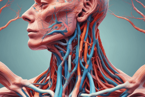
Forearm Anatomy
Cutaneous Innervation of the Forearm
- Lateral cutaneous nerve of forearm (lateral antebrachial cutaneous nerve) is a branch of the musculocutaneous nerve.
- Medial cutaneous nerve of forearm (medial antebrachial cutaneous nerve) is a branch of the medial cord of the brachial plexus.
- Posterior cutaneous nerve of forearm (posterior antebrachial cutaneous nerve) is a branch of the radial nerve.
Superficial Veins of the Forearm
- Cephalic vein (lateral) and basilic vein (medial) are the two main superficial veins.
- Median cubital vein is a communicating vein that connects the cephalic and basilic veins.
- Median antebrachial vein is a superficial vein that drains into the median cubital vein.
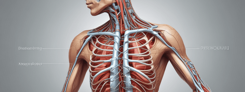
Compartments of the Forearm
- The forearm is divided into anterior and posterior compartments by the interosseous membrane.
- The anterior compartment contains flexor and pronator muscles, while the posterior compartment contains extensor and supinator muscles.
Anterior Compartment of the Forearm
- Muscles of the anterior compartment:
- Superficial layer: Pronator teres, Palmaris longus, Flexor carpi radialis, and Flexor carpi ulnaris.
- Intermediate layer: Flexor digitorum superficialis, Flexor pollicis longus, and Pronator quadratus.
- Innervation and arterial supply of each muscle:
- Pronator teres: Median nerve (C6, C7), Ulnar artery.
- Palmaris longus: Median nerve (C7, C8), Ulnar artery.
- Flexor carpi radialis: Median nerve (C6, C7), Ulnar artery.
- Flexor carpi ulnaris: Ulnar nerve, Ulnar artery.
- Flexor digitorum superficialis: Median nerve (C8, T1) for digits 2 & 3, Ulnar nerve for digits 4 & 5, Ulnar artery and Anterior interosseous artery.
- Flexor pollicis longus: Median nerve (C8, T1), Anterior Interosseous artery.
- Pronator quadratus: Median nerve (C6, C7), Anterior Interosseous artery.
Arteries and Veins of the Anterior Compartment
- Ulnar artery:
- Begins in the cubital fossa.
- Passes deep to the ulnar head of the pronator teres.
- In the upper forearm, it is deeply located and passes deep to the superficial and intermediate muscles.
- In the lower forearm, it is more superficial.
- Radial artery:
- Begins in the cubital fossa.
- In the upper forearm, it is covered by the brachioradialis and passes superficial to the pronator teres.
- In the lower forearm, it is superficial and lies between the tendon of brachioradialis and the tendon of flexor carpi radialis.
- Deep veins of the forearm:
- Radial and ulnar arteries are accompanied by two veins each (radial and ulnar veins or venae comitantes).
- There are numerous connections between the two radial veins and between the two ulnar veins.
Nerves of the Anterior Compartment
-
Median nerve:
- Exits the cubital fossa by passing between the humeral and ulnar heads of the pronator teres.
- Descends in the forearm between the flexor digitorum superficialis and flexor digitorum profundus.
- Enters the hand by passing through the carpal tunnel.
- Branches:
- Muscular branches: supply pronator teres, flexor carpi radialis, palmaris longus, and flexor digitorum superficialis.
- Articular branch: to the elbow joint.
- Palmar branch: supplies the skin of the central, depressed area of the palm of the hand.
- Anterior interosseous nerve: supplies the anterior surface of the carpus.### Posterior Compartment of Forearm
-
Extensor Digitorum Communis
- Arterial supply: Radial artery
- Action: Extends the wrist, abducts the hand
-
Extensor Digiti Minimi
- Bony attachments: Common extensor tendon on humerus, extensor digitorum tendon of the 5th digit and extensor expansion
- Innervation: Radial nerve (C7, C8)
- Arterial supply: Interosseous artery
- Action: Extends the metacarpophalangeal, proximal interphalangeal and distal interphalangeal joints of the 5th digit
-
Anconeus
- Considered by some authors as a muscle of the posterior compartment of the arm
- Origin: Lateral epicondyle of humerus
- Insertion: Olecranon and proximal part of posterior surface of shaft of ulna
- Nerve supply: Radial nerve
- Action: Assists triceps brachii with extension of forearm at elbow joint, may abduct ulna during pronation of forearm
Deep Layer
- Extensor Indicis
- Bony attachments: Interosseous membrane and distal ulna, extensor digitorum tendon of the 2nd digit and extensor expansion
- Innervation: Radial nerve (C7, C8)
- Arterial supply: Interosseous artery
- Action: Extends the metacarpophalangeal, proximal interphalangeal and distal interphalangeal joints of the index finger
- Extensor Pollicis Brevis
- Bony attachments: Interosseous membrane and distal radius, proximal phalanx of thumb
- Innervation: Radial nerve
- Arterial supply: Interosseous artery
- Action: Extends the metacarpophalangeal joint of the thumb
- Extensor Pollicis Longus
- Bony attachments: Interosseous membrane and mid shaft of the ulna, distal phalanx of thumb
- Innervation: Radial nerve
- Arterial supply: Interosseous artery
- Action: Extends the thumb at the interphalangeal joint
- Supinator
- Bony attachments: Distal lateral humerus, proximal ulna, radial ligament, proximal lateral radius
- Innervation: Radial nerve (C7, C8)
- Arterial supply: Recurrent interosseous artery
- Action: Supinates the forearm
Compartments (Tunnels) Under the Extensor Retinaculum
- 1st compartment: Tendons of abductor pollicis longus and extensor pollicis brevis
- 2nd compartment: Tendons of extensor carpi radialis longus and extensor carpi radialis brevis
- 3rd compartment: Tendon of extensor pollicis longus
- 4th compartment: Tendons of extensor digitorum and extensor indicis
- 5th compartment: Tendon of extensor digiti minimi
- 6th compartment: Tendon of extensor carpi ulnaris
Anatomical Snuffbox
- Triangular depression on the lateral side of the wrist
- Bounded laterally by tendons of abductor pollicis longus and extensor pollicis brevis
- Bounded medially by tendon of extensor pollicis longus
- Contains styloid process of radius, scaphoid, trapezium, and base of 1st metacarpal
- Radial artery crosses anatomical snuff box and passes deep to tendons that form its boundaries
- Superficial fascia over anatomical snuff box contains branches of superficial radial nerve and origin of cephalic vein from dorsal venous arch of hand
Radial Nerve
- Divides into superficial and deep terminal branches anterior to lateral epicondyle of humerus
- Before dividing into its terminal branches, radial nerve supplies three heads of triceps brachii, anconeus, brachioradialis, and extensor carpi radialis longus
- Also gives off cutaneous branches to arm and forearm
- Superficial branch of radial nerve:
- Descends in forearm deep to brachioradialis muscle and lateral to radial artery
- In distal forearm, it separates from artery and passes posteriorly, under tendon of brachioradialis, to reach posterior surface of wrist
- Divides into branches that supply skin of lateral part of dorsum of hand and dorsal aspects of lateral 2½ (or 3½) digits
- Deep branch of radial nerve:
- Pierces supinator and winds around lateral aspect of radius within supinator to reach posterior compartment of forearm
- After it emerges from supinator, it is sometimes referred to as posterior interosseous nerve
- Supplies extensor carpi radialis brevis, extensor digitorum, extensor digiti minimi, extensor carpi ulnaris, supinator, abductor pollicis longus, extensor pollicis brevis, extensor pollicis longus, and extensor indicis
- Also gives off articular branches to distal radioulnar and wrist joints
Blood Supply of the Posterior Compartment
- Posterior interosseous artery:
- Originates in anterior compartment of forearm from common interosseous artery
- Passes posteriorly, between ulna and radius, and above superior border of interosseous membrane, to reach posterior compartment
- Accompanies posterior interosseous nerve in posterior compartment
- Gives off interosseous recurrent artery - participates in arterial anastomosis around elbow joint
- Terminal part of anterior interosseous artery pierces interosseous membrane and anastomoses with posterior interosseous artery
Cutaneous Innervation of the Forearm
- The lateral cutaneous nerve of the forearm (lateral antebrachial cutaneous nerve) is a branch of the musculocutaneous nerve.
- The medial cutaneous nerve of the forearm (medial antebrachial cutaneous nerve) is a branch of the medial cord of the brachial plexus.
- The posterior cutaneous nerve of the forearm (posterior antebrachial cutaneous nerve) is a branch of the radial nerve.
Superficial Veins of the Forearm
- The cephalic vein is located laterally.
- The basilic vein is located medially.
- The median cubital vein is frequently used for venipuncture and lies superficial to the cubital fossa in the superficial fascia.
- The median cubital vein runs superiorly and medially from the cephalic vein to the basilic vein.
- The median antebrachial vein ascends along the anterior midline of the forearm and drains into the median cubital vein.
Compartments of the Forearm
- The forearm is enclosed in a sheath of deep fascia (antebrachial fascia).
- The interosseous membrane is a thin, strong fibrous membrane that attaches to the interosseous borders of the radius and ulna.
- The interosseous membrane, radius, and ulna divide the forearm into anterior and posterior compartments, which are surrounded by antebrachial fascia.
- Each compartment has its own muscles, nerves, and blood vessels.
- The anterior compartment contains flexor and pronator muscles.
- The posterior compartment contains extensor and supinator muscles.
Muscles of the Anterior Compartment
- Pronator Teres
- Bony attachments: medial epicondyle and adjacent medial supracondylar ridge of the humerus, and medial aspect of the coronoid process of the ulna.
- Innervation: Median nerve (C6, C7).
- Arterial supply: Ulnar artery.
- Action: Pronates the forearm.
- Important relationships: The median nerve passes between the two heads of the pronator teres as it leaves the cubital fossa (possible site of compression).
- Palmaris Longus
- Bony attachments: common flexor tendon on the humerus, palmar aponeurosis, and flexor retinaculum.
- Innervation: Median nerve (C7, C8).
- Arterial supply: Ulnar artery.
- Action: Flexes the wrist.
- Note: Absent in about 15% of the population.
- Flexor Carpi Radialis
- Bony attachments: common flexor tendon on the humerus, base of the 2nd metacarpal.
- Innervation: Median nerve (C6, C7).
- Arterial supply: Ulnar artery.
- Action: Flexes the wrist, abducts the hand.
- Flexor Carpi Ulnaris
- Bony attachments: medial epicondyle of the humerus, medial aspect of the olecranon and posterior border of the ulnar shaft, and pisiform, hook of hamate, and proximal 5th metacarpal.
- Innervation: Ulnar nerve.
- Arterial supply: Ulnar artery.
- Action: Flexes the wrist, adducts the hand.
- Important relationships: The ulnar nerve passes between the two heads of the flexor carpi ulnaris as it enters the anterior compartment of the forearm (cubital tunnel).
- Flexor Digitorum Superficialis
- Bony attachments: common flexor tendon, coronoid process of the ulna, and middle phalanges of the 2nd to 5th digits.
- Innervation: Median nerve (C8, T1) for digits 2 & 3, and Ulnar nerve for digits 4 & 5.
- Arterial supply: Ulnar artery and Anterior Interosseous artery.
- Action: Flexes the metacarpophalangeal, proximal interphalangeal, and distal interphalangeal joints.
- Flexor Pollicis Longus
- Bony attachments: interosseous membrane, and anterior radius, and base of the distal phalanx of the thumb.
- Innervation: Median nerve (C8, T1).
- Arterial supply: Anterior Interosseous artery.
- Action: Flexes the metacarpophalangeal and interphalangeal joints of the thumb.
- Pronator Quadratus
- Bony attachments: distal anteriomedial ulna, and distal anterior radius.
- Innervation: Median nerve (C6, C7).
- Arterial supply: Anterior Interosseous artery.
- Action: Pronates the forearm.
Arteries and Veins of the Anterior Compartment
- Ulnar Artery
- Begins in the cubital fossa as a terminal branch of the brachial artery.
- Leaves the cubital fossa by passing deep to the ulnar head of the pronator teres, which separates it from the median nerve.
- In the upper forearm, it is deeply located and passes deep to the superficial and intermediate muscles of the anterior compartment.
- In the lower forearm, it is more superficial.
- The tendon of the flexor carpi ulnaris and the ulnar nerve are medial to the ulnar artery, and the tendons of the flexor digitorum superficialis are lateral to it.
- Along most of its course, it is accompanied by the ulnar nerve (nerve lies medial to the artery).
- Enters the palm of the hand anterior (superficial) to the flexor retinaculum with the ulnar nerve and ends in the hand by forming the superficial palmar arch.
- Branches of the Ulnar Artery
- Muscular branches: supply neighboring muscles along its course.
- Anterior and posterior ulnar recurrent arteries: participate in arterial anastomosis around the elbow joint.
- Common interosseous artery: short branch that originates from the proximal part of the ulnar artery and divides into anterior and posterior interosseous arteries.
- Palmar and dorsal carpal branches: anastomose with corresponding branches of the radial artery and supply the wrist.
- Ulnar Nerve
- Enters the anterior forearm by passing between the humeral and ulnar heads of the flexor carpi ulnaris (cubital tunnel).
- Along most of its course in the forearm, it is accompanied by the ulnar artery (artery lies lateral to the nerve).
- In the upper forearm, it is more deeply located (between the flexor carpi ulnaris and flexor digitorum profundus).
- In the lower forearm, it is more superficial and lies between the tendon of the flexor carpi ulnaris (medial to the nerve) and the ulnar artery (lateral to the nerve).
- Enters the palm of the hand by passing anterior (superficial) to the flexor retinaculum.
Flexor Retinaculum (Transverse Carpal Ligament)
- Strong fibrous band on the anterior aspect of the wrist.
- Attaches medially to the pisiform and hook of hamate.
- Attaches laterally to the scaphoid and trapezium.
- Transforms the anterior, concave surface of the carpus into an osteofibrous tunnel (carpal tunnel/canal) and holds the median nerve and tendons of the long flexor muscles of the thumb and fingers (surrounded by their synovial sheaths).
- Compression of the median nerve at this level causes carpal tunnel syndrome.
Contents of the Carpal Tunnel
- 4 tendons of the flexor digitorum superficialis.
- 4 tendons of the flexor digitorum profundus.
- Tendon of the flexor pollicis longus.
- Median nerve.
- The tendon of the flexor pollicis longus has its own synovial sheath (radial bursa).
- The 4 tendons of the flexor digitorum superficialis and 4 tendons of the flexor digitorum profundus share the same synovial sheath (common flexor sheath or ulnar bursa).
The Posterior Compartment of the Forearm
- Extensor Retinaculum
- Thickening of the deep fascia of the forearm that stretches across the back of the wrist.
- Holds long extensor tendons in position and prevents them from bowstringing as they cross the wrist joint.
- Converts the grooves on the posterior aspects of the distal ends of the radius and ulna into 6 separate compartments (tunnels) for the long extensor tendons (surrounded by synovial sheaths).
Muscles of the Posterior Compartment
- Extensor Digitorum
- Bony attachments: common extensor tendon on the humerus, and extensor expansion of the 2nd-5th digits.
- Innervation: Radial nerve (C7
Studying That Suits You
Use AI to generate personalized quizzes and flashcards to suit your learning preferences.