Podcast
Questions and Answers
What is the primary function of the ependymal cells' cilia?
What is the primary function of the ependymal cells' cilia?
- To facilitate the movement of cerebrospinal fluid (correct)
- To facilitate the absorption of cerebrospinal fluid
- To facilitate the proliferation of astrocytes
- To facilitate the phagocytosis of apoptotic bodies
What is the main difference between ependymal cells and epithelial cells?
What is the main difference between ependymal cells and epithelial cells?
- Ependymal cells lack a basal lamina, while epithelial cells have one (correct)
- Ependymal cells have a basal lamina, while epithelial cells do not
- Ependymal cells are more numerous than epithelial cells
- Ependymal cells have apical junctional complexes, while epithelial cells do not
What is the term for the barrier layer formed by astrocytes at the external CNS surface?
What is the term for the barrier layer formed by astrocytes at the external CNS surface?
- Glial limiting membrane (correct)
- Ependymal layer
- Microglial membrane
- Astrocytic scar
What is the origin of microglia?
What is the origin of microglia?
What is the function of microglia in removing apoptotic bodies and debris?
What is the function of microglia in removing apoptotic bodies and debris?
What is the distribution of microglia in the CNS?
What is the distribution of microglia in the CNS?
What is unique about the shape of ependymal cells?
What is unique about the shape of ependymal cells?
Why are microglia not typically visualized with H&E staining?
Why are microglia not typically visualized with H&E staining?
What is the primary function of Schwann cells in the PNS?
What is the primary function of Schwann cells in the PNS?
What is the main component of white matter in the CNS?
What is the main component of white matter in the CNS?
Where are satellite cells typically found in the PNS?
Where are satellite cells typically found in the PNS?
What is the function of oligodendrocytes in the CNS?
What is the function of oligodendrocytes in the CNS?
What is the term for the lipid-rich myelin-producing glial cells in the CNS?
What is the term for the lipid-rich myelin-producing glial cells in the CNS?
Where is gray matter primarily found in the CNS?
Where is gray matter primarily found in the CNS?
What is the purpose of the meninges in the CNS?
What is the purpose of the meninges in the CNS?
What is the main difference between Schwann cells and oligodendrocytes?
What is the main difference between Schwann cells and oligodendrocytes?
Where are the neuronal cell bodies of preganglionic fibers located in the sympathetic nervous system?
Where are the neuronal cell bodies of preganglionic fibers located in the sympathetic nervous system?
What is the neurotransmitter released by preganglionic fibers?
What is the neurotransmitter released by preganglionic fibers?
Where are postganglionic neurons located?
Where are postganglionic neurons located?
What is the function of neurotrophins?
What is the function of neurotrophins?
Where are neural stem cells located in the adult CNS?
Where are neural stem cells located in the adult CNS?
What is the potential of neural stem cells?
What is the potential of neural stem cells?
What is the characteristic of the autonomic nervous system?
What is the characteristic of the autonomic nervous system?
What is the characteristic of ganglia in the autonomic nervous system?
What is the characteristic of ganglia in the autonomic nervous system?
What is the primary function of the epineurium in peripheral nerves?
What is the primary function of the epineurium in peripheral nerves?
What type of neurons are typically found in autonomic ganglia?
What type of neurons are typically found in autonomic ganglia?
What is the main difference between sensory ganglia and autonomic ganglia?
What is the main difference between sensory ganglia and autonomic ganglia?
What is the role of the blood-nerve barrier in peripheral nerves?
What is the role of the blood-nerve barrier in peripheral nerves?
What is the primary function of intramural ganglia?
What is the primary function of intramural ganglia?
What is the characteristic of nerves that are classified as mixed?
What is the characteristic of nerves that are classified as mixed?
What is the role of the capsule surrounding ganglia?
What is the role of the capsule surrounding ganglia?
What is the function of the neurons in sensory ganglia?
What is the function of the neurons in sensory ganglia?
What is the consequence of astrocytes proliferating at injured sites in spinal cord tracts?
What is the consequence of astrocytes proliferating at injured sites in spinal cord tracts?
What is the term for the changes in the perikaryon that signal the onset of regeneration?
What is the term for the changes in the perikaryon that signal the onset of regeneration?
What is the role of new Schwann cells in regeneration?
What is the role of new Schwann cells in regeneration?
What is the result of the proximal segment of the axon degenerating after injury?
What is the result of the proximal segment of the axon degenerating after injury?
What is the final outcome of axonal regeneration in peripheral nerves?
What is the final outcome of axonal regeneration in peripheral nerves?
What occurs to the Nissl substance during chromatolysis?
What occurs to the Nissl substance during chromatolysis?
Flashcards are hidden until you start studying
Study Notes
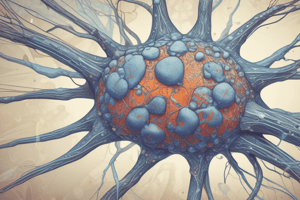
Glial Cells
- Astrocytes:
- Extend fibrous processes with expanded perivascular feet that cover capillary endothelial cells and modulate blood flow
- Help move nutrients, wastes, and other metabolites between neurons and capillaries
- Form a barrier layer of expanded protoplasmic processes called the glial limiting membrane, which lines the meninges at the external CNS surface
- Fill tissue defects after CNS injury by proliferation to form an astrocytic scar
- Ependymal cells:
- Line the fluid-filled ventricles of the brain and the central canal of the spinal cord
- Have cilia and long microvilli that facilitate the movement of cerebrospinal fluid and absorption
- Joined apically by apical junctional complexes, but lack a basal lamina
- Have elongated basal ends that extend branching processes into the adjacent neuropil
- Microglia:
- Origin: From circulating blood monocytes, belonging to the same family as macrophages and other antigen-presenting cells
- Small cell bodies with many long, branched processes
- Relatively static, but their processes continuously probe and interact with neuropil, synapses, and other cells in an area up to 10-fold that of the cell body
- Function: Remove apoptotic bodies and debris from damaged or remodeled synapses by phagocytosis, and constitute the major mechanism of immune defense in the CNS
Glial Cells of the PNS
- Schwann cells (neurolemmocytes):
- Origin: From precursors in the neural crest
- Found only in the PNS, considered as the counterpart to oligodendrocytes of the CNS
- Have trophic interactions with axons and form their myelin sheathes
- Unlike oligodendrocytes, each Schwann cell forms myelin around a portion of only one axon
- Satellite cells of ganglia:
- Origin: From the embryonic neural crest
- Form a thin, intimate glial layer around each large neuronal cell body in the ganglia of the PNS
- Exert a trophic or supportive effect on these neurons, insulating, nourishing, and regulating their microenvironments
Central Nervous System (CNS)
- Composed of three major structures: cerebrum, cerebellum, and spinal cord
- CNS tissue is covered with a connective tissue layer called meninges, which is relatively soft and easily damaged by injuries
- Arranged into two areas: white matter and gray matter
- White matter: Main components are myelinated axons, myelin-producing oligodendrocytes, astrocytes, and microglia
- Gray matter: Contains abundant neuronal cell bodies, dendrites, astrocytes, and microglial cells, and is where most synapses occur
Nerve Organization
- Peripheral nerves have a dense, irregular fibrous coat called the epineurium, which extends deeply to fill the space between fascicles
- Nerves can be afferent/sensory or efferent/motor, and can be motor, sensory, or mixed
- Ganglia:
- Ovoid structures containing neuronal cell bodies and their surrounding glial satellite cells
- Serve as relay stations to transmit nerve impulses
- Divided into sensory or autonomic ganglia based on the direction of the nerve impulse
- Associated with both cranial nerves (cranial ganglia) and the dorsal roots of the spinal nerves (spinal ganglia)
Autonomic Ganglia
- Autonomic nerves control the activity of smooth muscle, secretion of some glands, heart rate, and involuntary activities that maintain a constant internal environment (homeostasis)
- Autonomic ganglia:
- Contain multipolar neurons
- Located within certain organs, especially in the walls of the digestive tract, where they constitute the intramural ganglia
- Have two neuron circuits: preganglionic and postganglionic fibers
- Autonomic nervous system has two parts: sympathetic and parasympathetic nervous systems
Neural Plasticity and Regeneration
- The nervous system exhibits neuronal differentiation and formation of new synapses even in adults
- Neural stem cells are present in the adult CNS, located in part among the cells of the ependymal
- Astrocytes can proliferate at injured sites, but can interfere with successful axonal regeneration
- In peripheral nerves, injured axons have a much greater potential for regeneration and return of function
- The onset of regeneration is signaled by changes in the perikaryon, including chromatolysis
- The new Schwann cells align to serve as guides for the regrowing axons and produce polypeptide factors that promote axonal outgrowth
Studying That Suits You
Use AI to generate personalized quizzes and flashcards to suit your learning preferences.