Podcast
Questions and Answers
Which type of epithelial tissue is characterized by a single layer of tall, column-like cells?
Which type of epithelial tissue is characterized by a single layer of tall, column-like cells?
- Simple columnar epithelium (correct)
- Pseudostratified columnar ciliated epithelium
- Stratified squamous keratinized epithelium
- Simple squamous epithelium
Which type of connective tissue is primarily responsible for providing insulation and energy storage?
Which type of connective tissue is primarily responsible for providing insulation and energy storage?
- Reticular tissue
- Areolar connective tissue
- Hyaline cartilage
- Adipose tissue (correct)
What is the primary function of mitochondria within a cell?
What is the primary function of mitochondria within a cell?
- Cell division
- Membrane transport
- Energy production (correct)
- Protein synthesis
Which type of muscle tissue is under involuntary control and is found in the heart?
Which type of muscle tissue is under involuntary control and is found in the heart?
What structure is responsible for processing and packaging proteins in the cell?
What structure is responsible for processing and packaging proteins in the cell?
Flashcards
Mitochondria
Mitochondria
The powerhouse of the cell, responsible for generating energy (ATP) through cellular respiration.
Endoplasmic Reticulum
Endoplasmic Reticulum
A network of interconnected membranes involved in protein synthesis, lipid metabolism, and detoxification.
Areolar connective tissue
Areolar connective tissue
A type of connective tissue that forms a flexible and supportive framework for many organs.
Adipose tissue
Adipose tissue
Signup and view all the flashcards
Haversian system
Haversian system
Signup and view all the flashcards
Study Notes
General Histology Practical Revision
- This set of notes covers various histological samples, including cell structures and different tissue types.
Cell Membrane
- Images show a cell membrane's structure, including its key components.
Mitochondria
- Images display the distinctive structure of mitochondria, including inner and outer membranes.
Golgi Complex
- Images depict the Golgi apparatus’s structural arrangement as stacks of flattened sacs.
Nucleus
- Images show different views of the nucleus, highlighting its key parts and structure.
- Diagrams may include labels for different components within the nucleus.
Endoplasmic Reticulum
- Images of both rough and smooth endoplasmic reticulums (ER).
- Rough ER is characterized by ribosomes, while smooth ER lacks them.
Centriole
- Images show the centriole's characteristic arrangement.
Simple Squamous Epithelium
- Micrograph of simple squamous epithelium, showing flattened cells.
- Scale bar (50 µm) provided in the image.
Simple Cuboidal Epithelium
- Micrograph of simple cuboidal epithelium, showing cube-shaped cells.
- Scale bar (40 µm) provided in the image.
Simple Columnar Epithelium
- Image of simple columnar epithelium, showing column-shaped cells.
- Scale bar (70 µm) provided in the image
Pseudostratified Columnar Ciliated Epithelium
- Micrograph of pseudostratified columnar ciliated epithelium.
Stratified Squamous Non-Keratinized Epithelium
- Micrograph of stratified squamous non-keratinized epithelium, showing multiple layers of cells.
Stratified Squamous Keratinized Epithelium
- Micrograph of stratified squamous keratinized epithelium, showing a stratified layer with a keratinized surface.
Transitional Epithelium
- Image highlighting the unique adaptation of transitional epithelium, capable of stretching.
Neutrophil
- Image of a neutrophil, a type of white blood cell.
Eosinophil
- Image of an eosinophil, a type of white blood cell.
Basophil
- Image of a basophil, a type of white blood cell.
Lymphocyte
- Image of a lymphocyte, a type of white blood cell.
Monocyte
- Image of a monocyte, a type of white blood cell.
Nucleated RBCs of Toad (Frog)
- Images of nucleated red blood cells (RBCs) from a toad or frog.
Red Blood Corpuscles (RBCs)
- Image of mature mammalian red blood cells (RBCs).
Plasma Cell
- Images of plasma cells, showing their characteristic morphology.
Fibroblast and Fibrocyte
- Images differentiated fibroblasts and fibrocytes, showing differences in structure.
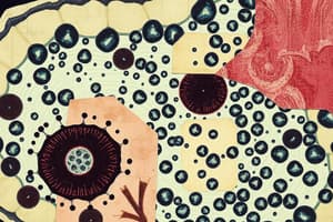
Areolar Connective Tissue
- Micrograph of areolar connective tissue, highlighting its diverse cell types and extracellular fibers.
Adipose Tissue, Sudan III
- Images show adipose tissue stained using Sudan III, highlighting fat storage.
Adipose Tissue, Sudan Black
- Image shows the effect of the Sudan black stain, again emphasising the fat storage.
Mucoid Connective Tissue, Umbilical Cord
- Micrograph of mucoid connective tissue like in the umbilical cord, showcasing unique features.
Reticular Tissue, Silver Stain
- Image of reticular tissue stained using a silver stain, highlighting reticular fibers.
White Fibrous Connective Tissue
- Image of white fibrous connective tissue, demonstrating collagen fibers.
Elastic Connective Tissue
- Image of elastic connective tissue, with visible elastic fibers.
Hyaline Cartilage
- Micrograph of hyaline cartilage, showing chondrocytes within the cartilage matrix.
- Label identifying perichondrium.
Elastic Cartilage
- Image highlights elastic cartilage, with a focus on elastic fibers within the matrix.
Fibrocartilage
- Micrograph highlighting the distinctive features of fibrocartilage.
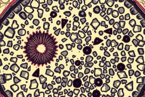
Haversian System
- Image of the Haversian system in compact bone.
Ground of Compact Bone
- Image showcasing the ground or substance of compact bone.
Spongy Bone
- Image of spongy bone showing the trabecular structure.
Cardiac Muscle
- Image representing the histologic appearance of cardiac muscle.
Skeletal Muscle
- Image of skeletal muscle tissue, displaying its characteristic striations and fibers.
Nerve Trunk
- Image showcasing the structure of a nerve trunk.
Nerve Trunk (Osmic Acid)
- Image of nerve trunk stained with osmic acid.
Studying That Suits You
Use AI to generate personalized quizzes and flashcards to suit your learning preferences.