Podcast
Questions and Answers
What is the primary function of smooth muscle in the colon wall?
What is the primary function of smooth muscle in the colon wall?
- Storage of waste
- Movement of food (correct)
- Secretion of enzymes
- Absorption of nutrients
The pouch of peritoneum filled with fat is a major organ of digestion.
The pouch of peritoneum filled with fat is a major organ of digestion.
False (B)
What structure in the colon is described as sac-like?
What structure in the colon is described as sac-like?
outpouching
The smooth muscle in the colon is responsible for _____ food.
The smooth muscle in the colon is responsible for _____ food.
Match the following components of the colon with their descriptions:
Match the following components of the colon with their descriptions:
What is the structure in the colon known for its sac-like appearance?
What is the structure in the colon known for its sac-like appearance?
Smooth muscle assists in the movement of food through the colon.
Smooth muscle assists in the movement of food through the colon.
What is the primary role of the pouch of peritoneum in relation to the colon?
What is the primary role of the pouch of peritoneum in relation to the colon?
The _____ of peritoneum is filled with fat and assists in the colon's structure.
The _____ of peritoneum is filled with fat and assists in the colon's structure.
Match the following components of the colon with their characteristics:
Match the following components of the colon with their characteristics:
Flashcards
Colon
Colon
A large intestine responsible for absorbing water and forming waste.
Smooth Muscle
Smooth Muscle
Involuntary muscle found in the walls of organs like the colon, responsible for contractions and movement.
Peritenium
Peritenium
A protective membrane that surrounds the abdomen and its organs, including the colon.
Outpouchings
Outpouchings
Signup and view all the flashcards
Move Food
Move Food
Signup and view all the flashcards
Colon's Role
Colon's Role
Signup and view all the flashcards
Smooth Muscle in the Colon
Smooth Muscle in the Colon
Signup and view all the flashcards
Outpouchings on the Colon
Outpouchings on the Colon
Signup and view all the flashcards
What is 'Poouch of penitenium' ?
What is 'Poouch of penitenium' ?
Signup and view all the flashcards
What does 'sac Like outpoutching the colon' mean?
What does 'sac Like outpoutching the colon' mean?
Signup and view all the flashcards
Study Notes
Gastrointestinal System Outline
- The gastrointestinal system is responsible for digestion, absorption of food, and excretion of waste.
- The digestive system consists of:
- Oral cavity (tongue, teeth)
- Gastrointestinal Tract (GIT): begins at the oropharynx, travels through the esophagus, and terminates at the anal canal.
- Accessory glands (liver, pancreas, salivary glands) that aid in digestive processes.
Learning Objectives
- Understand the anatomical divisions of the digestive system.
- Define the gastrointestinal tract (GIT) and its component parts.
- Differentiate between the large and small intestines.
- Recognize the general structure of the GIT wall.
Anatomical Divisions of the GIT
- Oropharynx: A portion of the pharynx located behind the oral cavity
- Esophagus: A 25cm muscular tube extending from the oropharynx to the stomach, with cervical, thoracic, and abdominal sections. The thoracic portion passes through the esophageal hiatus of the diaphragm.
- Stomach: The most expansive part of the GIT, shaped like a "J," that serves as a food reservoir. Consists of four regions: Cardia, Fundus, Body, and Pylorus. The stomach is the most dilated part of the GIT.
Small Intestine
- 6 meters in length, divided into three parts: Duodenum, Jejunum, and Ileum.
Large Intestine
- 1.5 meters in length, composed of:
- Cecum
- Ascending Colon
- Transverse Colon
- Descending Colon
- Sigmoid Colon
- Rectum
- Anal Canal
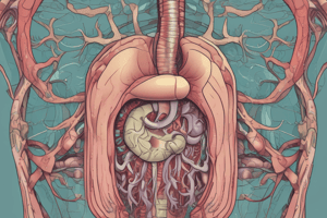
Small Intestine Structure (Image)
- Shown as a series of coiled structures in various diagrams.
- Parts labeled as duodenum, jejunum, and ileum.
Large Intestine Structure (Image)
- Distinguishable from small intestine by external features like Taenia Coli (bands of smooth muscle), Omental appendices (fat pouches), and sacculations (haustrations).
Wall of the GIT
- Consists of four layers:
- Mucosa: The innermost layer contacting food; containing epithelium, lamina propria and muscularis mucosae.
- Submucosa: A connective tissue layer containing blood vessels, lymphatics, and Meissner's nerve plexus.
- Muscularis: A smooth muscle layer consisting of inner circular and outer longitudinal layers, with the myenteric (Auerbach's) nerve plexus in between.
- Adventitia/Serosa: The outermost layer; serosa in the abdominal cavity, adventitia elsewhere.
Liver Anatomy
- The largest intra-abdominal organ.
- Located in the right upper quadrant of the abdominal cavity, below the diaphragm.
- Composed of four lobes: right, left, quadrate, and caudate. The liver's position is described as being in the right hypochondrium.
Studying That Suits You
Use AI to generate personalized quizzes and flashcards to suit your learning preferences.