Podcast
Questions and Answers
Which statement accurately describes the location affected by acute compromise of major vessels?
Which statement accurately describes the location affected by acute compromise of major vessels?
- Anemia primarily affects the jejunum and ileum.
- Watershed zones include the splenic flexure and sigmoid colon. (correct)
- The inferior mesenteric arterial circulation primarily supplies the upper rectum.
- Transmural infarction is typical in the duodenum.
What primary feature distinguishes Hirschsprung disease in its pathology?
What primary feature distinguishes Hirschsprung disease in its pathology?
- Increased tone of the lower esophageal sphincter
- Absence of ganglion cells in the affected segment (correct)
- Inflammation of the rectal mucosa
- Proliferation of smooth muscle cells
What is the primary cause of transmural infarction in the intestine?
What is the primary cause of transmural infarction in the intestine?
- Acute vascular obstruction of major blood vessels. (correct)
- Chronic hypoperfusion due to systemic diseases.
- Defective absorption in malabsorption syndromes.
- Localized bacterial infection causing inflammation.
What is the primary mechanism causing symptoms in achalasia?
What is the primary mechanism causing symptoms in achalasia?
Which feature is characteristic of the histology observed in acute ischemic intestine?
Which feature is characteristic of the histology observed in acute ischemic intestine?
Which diagnostic method is important for confirming Hirschsprung disease?
Which diagnostic method is important for confirming Hirschsprung disease?
Which factor contributes to the vulnerability of the intestinal surface epithelium to ischemic injury?
Which factor contributes to the vulnerability of the intestinal surface epithelium to ischemic injury?
What is the primary risk associated with pyloric stenosis?
What is the primary risk associated with pyloric stenosis?
In chronic ischemia, which complication can occur despite being uncommon?
In chronic ischemia, which complication can occur despite being uncommon?
Which symptom is commonly associated with achalasia?
Which symptom is commonly associated with achalasia?
What indicates the presence of malabsorption in an individual?
What indicates the presence of malabsorption in an individual?
What characterizes the morphology of Hirschsprung disease?
What characterizes the morphology of Hirschsprung disease?
What role do neutrophils play in acute ischemic episodes upon reperfusion?
What role do neutrophils play in acute ischemic episodes upon reperfusion?
What is the incidence of Hirschsprung disease in live births?
What is the incidence of Hirschsprung disease in live births?
Which statement best characterizes the impact of acute ischemic disease on the intestinal mucosa?
Which statement best characterizes the impact of acute ischemic disease on the intestinal mucosa?
Which condition is characterized by functional obstruction and proximal dilation of intestine?
Which condition is characterized by functional obstruction and proximal dilation of intestine?
What is the underlying cause of secondary achalasia in Chagas disease?
What is the underlying cause of secondary achalasia in Chagas disease?
Which treatment is NOT indicated for achalasia?
Which treatment is NOT indicated for achalasia?
What characterizes the esophageal changes in reflux esophagitis?
What characterizes the esophageal changes in reflux esophagitis?
What is a significant risk associated with Barrett esophagus?
What is a significant risk associated with Barrett esophagus?
Which of the following describes a common histological finding in esophageal varices?
Which of the following describes a common histological finding in esophageal varices?
The most common symptom of gastroesophageal reflux disease (GERD) is:
The most common symptom of gastroesophageal reflux disease (GERD) is:
What physiological mechanism primarily prevents reflux of gastric contents into the esophagus?
What physiological mechanism primarily prevents reflux of gastric contents into the esophagus?
Which feature distinguishes Barrett esophagus from normal esophageal mucosa?
Which feature distinguishes Barrett esophagus from normal esophageal mucosa?
Mallory-Weiss syndrome is primarily caused by:
Mallory-Weiss syndrome is primarily caused by:
What primary condition often leads to the development of esophageal varices?
What primary condition often leads to the development of esophageal varices?
Which statement about the esophagus is false?
Which statement about the esophagus is false?
What is a key characteristic of the epithelial changes in Barrett esophagus?
What is a key characteristic of the epithelial changes in Barrett esophagus?
What can severe, chronic gastroesophageal reflux lead to in certain patients?
What can severe, chronic gastroesophageal reflux lead to in certain patients?
In the context of chronic GERD, what happens to the esophageal tissue?
In the context of chronic GERD, what happens to the esophageal tissue?
Which inflammatory cell type is NOT typically found in the epithelial layer of esophageal lesions?
Which inflammatory cell type is NOT typically found in the epithelial layer of esophageal lesions?
What is the primary mechanism by which portal hypertension leads to esophageal varices?
What is the primary mechanism by which portal hypertension leads to esophageal varices?
What histological feature is characterized by increased numbers of specific immune cells in the lamina propria in conditions such as celiac disease?
What histological feature is characterized by increased numbers of specific immune cells in the lamina propria in conditions such as celiac disease?
Which condition is primarily associated with the overgrowth of Clostridium difficile following antibiotic use?
Which condition is primarily associated with the overgrowth of Clostridium difficile following antibiotic use?
Which of the following symptoms is NOT typically associated with enterocolitis?
Which of the following symptoms is NOT typically associated with enterocolitis?
What is a common consequence of untreated celiac disease?
What is a common consequence of untreated celiac disease?
What characterizes the histopathology of fully developed Clostridium difficile-associated colitis?
What characterizes the histopathology of fully developed Clostridium difficile-associated colitis?
In celiac disease, the histological finding of villous atrophy suggests which of the following?
In celiac disease, the histological finding of villous atrophy suggests which of the following?
Which of the following immune cells is typically increased in the lamina propria in response to intestinal inflammation?
Which of the following immune cells is typically increased in the lamina propria in response to intestinal inflammation?
In the context of antibiotic-associated diarrhea, how does Clostridium difficile affect the colonic microbiota?
In the context of antibiotic-associated diarrhea, how does Clostridium difficile affect the colonic microbiota?
What is the distinguishing feature of collagenous colitis?
What is the distinguishing feature of collagenous colitis?
Which statement correctly describes lymphocytic colitis?
Which statement correctly describes lymphocytic colitis?
In the context of diverticular disease, what does diverticulosis refer to?
In the context of diverticular disease, what does diverticulosis refer to?
Which demographic is most commonly affected by collagenous colitis?
Which demographic is most commonly affected by collagenous colitis?
What is a common characteristic of both collagenous and lymphocytic colitis?
What is a common characteristic of both collagenous and lymphocytic colitis?
What is the primary cause of colonic diverticula formation?
What is the primary cause of colonic diverticula formation?
At what age does the prevalence of diverticulosis begin to significantly increase?
At what age does the prevalence of diverticulosis begin to significantly increase?
Which autoimmune diseases are strongly associated with lymphocytic colitis?
Which autoimmune diseases are strongly associated with lymphocytic colitis?
Flashcards
Pyloric Stenosis
Pyloric Stenosis
A condition where the pyloric sphincter, the muscle between the stomach and the small intestine, is narrowed, causing difficulty in emptying the stomach. This leads to vomiting, often projectile, in infants.
Megacolon
Megacolon
A condition where the descending colon becomes dilated due to an obstruction. This often occurs in connection with Hirschsprung's Disease.
Hirschsprung's Disease
Hirschsprung's Disease
A rare condition where the colon lacks nerve cells, causing constipation and an inability to pass waste effectively.
Achalasia
Achalasia
Signup and view all the flashcards
Lower Esophageal Sphincter (LES)
Lower Esophageal Sphincter (LES)
Signup and view all the flashcards
Symptoms of Achalasia
Symptoms of Achalasia
Signup and view all the flashcards
Peristalsis
Peristalsis
Signup and view all the flashcards
Ganglionosis
Ganglionosis
Signup and view all the flashcards
Secondary Achalasia
Secondary Achalasia
Signup and view all the flashcards
Mallory-Weiss Syndrome
Mallory-Weiss Syndrome
Signup and view all the flashcards
Reflux Esophagitis
Reflux Esophagitis
Signup and view all the flashcards
Esophageal Lining
Esophageal Lining
Signup and view all the flashcards
Gastroesophageal Reflux Disease (GERD)
Gastroesophageal Reflux Disease (GERD)
Signup and view all the flashcards
Transient LES Relaxation
Transient LES Relaxation
Signup and view all the flashcards
Esophagitis
Esophagitis
Signup and view all the flashcards
Barrett's Esophagus
Barrett's Esophagus
Signup and view all the flashcards
Esophageal Varices
Esophageal Varices
Signup and view all the flashcards
Portal Hypertension
Portal Hypertension
Signup and view all the flashcards
Dysplasia
Dysplasia
Signup and view all the flashcards
Intestinal Metaplasia
Intestinal Metaplasia
Signup and view all the flashcards
Lamina propria inflammation in celiac disease
Lamina propria inflammation in celiac disease
Signup and view all the flashcards
Villous atrophy
Villous atrophy
Signup and view all the flashcards
Crypt hyperplasia
Crypt hyperplasia
Signup and view all the flashcards
Reversibility of celiac disease
Reversibility of celiac disease
Signup and view all the flashcards
Pseudomembranous colitis
Pseudomembranous colitis
Signup and view all the flashcards
Clostridium difficile
Clostridium difficile
Signup and view all the flashcards
Disruption of colonic microbiota
Disruption of colonic microbiota
Signup and view all the flashcards
Pseudomembranes
Pseudomembranes
Signup and view all the flashcards
Watershed Zones
Watershed Zones
Signup and view all the flashcards
Transmural Infarction
Transmural Infarction
Signup and view all the flashcards
Mural Infarction
Mural Infarction
Signup and view all the flashcards
Mucosal Infarction
Mucosal Infarction
Signup and view all the flashcards
Intestinal Capillary Arrangement
Intestinal Capillary Arrangement
Signup and view all the flashcards
Microscopic Examination of Ischemic Intestine
Microscopic Examination of Ischemic Intestine
Signup and view all the flashcards
Chronic Ischemia
Chronic Ischemia
Signup and view all the flashcards
Malabsorption
Malabsorption
Signup and view all the flashcards
Collagenous Colitis
Collagenous Colitis
Signup and view all the flashcards
Lymphocytic Colitis
Lymphocytic Colitis
Signup and view all the flashcards
Colonic Diverticula
Colonic Diverticula
Signup and view all the flashcards
Diverticulosis
Diverticulosis
Signup and view all the flashcards
Diverticulitis
Diverticulitis
Signup and view all the flashcards
Sigmoid Diverticular Disease
Sigmoid Diverticular Disease
Signup and view all the flashcards
True Diverticula
True Diverticula
Signup and view all the flashcards
Study Notes
Esophageal Atresia and Tracheoesophageal Fistula
- Discovered shortly after birth, usually due to regurgitation during feeding
- Esophageal atresia: a thin, non-canalized cord replaces a segment of esophagus, causing mechanical obstruction
- Atresia most common at or near the tracheal bifurcation, often associated with a fistula
- Fistula connects the upper or lower esophageal pouches to a bronchus or trachea
- Fistula can occur without atresia
- Complications include aspiration, suffocation, pneumonia, and severe fluid and electrolyte imbalances
Diaphragmatic Hernia
- Occurs when incomplete diaphragm formation allows abdominal viscera to herniate into the thoracic cavity
- Severe cases cause pulmonary hypoplasia, which is incompatible with life
Omphalocele
- Closure of the abdominal musculature is incomplete, leading to abdominal viscera herniating into a ventral membranous sac.
- Location: Centered
- Content: Primarily intestines, frequently including colon, bladder, and sometimes gonads.
- Associated with other congenital anomalies (15%)
Gastroschisis
- Similar to omphalocele, but involves all layers of the abdominal wall (peritoneum to skin).
- Location: Right side
- Content: Intestines, liver, and sometimes spleen, colon or bladder
- Frequently associated with other congenital anomalies (40-80%)
Meckel Diverticulum
- A blind pouch of the ileum, usually within 2 feet of the ileocecal valve
- Approximately 2 inches long
- More common in males
- Often asymptomatic, but symptomatic cases usually present before age 2 (approximately 4%).
- "Rule of 2s": 2% of population, 2 ft from ileocecal valve, 2 in long
Pyloric Stenosis
- Congenital hypertrophy of the pyloric sphincter
- Infants feed well initially but progressively develop projectile vomiting
- Vomiting usually begins at 2–3 weeks old, or even earlier
- Projectile vomiting occurs after feeding
- Infants are frequently hungry and voracious eaters
Hirschsprung Disease
- Also called congenital aganglionic megacolon
- Absence of ganglion cells in the distal portion of the intestine
- Results in functional obstruction
- Occurs in approximately one in 5,000 live births
Achalasia
- Increased tone of the lower esophageal sphincter (LES) due to impaired smooth muscle relaxation
- Characterized by incomplete LES relaxation, increased LES tone, and esophageal aperistalsis
- Symptoms include dysphagia (difficulty swallowing, for both solids and liquids), chest pain, and difficulty with belching
- Secondary achalasia may occur with Chagas disease, where Trypanosoma cruzi infection damages the myenteric plexus
Mallory-Weiss Syndrome
- Longitudinal mucosal tears near the gastroesophageal junction, often associated with severe retching or vomiting, frequently related to alcohol abuse.
- Usually presents with hematemesis (vomiting blood)
- Risk factors include alcohol abuse and hiatal hernias.
Boerhaave's Syndrome
- Complete rupture of the lower esophageal sphincter, often associated with severe retching or vomiting, usually unrelated to alcohol.
- May cause chest pain, and hypotension
- Presents with subq emphysema, and often with crepitus upon auscultation.
Reflux Esophagitis
- Stratified squamous epithelium of esophagus is resistant to abrasion but sensitive to acid.
- Submucosal glands in the proximal and distal esophagus secrete mucin and bicarbonate.
- Lower esophageal sphincter (LES) tone prevents reflux of highly acidic gastric contents into the esophagus.
- Reflux of gastric contents into the esophagus is the most frequent cause of esophagitis and the associated condition: Gastroesophageal Reflux Disease (GERD).
Histology of Reflux Esophagitis
- Inflammatory cells, eosinophils, neutrophils, and T cells
- Acanthosis and papillomatosis
- Basal cell hyperplasia
- Ballooned squamous cells
- Vascular ectasia in lamina propria
Esophageal Varices
- Venous blood from the GI tract passes through the liver, via the portal vein, before returning to the heart.
- Portal hypertension results in the development of collateral channels.
- These collateral veins lead to the development of varices in the distal esophagus and proximal stomach.
- Varices commonly develop in patients with cirrhosis (especially alcoholic liver disease)
How Liver Disease Leads to Bleeding Varices
- Cirrhosis increases intrahepatic vascular resistance, potentially due to architectural distortion and inadequate nitric oxide
- Portal hypertension results from increased intrahepatic resistance and splanchnic arteriolar vasodilation
- Increased pressure in these vessels can cause varices to form and rupture, leading to bleeding
Barrett's Esophagus
- Complication of chronic gastroesophageal reflux disease (GERD)
- Characterized by intestinal metaplasia replacing the normal esophageal squamous mucosa
- Increased risk of esophageal adenocarcinoma, related to preinvasive change of dysplasia, prolonged symptoms, length of affected esophageal segments, and Caucasian race
Morphology of Barrett Esophagus
- Red, velvety mucosa extending upward from the gastroesophageal junction
- Alternating with light-brown, columnar (gastric) mucosa
- Length of affected esophagus associated with dysplasia risk
Histology of Barrett's Esophagus
- Intestinal-type metaplasia manifested as squamous esophageal epithelium replacement by goblet cells
Chronic Gastritis (H. pylori)
- Common cause of chronic gastritis
- Spiral-shaped or curved bacilli, frequently present in gastric biopsies (of almost all gastric ulcer and/or gastritis patients)
- Primarily transmitted through the fecal-oral route in childhood
- The most common clinical presentation is predominantly antral gastritis with normal or increased acid production.
Autoimmune Gastritis
- Accounts for less than 10% of chronic gastritis cases
- Typically spares the antrum and is associated with hypergastrinemia
- Marked by antibodies to parietal cells and intrinsic factor
Gastric Intestinal Metaplasia
- Presence of goblet cells replacing the squamous esophageal epithelium
- Result of failed involution of the vitelline duct
Mucosal Atrophy and Intestinal Metaplasia
- Long-standing chronic gastritis, particularly affecting the body and fundus, can lead to significant loss of parietal cell mass
- Associated with intestinal metaplasia (goblet cells) and increased risk of gastric adenocarcinoma
Intestinal Obstruction
- Hernias- Protrusion of serosa-lined pouch of peritoneum through a defect in abdominal wall
- Adhesions- Fibrous bridges forming between organs, possibly leading to internal herniation
- Volvulus- Twisting of a bowel loop about its mesenteric point of attachment.
- Intussusception- A segment of bowel telescoping into another segment, potentially leading to obstruction, compression of mesenteric vessels, and infarction
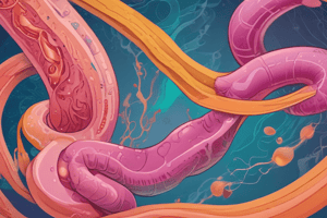
Ischemic Bowel Disease
- Majority of the GI tract is supplied by the celiac, superior mesenteric, and inferior mesenteric arteries. Interconnections result in the small intestine and colon’s ability to withstand decreased blood flow from one artery.
- Acute compromise of a major vessel can lead to infarction of meters of intestine
- Infarction can range from mucosal to transmural.
- Watershed zones (e.g., splenic flexure, rectosigmoid colon), where arterial supplies meet, are highly susceptible.
Infectious Enterocolitis
- Can present with a wide range of symptoms: Diarrhea, abdominal pain, urgency, perianal discomfort, incontinence, hemorrhage, and/or fever
- Viral, bacterial, and parasitic infections.
Pseudomembranous Colitis
- Caused primarily by Clostridium difficile
- Associated with disruption of normal colonic microbiota by antibiotic use (C. difficile overgrowth, most often)
- Presentation is characterized by pseudomembranes, made of inflammatory cells and debris at sites of mucosal injury
- While pseudomembranes can occur with infections or ischemia, the histopathology of C. difficile is distinctive.
Whipple Disease
- Rare, multi-visceral chronic disease
- Characterized by malabsorption and/or lymphatic obstruction, as well as arthritis, and diarrhea
- Microbial infection caused by Tropheryma whipplei, a gram-positive actinomycete.
- Morphologically characterized by macrophages containing PAS-positive, diastase-resistant granules, which are lysosomes containing partially digested bacteria
- The macrophages can accumulate in the small intestinal lamina propria, mesenteric lymph nodes, joints, cardiac valves, and other locations.
Inflammatory Bowel Disease (IBD)
- Chronic inflammatory condition caused by inappropriate mucosal immune activation
- Two main forms:
- Crohn's disease: Can affect any area of the gastrointestinal tract (typically transmural). Presenting symptoms: diarrhea, abdominal pain, abdominal cramping.
- Ulcerative colitis: Involves the rectum and colon, limited to the mucosal and submucosal layer, in a continuous fashion
- Distal ileum, ileocecal valve and cecum are frequently affected sites
Pathogenesis of IBD
- Altered gut microbial composition is thought to play a role, triggering pro-inflammatory cytokines, including TNF-α, IL-12, and IL-23.
- Loss of tolerance to commensal bacteria
Microscopic Colitis
- Two forms:
- Collagenous colitis: characterized by dense subepithelial collagen layer, increased numbers of intraepithelial lymphocytes, and a mixed inflammatory infiltrate.
- Lymphocytic colitis: characterized by increased intraepithelial lymphocytes.
Sigmoid Diverticular Disease
- Acquired outpouchings of the colonic mucosa
- Common in adults over 60, especially the sigmoid colon
- May lead to perforation if obstructed
Malabsorption
- Presence of diarrhea and steatorrhea, a sign of fat malabsorption
- Deficiencies in fat, fat-soluble vitamins, proteins, carbohydrates, electrolytes and minerals, and water. Multiple causes (e.g., lymphatic obstruction, infections) contributing to malabsorption
Studying That Suits You
Use AI to generate personalized quizzes and flashcards to suit your learning preferences.