Podcast
Questions and Answers
What is the definition of a rash?
What is the definition of a rash?
- A skin condition that only appears in patches.
- An area of skin with a decrease in moisture content.
- An area of the skin that has changes in texture or color. (correct)
- A skin area that is permanently discolored.
Which of the following describes a macule?
Which of the following describes a macule?
- A flat lesion that is perceptible only as an area of color difference and less than 1 cm in size. (correct)
- A lesion that is palpable and more than 1 cm in size.
- A raised lesion that appears inflamed and less than 1 cm.
- A lesion that is characterized by scaling and is more than 1 cm.
What is the main difference between a macule and a patch?
What is the main difference between a macule and a patch?
- A patch is raised, while a macule is flat.
- A macule is a type of primary lesion caused by infection, whereas a patch is not.
- A macule is always less than 1 cm, while a patch is larger than 1 cm. (correct)
- A macule is colored, while a patch is clear.
Which term refers to skin lesions that are directly caused by a condition?
Which term refers to skin lesions that are directly caused by a condition?
What is the size characteristic of a papule?
What is the size characteristic of a papule?
Which of the following describes a wheal?
Which of the following describes a wheal?
What is a nodule's size and shape?
What is a nodule's size and shape?
What distinguishes a plaque from other raised lesions?
What distinguishes a plaque from other raised lesions?
Which statement about palpable lesions is correct?
Which statement about palpable lesions is correct?
What is the defining characteristic of a bullae?
What is the defining characteristic of a bullae?
What does a pustule contain?
What does a pustule contain?
What does an ulcer refer to in dermatology?
What does an ulcer refer to in dermatology?
What type of lesion is defined by a size of less than 1 cm?
What type of lesion is defined by a size of less than 1 cm?
Which condition results in partial loss of the epidermis?
Which condition results in partial loss of the epidermis?
What does the presence of scale on the skin indicate?
What does the presence of scale on the skin indicate?
What skin lesion is characterized by a linear loss of continuity caused by excessive tension or decreased elasticity?
What skin lesion is characterized by a linear loss of continuity caused by excessive tension or decreased elasticity?
Which secondary lesion exhibits a shiny quality with 'cigarette-paper' wrinkling?
Which secondary lesion exhibits a shiny quality with 'cigarette-paper' wrinkling?
What describes the condition caused by repeated scratching leading to thickened skin lines?
What describes the condition caused by repeated scratching leading to thickened skin lines?
What defines the appearance of a crust on the skin?
What defines the appearance of a crust on the skin?
Which secondary lesion represents exogenous injury to the epidermis, possibly appearing linear or punctate?
Which secondary lesion represents exogenous injury to the epidermis, possibly appearing linear or punctate?
What condition is associated with brown pigmented lesions?
What condition is associated with brown pigmented lesions?
Which color of skin lesion is primarily linked to the absence of melanocytes?
Which color of skin lesion is primarily linked to the absence of melanocytes?
Post-inflammatory hyperpigmentation - dermal is characterized by which color of lesions?
Post-inflammatory hyperpigmentation - dermal is characterized by which color of lesions?
What condition could cause pink/red skin lesions?
What condition could cause pink/red skin lesions?
Which color lesion indicates conditions like dermal melanocytosis?
Which color lesion indicates conditions like dermal melanocytosis?
Which condition is primarily associated with a black color in skin lesions?
Which condition is primarily associated with a black color in skin lesions?
Which skin lesion color is mainly associated with purpura or lichen planus?
Which skin lesion color is mainly associated with purpura or lichen planus?
Which color of skin lesions is linked to xanthogranulomas and solar elastosis?
Which color of skin lesions is linked to xanthogranulomas and solar elastosis?
What skin lesion color is characteristic of a pseudomonas nail infection?
What skin lesion color is characteristic of a pseudomonas nail infection?
Which condition is predominantly represented by a lesion that appears orange in color?
Which condition is predominantly represented by a lesion that appears orange in color?
What shape is characterized as snake-like in appearance?
What shape is characterized as snake-like in appearance?
Which configuration describes a lesion that appears as a solid circle or oval?
Which configuration describes a lesion that appears as a solid circle or oval?
What term is used to describe a target-like lesion with a darker center?
What term is used to describe a target-like lesion with a darker center?
Which shape is likely to indicate external contact or Koebner phenomenon?
Which shape is likely to indicate external contact or Koebner phenomenon?
What shape or configuration signifies an edge with more prominent color or texture than the center?
What shape or configuration signifies an edge with more prominent color or texture than the center?
What term describes lesions that appear together in a grouped formation?
What term describes lesions that appear together in a grouped formation?
Which shape is characterized by having many angulated edges, resembling a star?
Which shape is characterized by having many angulated edges, resembling a star?
What term is used to describe skin lesions that have a net-like or lacy appearance?
What term is used to describe skin lesions that have a net-like or lacy appearance?
Which term refers to skin lesions that are widespread and can also be termed exanthems?
Which term refers to skin lesions that are widespread and can also be termed exanthems?
What term describes lesions that occur in a mirror-image pattern on both sides of the body?
What term describes lesions that occur in a mirror-image pattern on both sides of the body?
Which type of distribution is characterized by involvement of the entire cutaneous surface?
Which type of distribution is characterized by involvement of the entire cutaneous surface?
What does the term 'Acral' refer to in terms of lesion distribution?
What does the term 'Acral' refer to in terms of lesion distribution?
Which term describes skin lesions that are confined to a single body location?
Which term describes skin lesions that are confined to a single body location?
Which area is typically involved in sun-exposed skin lesions?
Which area is typically involved in sun-exposed skin lesions?
What characterizes photo-accentuated skin lesions?
What characterizes photo-accentuated skin lesions?
Which type of dermatosis generally improves with sun exposure?
Which type of dermatosis generally improves with sun exposure?
Where do seborrheic lesions predominantly occur?
Where do seborrheic lesions predominantly occur?
What describes follicular lesions?
What describes follicular lesions?
Which type of skin distribution is characterized by lesions occurring in skin folds where two surfaces are in contact?
Which type of skin distribution is characterized by lesions occurring in skin folds where two surfaces are in contact?
What type of distribution refers to lesions that are unilateral and lie in the distribution of a single spinal afferent nerve root?
What type of distribution refers to lesions that are unilateral and lie in the distribution of a single spinal afferent nerve root?
Lesions that occur around hair follicles are best classified as which type?
Lesions that occur around hair follicles are best classified as which type?
In which distribution would you commonly find skin lesions occurring over the dorsal extremities, such as the knees or elbows?
In which distribution would you commonly find skin lesions occurring over the dorsal extremities, such as the knees or elbows?
Which type of skin distribution is associated with lesions located in the antecubital and popliteal fossae?
Which type of skin distribution is associated with lesions located in the antecubital and popliteal fossae?
What does the term 'Blaschkoid' imply regarding skin cell migration?
What does the term 'Blaschkoid' imply regarding skin cell migration?
What is the characteristic distribution associated with lymphangitic conditions?
What is the characteristic distribution associated with lymphangitic conditions?
Flashcards are hidden until you start studying
Study Notes
Understanding Rashes
- A rash refers to a skin area exhibiting changes in texture or color, often appearing inflamed or irritated.
Primary Lesions
- Primary lesions are the initial skin manifestations directly resulting from a dermatological condition.
Nonpalpable (Flat) Primary Lesions
-
These lesions are only distinguishable by a difference in color rather than texture.
-
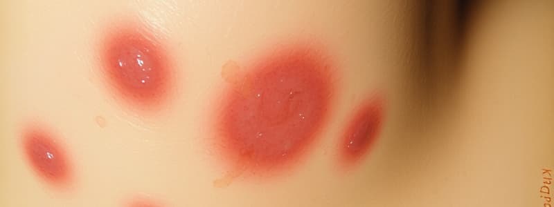
Macule
- Defined as a flat, discolored spot on the skin.
- Size is less than 1 cm in diameter.
-
Patch
- Similar to a macule but larger in size.
- Size is greater than 1 cm in diameter.
Palpable Lesions
- Palpable lesions are classified as either raised or depressed formations on the skin.
Types of Palpable Lesions
-
Papule:
- Size: Less than 1 cm.
- Appearance: Small, solid, raised spot on the skin.
-
Plaque:
- Size: More than 1 cm.
- Appearance: Larger, elevated area often formed by a group of papules.
-
Nodule:
- Size: More than 1 cm.
- Shape: Can be domed, spherical, or ovoid.
- Configuration: May be solid or cystic, indicating the internal structure or fluid content.
-
Wheal:
- Nature: Transient elevation caused by dermal edema.
- Characteristics: Typically associated with allergic reactions, presenting as raised welts on the skin that can change quickly.
Fluid-filled Lesions
- Vesicle: Small fluid-filled lesion, measuring less than 1 cm in diameter.
- Bullae: Larger fluid-filled lesion, exceeding 1 cm in diameter.
- Pustule: Contains visible pus, indicating infection or inflammation.
Subcorneal Pustular Dermatosis
- Erosion: Refers to the partial loss of the epidermis, often resulting in moist, exposed skin.
- Ulcer: Involves full-thickness loss of the epidermis, which may also include damage to the dermis or even deeper subcutis layers, leading to significant tissue loss.
Secondary Lesions
- Scale: Indicates changes in the epidermis, primarily affecting the stratum corneum, often appearing as flakes or patches.
- Crust: Forms from dried fluids (serum, blood, or pus) on the skin surface; can indicate underlying conditions; eschar signifies skin necrosis, suggesting tissue death.
- Fissure: Characterized by a linear break in the skin's surface, often resulting from excessive tension or a decrease in skin elasticity, leading to painful splits or cracks.
- Excoriation: Refers to external injuries to the epidermis, which may manifest as linear scratches or punctate lesions, typically caused by scratching or trauma.
- Atrophy: Involves thinning of the skin layers; epidermis may appear shiny with "cigarette-paper" like wrinkles, while dermal atrophy presents as depressed lesions.
Secondary Lesions
- Scale: Represents a transformation in the epidermis, predominantly affecting the stratum corneum, often visible as flakes or patches on the skin surface.
- Crust: Composed of dried fluid from serum, blood, or pus, indicating an area of inflammation or infection. Presence of eschar can signify skin necrosis.
- Fissure: Characterized by a linear break in the skin's surface, occurring due to excessive tension or a lack of elasticity, often found in areas subjected to movement.
- Excoriation: Refers to an external injury that damages part or all of the epidermis, presenting as linear or punctate marks resulting from scratching or trauma.
- Atrophy: Manifested in the epidermis as a smooth, shiny appearance with “cigarette-paper” wrinkles; in the dermis, it appears as a sunken or depressed lesion, indicating tissue loss.
- Lichenification: Thickening of the skin and enhancement of skin lines due to chronic scratching, commonly associated with long-term skin conditions or neurogenic factors.
Color Classification in Dermatology
-
Brown: Associated with pigmented lesions such as post-inflammatory hyperpigmentation, which occurs in the epidermis; includes melasma, phytophotodermatitis, and drug-induced hyperpigmentation. May also indicate metabolic disorders.
-
Pink/Red: Reflects conditions like viral exanthems and morbilliform drug eruptions; any insult to the skin that causes vasodilation, leading to redness.
-
White: Characterized by an absence of melanocytes or melanin production, which can result from scarring, vasospasm, or deposits in the skin.
-
Gray: Typically arises from post-inflammatory hyperpigmentation located in the dermis; can be caused by drugs or deposits, as well as combined melanocytic nevi and traumatic tattoos.
-
Blue: Indicates dermal melanocytosis, dermal melanocytomas, or conditions involving venous congestion.
-
Black: Can signify serious conditions such as cutaneous melanoma or eschar; may also relate to skin necrosis due to vasculitis or vascular compromise.
-
Violet: Associated with purpura (non-palpable), lichen planus, lymphoma cutis, and pyoderma gangrenosum.
-
Yellow: Often points to xanthogranulomas, solar elastosis, xanthomas, and adnexal tumors or hyperplasias with sebaceous differentiation.
-
Orange: Represents conditions like pityriasis rubra pilaris.
-
Green: Indicates a pseudomonas nail infection, often characterized by green pigment in nail pathology.
Color Associations in Skin Conditions
-
Black: Indicative of severe skin issues such as:
- Cutaneous melanoma: A dangerous skin cancer originating in pigment-producing melanocytes.
- Eschar: A dry, necrotic tissue often due to injury or infection.
- Necrosis of the skin can arise from:
- Vasculitis: Inflammation of blood vessels affecting skin circulation.
- Vascular compromise: Reduced blood flow leading to tissue death.
-
Violet: Associated with conditions characterized by purple spots or lesions:
- Purpura: Non-palpable purple rash caused by bleeding beneath the skin.
- Lichen planus: An inflammatory condition that affects skin, hair, nails, and mucous membranes, causing lichenoid eruptions.
- Lymphoma cutis: A skin manifestation of lymphatic system cancers.
- Pyoderma gangrenosum: A rare inflammatory skin condition that causes painful ulcers.
-
Yellow: Linked to various skin abnormalities:
- Xanthogranulomas: Yellowish nodules that can appear on the skin, often associated with lipid metabolism disorders.
- Solar elastosis: Sun-damaged skin characterized by yellowish discoloration and loss of elasticity.
- Xanthomas: Yellowish lesions that indicate cholesterol deposits under the skin.
- Adnexal tumors and hyperplasias: Growths in skin appendages (hair follicles, sebaceous glands) with sebaceous differentiation can present yellowish discoloration.
-
Orange: Represented by:
- Pityriasis rubra pilaris: A chronic skin disorder characterized by red, scaly patches and often orange appearance, with potential hair follicle involvement.
-
Green: Suggestive of specific infections or conditions:
- Pseudomonas nail infection: Green discoloration under nails resulting from bacterial colonization, often linked to moisture and trauma.
Color Categories in Pigmentation
-
Brown Pigmentation
- Associated with various conditions like pigmented lesions and post-inflammatory hyperpigmentation.
- Common types include epidermal hyperpigmentation, melasma, and phytophotodermatitis.
- Can also be drug-induced or due to metabolic factors.
-
Pink/Red Pigmentation
- Indicators of viral exanthems and morbilliform drug eruptions.
- Result from insults that cause vasodilation, leading to increased blood flow and redness in the skin.
-
White Pigmentation
- Indicates absence of melanocytes or melanin production.
- Can occur due to scarring or vasospasm.
- The presence of deposits may also lead to white patches on the skin.
-
Gray Pigmentation
- Often a result of post-inflammatory hyperpigmentation in the dermis.
- Associated with drug reactions or deposits in the skin.
- Can occur with combined melanocytic nevus or traumatic tattoos.
-
Blue Pigmentation
- Seen in conditions like dermal melanocytosis and dermal melanocytomas.
- May result from venous congestion, presenting a bluish tint in areas of the skin.
Shape or Configuration
- Annular (Ring-shaped): Features a distinct edge with more pronounced color or texture compared to the center, suggesting a ring-like appearance.
- Round/Nummular/Discoid: Describes lesions that are coin-shaped, featuring a solid circle or oval with consistent morphology throughout.
- Targetoid: Displays a target-like structure characterized by a darker center contrasted against a lighter periphery, resembling a bullseye.
- Serpiginous: Exhibits a snake-like, wavy configuration, indicating a pattern that resembles the movement or shape of a snake.
- Linear: Suggests a straight line formation, often indicative of an external irritant or contactant, including associations with the Koebner phenomenon, where lesions develop along lines of injury.
Rash Definition
- A rash is characterized by changes in skin texture or color, often appearing inflamed or irritated.
Primary Lesions
-
Nonpalpable (Flat): Only perceptible through color difference.
- Macule: Size less than 1 cm.
- Patch: Size more than 1 cm.
-
Palpable (Raised): Elevated or depressed lesions.
- Papule: Size less than 1 cm.
- Plaque: Size more than 1 cm.
- Nodule: Size more than 1 cm, shapes include domed, spherical, or ovoid; can be solid or cystic.
- Wheal: Transient elevation due to dermal edema.
-
Fluid-filled Lesions:
- Vesicle: Size less than 1 cm.
- Bullae: Size more than 1 cm.
- Pustule: Contains visible pus.
Loss of Skin Integrity
- Erosion: Partial loss of the epidermis.
- Ulcer: Full-thickness loss of the epidermis, possibly involving the dermis or subcutis.
Secondary Lesions
- Scale: Indicates changes in the stratum corneum of the epidermis.
- Crust: Dried fluid on the surface due to serum, blood, or pus.
- Eschar: Signals skin necrosis.
- Fissure: Linear loss of skin continuity due to excessive tension or decreased elasticity.
- Excoriation: Injury to the epidermis, linear or punctate.
- Atrophy:
- Epidermis: Shiny with “cigarette-paper” wrinkling.
- Dermis: Depressed lesions.
- Lichenification: Thickening of the skin, accentuating lines from chronic scratching.
Color Variations
- Brown: Pigmented lesions; conditions include post-inflammatory hyperpigmentation, melasma.
- Pink/Red: Associated with viral exanthems or any insult causing vasodilation.
- White: Indicates absence of melanocytes/melanin; may arise from scarring or vasospasm.
- Gray: Includes post-inflammatory hyperpigmentation and traumatic tattoos.
- Blue: Linked to conditions like dermal melanocytosis or venous congestion.
- Black: Signifies conditions like cutaneous melanoma or necrosis.
- Violet: Associated with conditions including purpura and lymphoma cutis.
- Yellow: Conditions like xanthomas and solar elastosis.
- Orange: Observed in pityriasis rubra pilaris.
- Green: Often related to pseudomonas nail infections.
Shape or Configuration of Lesions
- Annular: Ring-shaped with a more prominent edge.
- Round/Nummular/Discoid: Coin-shaped and uniform in morphology.
- Targetoid: Target-like shape with a darker center.
- Serpiginous: Snake-like configuration.
- Linear: Often suggests external contact.
- Stellate: Star-like with multiple angulated edges.
- Reticular or Retiform: Lacy or net-like pattern.
- Grouped/Herpetiform: Lesions appearing together.
Skin Distribution Patterns
- Localized: Refers to skin conditions or lesions that are restricted to a specific area of the body.
- Generalized (Exanthems): Conditions that are widely spread across the body, typically associated with systemic diseases and infections, resulting in rashes or eruptive lesions.
- Bilateral Symmetric: Observations that show a mirrored appearance of lesions on both sides of the body, often indicative of certain dermatological conditions.
- Universal: Characterizes skin conditions that affect the entire surface of the skin, indicating severe systemic involvement.
- Acral: Describes skin conditions appearing in the distal areas such as the hands, feet, wrists, ankles, and ears, commonly associated with various dermatological diseases.
Distribution of Skin Lesions
- Sun Exposed/Photodistributed: Skin areas commonly affected include the face, dorsal hands, and upper chest, where lesions are frequently seen due to sun exposure.
- Photo-accentuated: Lesion density is significantly higher on sun-exposed skin compared to non-sun-exposed skin, indicating that UV exposure can exacerbate skin conditions.
- Sun Protected: Certain dermatoses show improvement with sunlight exposure, suggesting that some skin conditions may benefit from UV light rather than worsen with it.
- Seborrheic Distribution: Affects hair-bearing regions such as scalp, eyebrows, nasolabial folds, post-auricular crease, beard area, central chest, axillae, and genitals, indicating where seborrheic conditions are prevalent.
- Follicular Presentation: Characterized by the presence of papules that are specifically centered around hair follicles, highlighting a distinct pattern of lesion distribution.
Distribution Types of Skin Lesions
- Extensor Distribution: Lesions appear over dorsal extremities, especially over the extensor muscles such as the knees and elbows.
- Flexor Distribution: Lesions are found in the antecubital (inner elbow) and popliteal (behind the knee) fossae.
- Intertriginous Distribution: Occurs in skin folds where two surfaces touch, notably in areas like axillae, inguinal folds, inner thighs, inframammary skin, and beneath an abdominal pannus. Associated with moisture and heat accumulation.
- Dermatomal/Zosteriform Distribution: Characterized by unilateral lesions that follow the pathway of a single spinal afferent nerve root.
- Follicular Distribution: Involves papules that are concentrated around hair follicles, indicating a localized reaction in these areas.
Skin Cell Migration and Distribution Patterns
-
Blaschkoid Distribution:
- Refers to patterns formed by skin cell migration during embryonic development.
- Indicative of a mosaic disorder, highlighting that different areas of the skin can have varying genetic makeup.
-
Lymphangitic Distribution:
- Represents a pattern following lymph vessel pathways.
- Suggests the presence of an infectious agent that spreads in a centralized manner, following the lymphatic system's network.
Studying That Suits You
Use AI to generate personalized quizzes and flashcards to suit your learning preferences.


