Podcast
Questions and Answers
What is one of the primary functions of the nervous system?
What is one of the primary functions of the nervous system?
- Absorbs nutrients
- Produces hormones
- Transmits signals throughout the body (correct)
- Regulates body temperature
Which of the following is NOT a component of the central nervous system (CNS)?
Which of the following is NOT a component of the central nervous system (CNS)?
- Ganglia
- Brain
- Spinal cord
- Nerves (correct)
What is the primary role of neuroglial cells?
What is the primary role of neuroglial cells?
- Transmit electrical signals
- Facilitate muscle contractions
- Support and protect neurons (correct)
- Generate sensory information
Which type of neuroglial cell is responsible for forming the myelin sheath in the central nervous system (CNS)?
Which type of neuroglial cell is responsible for forming the myelin sheath in the central nervous system (CNS)?
What is the function of the nodes of Ranvier in myelinated axons?
What is the function of the nodes of Ranvier in myelinated axons?
Which division of the nervous system is responsible for controlling voluntary movements?
Which division of the nervous system is responsible for controlling voluntary movements?
What type of neuron is primarily responsible for carrying sensory information to the central nervous system?
What type of neuron is primarily responsible for carrying sensory information to the central nervous system?
What is the function of Schwann cells in the peripheral nervous system?
What is the function of Schwann cells in the peripheral nervous system?
What is a nerve primarily composed of?
What is a nerve primarily composed of?
Which structure surrounds individual nerve fibers within a fascicle?
Which structure surrounds individual nerve fibers within a fascicle?
What distinguishes white matter from gray matter in the central nervous system?
What distinguishes white matter from gray matter in the central nervous system?
What type of neuron carries information from sensory receptors to the central nervous system?
What type of neuron carries information from sensory receptors to the central nervous system?
Which part of the brain is responsible for regulating balance and coordination?
Which part of the brain is responsible for regulating balance and coordination?
Which structure connects the two cerebral hemispheres?
Which structure connects the two cerebral hemispheres?
What is the function of the dorsal root in spinal cord anatomy?
What is the function of the dorsal root in spinal cord anatomy?
Which structure is involved in processing and reflex actions between sensory and motor neurons?
Which structure is involved in processing and reflex actions between sensory and motor neurons?
Which layer of the meninges is the outermost and tough?
Which layer of the meninges is the outermost and tough?
What is the primary function of cerebrospinal fluid (CSF)?
What is the primary function of cerebrospinal fluid (CSF)?
Where are the choroid plexuses located?
Where are the choroid plexuses located?
What function is primarily associated with the basal nuclei within the cerebrum?
What function is primarily associated with the basal nuclei within the cerebrum?
Which cranial nerve function is primarily linked to vision?
Which cranial nerve function is primarily linked to vision?
What is the primary role of the thalamus within the diencephalon?
What is the primary role of the thalamus within the diencephalon?
What separates the lateral ventricles from each other?
What separates the lateral ventricles from each other?
Where is the cerebral cortex located and what is its primary composition?
Where is the cerebral cortex located and what is its primary composition?
What does the subarachnoid space contain?
What does the subarachnoid space contain?
Which structure helps to separate the frontal and parietal lobes?
Which structure helps to separate the frontal and parietal lobes?
What is the main function of the blood-brain barrier?
What is the main function of the blood-brain barrier?
What function is associated with the pineal gland found in the epithalamus?
What function is associated with the pineal gland found in the epithalamus?
Which structure connects the third and fourth ventricles?
Which structure connects the third and fourth ventricles?
What is the role of the medulla oblongata within the brainstem?
What is the role of the medulla oblongata within the brainstem?
What separates the cerebrum from the cerebellum?
What separates the cerebrum from the cerebellum?
Which part of the spinal cord contains sensory neurons?
Which part of the spinal cord contains sensory neurons?
Flashcards
Nervous System Function
Nervous System Function
Transmits signals, coordinates actions, regulates internal state, facilitates responses to changes & enables thought processes.
Nervous System Structure
Nervous System Structure
Made up of neurons (signal transmitters) and neuroglia (support cells).
Central Nervous System (CNS)
Central Nervous System (CNS)
Brain and spinal cord that process information and control actions.
Peripheral Nervous System (PNS)
Peripheral Nervous System (PNS)
Signup and view all the flashcards
Neuron Structure (cell body)
Neuron Structure (cell body)
Signup and view all the flashcards
Myelin Sheath Function
Myelin Sheath Function
Signup and view all the flashcards
Neuroglia Function
Neuroglia Function
Signup and view all the flashcards
Neuron Parts (dendrites)
Neuron Parts (dendrites)
Signup and view all the flashcards
Nerve
Nerve
Signup and view all the flashcards
Fascicle
Fascicle
Signup and view all the flashcards
Sensory Neuron
Sensory Neuron
Signup and view all the flashcards
Motor Neuron
Motor Neuron
Signup and view all the flashcards
Cerebrum
Cerebrum
Signup and view all the flashcards
Dorsal Root
Dorsal Root
Signup and view all the flashcards
White Matter
White Matter
Signup and view all the flashcards
Longitudinal Fissure
Longitudinal Fissure
Signup and view all the flashcards
Gyri and Sulci
Gyri and Sulci
Signup and view all the flashcards
Cerebral Cortex
Cerebral Cortex
Signup and view all the flashcards
Spinal Cord Length
Spinal Cord Length
Signup and view all the flashcards
Ventral Horn
Ventral Horn
Signup and view all the flashcards
Spinal Cord Regions
Spinal Cord Regions
Signup and view all the flashcards
Dorsal Root Ganglion
Dorsal Root Ganglion
Signup and view all the flashcards
Cranial Nerves vs. Spinal Nerves
Cranial Nerves vs. Spinal Nerves
Signup and view all the flashcards
Meninges: Dura Mater
Meninges: Dura Mater
Signup and view all the flashcards
Meninges: Arachnoid Mater
Meninges: Arachnoid Mater
Signup and view all the flashcards
Meninges: Pia Mater
Meninges: Pia Mater
Signup and view all the flashcards
Cerebrospinal Fluid (CSF)
Cerebrospinal Fluid (CSF)
Signup and view all the flashcards
Ventricles: Lateral Ventricles
Ventricles: Lateral Ventricles
Signup and view all the flashcards
Blood-Brain Barrier
Blood-Brain Barrier
Signup and view all the flashcards
Study Notes
Overall Functions of the Nervous System
- Transmits signals throughout the body
- Integrates sensory input and motor outputs
- Regulates internal conditions (homeostasis)
- Facilitates reactions to environmental changes (response)
- Enables thought processes, memory, and learning (cognition)
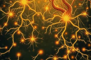
Structure and Function of Nervous Tissue
- Composed of neurons and neuroglia (glial cells)
- Neurons transmit electrical signals
- Neuroglia support and protect neurons
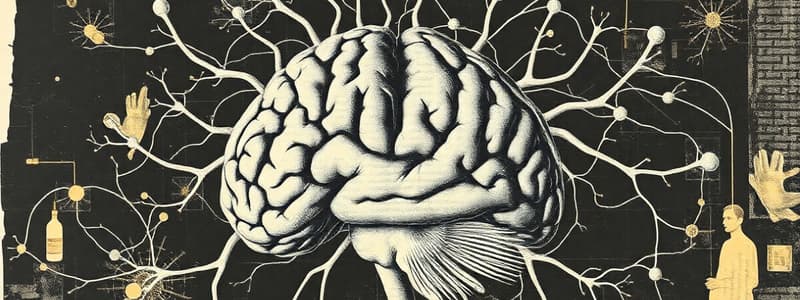
Organs of the Nervous System
- Brain
- Spinal cord
- Nerves
- Ganglia
Divisions of the Nervous System
- Central Nervous System (CNS): Includes the brain and spinal cord; processes information and coordinates activity
- Processes information
- Coordinates activity
- Peripheral Nervous System (PNS): Connects the CNS to limbs and organs; facilitates communication
- Afferent fibers: Carry sensory information to the CNS
- Efferent fibers: Carry information from the CNS
- Somatic Nervous System: Controls voluntary movements via skeletal muscles
- Autonomic Nervous System (ANS): Regulates involuntary functions (e.g., heart rate, digestion)
- Sympathetic Nervous System: Prepares the body for "fight or flight" responses
- Parasympathetic Nervous System: Promotes "rest and digest" activities
Cells of the Nervous System
- Neurons: Transmit signals
- Neuroglia: Support, protect, and nourish neurons
Neuroglial Cell Functions
- Astrocytes: Maintain blood-brain barrier, provide nutrients, and repair tissue
- Microglial: Act as immune defense in the CNS
- Oligodendrocytes: Form myelin sheath in CNS
- Ependymal: Line ventricles and produce cerebrospinal fluid (CSF)
- Schwann cells: Form myelin sheath in PNS
Neuron Structure
- Cell body: Contains the nucleus and organelles
- Dendrites: Receive signals from other neurons
- Axons: Transmit impulses away from the cell body to other cells
- Schwann cells: Wrap around axons in PNS
- Myelin sheath: Insulates axons to speed up signal transmission
- Nodes of Ranvier: Gaps in the myelin sheath that facilitate rapid conduction of nerve impulses
Nerves
- Nerves: Bundles of axons (nerve fibers) wrapped in connective tissue, transmitting signals between the CNS and PNS
- Fascicles: Groups of nerve fibers bundled together, each surrounded by a perineurium.
- Epineurium: The outermost layer of connective tissue enclosing the entire nerve, providing protection and support.
- Perineurium: A protective sheath surrounding each fascicle; it helps maintain the internal environment of the nerve.
- Endoneurium: The delicate connective tissue surrounding each individual nerve fiber, providing nourishment and support.
White vs. Gray Matter
- White matter: Composed of myelinated axons
- Gray matter: Contains neuronal cell bodies and unmyelinated fibers
- Tracts: Bundles of axons in the CNS that carry specific types of information
Sensory Receptors
- Structures that detect stimuli and convert them into neural signals
Sensory Neurons
- Neurons that carry information from sensory receptors to the CNS
Motor Neurons
- Neurons that transmit signals from the CNS to muscles or glands
Spinal Cord Parts
- Dorsal Root: Sensory information to the spinal cord
- Ventral Root: Motor commands away from the spinal cord
- Dorsal Root Ganglion: Cluster of sensory neuron cell bodies located just outside the spinal cord
- Synapse: The meeting point between two neurons via neurotransmitters
- Interneuron: Neuron that connects sensory and motor neurons within the CNS, allowing for processing and reflex actions
- Motor Neuron: Transmits signals to effector organs (muscles and glands)
Main Parts of the CNS
- Brain
- Spinal cord
Brain Regions
- Cerebrum
- Cerebellum
- Brainstem (midbrain, pons, medulla oblongata)
- Diencephalon (thalamus, hypothalamus, epithalamus)
Cerebral Cortex
- Outer layer of the cerebrum
- Composed of gray matter
- Responsible for higher brain functions (thought, perception, voluntary movement)
Basal Nuclei
- Located deep within cerebral hemispheres
- Regulate voluntary motor control, procedural learning, and emotional responses
Diencephalon
- Contains the thalamus and hypothalamus
- Thalamus: Relay station for sensory information
- Hypothalamus: Regulates homeostasis, hunger, temperature, and circadian rhythms
- Pineal gland: Produces melatonin, regulating sleep-wake cycles
Brainstem
- Controls reflexes and pathways for visual and auditory information
- Connects different parts of the brain
- Regulates sleep and autonomic functions (e.g., breathing, heart rate)
Cerebellum
- Coordinates voluntary movements, balance, and posture
Spinal Cord Dimensions
- Approximate length: 42-45 cm in adults
- Starts at the base of the skull (foramen magnum)
- Ends around L1-L2 vertebrae
- Regions: Cervical, thoracic, lumbar, sacral, and coccygeal
- Cauda equina: Bundle of spinal nerves below the end of the spinal cord
Spinal Cord Parts (cont.)
- Dorsal Horn: Contains sensory neurons
- Ventral Horn: Contains motor neurons
- Lateral Horn: Found in thoracic and upper lumbar regions, contains autonomic neurons
- Central Canal: Contains cerebrospinal fluid (CSF)
Meninges
- Protective membranes surrounding the brain and spinal cord
- Dura mater: Outermost layer, tough
- Arachnoid mater: Middle layer, web-like
- Pia mater: Innermost layer, adheres to brain/spinal cord
- Subdural space: Between dura and arachnoid mater
- Subarachnoid space: Contains cerebrospinal fluid (CSF)
- Cerebrospinal fluid (CSF): Cushions the brain and spinal cord, provides buoyancy, and transports nutrients to the nervous tissues
- Dural sinuses: Venous channels that drain blood from the brain
Ventricles
- Cavities within the brain filled with cerebrospinal fluid
- Lateral ventricles: Two large cavities in each hemisphere
- Septum pellucidum: Thin membrane separating lateral ventricles
- Interventricular foramen: Connects lateral ventricles to the third ventricle
- Third ventricle: Connects to the fourth ventricle via the cerebral aqueduct
- Fourth ventricle: Located between the brainstem and cerebellum
- Medial/Lateral Apertures: Openings allowing CSF to flow into the subarachnoid space
Cerebrospinal Fluid (CSF)
- Produced by choroid plexuses in the ventricles
- Clear fluid containing glucose, electrolytes, and proteins
- Circulates through ventricles and subarachnoid space
- Absorbed into venous system via arachnoid villi
- Provides cushioning, buoyancy, and nutrient transport for the brain and spinal cord
Blood- Brain Barrier
- A selective permeability barrier that protects the brain from harmful substances.
- Composed of tightly packed endothelial cells and astrocytic end-feet.
Studying That Suits You
Use AI to generate personalized quizzes and flashcards to suit your learning preferences.