Podcast
Questions and Answers
Which type of fracture is characterized by the bone being broken into multiple fragments?
Which type of fracture is characterized by the bone being broken into multiple fragments?
- Compound
- Comminuted (correct)
- Displaced
- Simple
In the context of fracture repair, what is the primary role of the hematoma that forms immediately after the injury?
In the context of fracture repair, what is the primary role of the hematoma that forms immediately after the injury?
- To activate osteoprogenitor cells in the periosteum
- To stimulate osteoclastic activity for bone remodeling
- To provide a fibrin mesh for fracture sealing and initial stabilization (correct)
- To directly transform into bony callus for permanent fixation
What is the potential outcome if a fracture is inadequately immobilized?
What is the potential outcome if a fracture is inadequately immobilized?
- Immediate activation of endochondral ossification
- Delayed union or nonunion, potentially leading to pseudoarthrosis (correct)
- Complete restoration of the medullary cavity
- Rapid formation of strong bony callus
Which of the following is the most common causative organism in pyogenic osteomyelitis?
Which of the following is the most common causative organism in pyogenic osteomyelitis?
What is a sequestrum in the context of osteomyelitis?
What is a sequestrum in the context of osteomyelitis?
In seronegative spondyloarthropathies, what tissue is primarily affected by pathologic changes?
In seronegative spondyloarthropathies, what tissue is primarily affected by pathologic changes?
What is a common clinical presentation of ankylosing spondylitis?
What is a common clinical presentation of ankylosing spondylitis?
Which of the following is a characteristic extra-articular manifestation of reactive arthritis (Reiter's syndrome)?
Which of the following is a characteristic extra-articular manifestation of reactive arthritis (Reiter's syndrome)?
What is the typical presentation of the skin rash associated with Lyme disease?
What is the typical presentation of the skin rash associated with Lyme disease?
Histologically, what is a key characteristic of fibrous dysplasia?
Histologically, what is a key characteristic of fibrous dysplasia?
A patient presents with pain, swelling, and pathologic fractures. Imaging reveals café-au-lait spots. Which condition is most likely?
A patient presents with pain, swelling, and pathologic fractures. Imaging reveals café-au-lait spots. Which condition is most likely?
What is the typical radiographic appearance of an aneurysmal bone cyst (ABC)?
What is the typical radiographic appearance of an aneurysmal bone cyst (ABC)?
How does a synovial cyst typically form?
How does a synovial cyst typically form?
Which of the following factors is considered the most important clinical clue in the diagnosis of bone tumors?
Which of the following factors is considered the most important clinical clue in the diagnosis of bone tumors?
Which of the following is a characteristic of osteoid osteoma?
Which of the following is a characteristic of osteoid osteoma?
How does osteoblastoma differ from osteoid osteoma?
How does osteoblastoma differ from osteoid osteoma?
What is the descriptive term for the bony overgrowth seen radiographically with osteochondroma?
What is the descriptive term for the bony overgrowth seen radiographically with osteochondroma?
A patient is found to have multiple enchondromas and spindle cell hemangiomas. Which syndrome is most likely?
A patient is found to have multiple enchondromas and spindle cell hemangiomas. Which syndrome is most likely?
In a giant cell tumor, what cell types are predominantly found?
In a giant cell tumor, what cell types are predominantly found?
What radiographic finding is associated with osteosarcoma?
What radiographic finding is associated with osteosarcoma?
Which genetic change is commonly associated with Ewing sarcoma?
Which genetic change is commonly associated with Ewing sarcoma?
What histological feature is characteristic of Ewing sarcoma?
What histological feature is characteristic of Ewing sarcoma?
What is a typical characteristic of chondrosarcomas on radiographic imaging?
What is a typical characteristic of chondrosarcomas on radiographic imaging?
Which type of bone tumor is known for spotty calcifications?
Which type of bone tumor is known for spotty calcifications?
Which type of cancer commonly metastasizes to bone with blastic lesions?
Which type of cancer commonly metastasizes to bone with blastic lesions?
What is the effect of tumor cells on bone in metastatic bone disease?
What is the effect of tumor cells on bone in metastatic bone disease?
What is the primary treatment for metastatic bone tumors focused on?
What is the primary treatment for metastatic bone tumors focused on?
Which of the following fracture types is typically seen in infants due to their softer bones?
Which of the following fracture types is typically seen in infants due to their softer bones?
How does a compound fracture differ from a simple fracture?
How does a compound fracture differ from a simple fracture?
In the process of fracture repair, what is the role of osteoclastic activity?
In the process of fracture repair, what is the role of osteoclastic activity?
What is the difference between Involucrum and Sequestrum?
What is the difference between Involucrum and Sequestrum?
Which of the following features is characteristic of seronegative spondyloarthropathies?
Which of the following features is characteristic of seronegative spondyloarthropathies?
What is the role of HLA-B27?
What is the role of HLA-B27?
Which is a common symptom of joints between tuberosities?
Which is a common symptom of joints between tuberosities?
How do bacterium infect when the infection is infectious arthritis?
How do bacterium infect when the infection is infectious arthritis?
A skin rash with pain and stiffness in knees might be a symptom of?
A skin rash with pain and stiffness in knees might be a symptom of?
Conjunctivitis, dysuria and urethritis are all symptoms of?
Conjunctivitis, dysuria and urethritis are all symptoms of?
Classic Lyme disease has three clinical phases, those phases are?
Classic Lyme disease has three clinical phases, those phases are?
Lyme arthritis is associated with which type of tick?
Lyme arthritis is associated with which type of tick?
What is the result of fibrous dysplasia?
What is the result of fibrous dysplasia?
Fibrous dysplasia is detected by?
Fibrous dysplasia is detected by?
Flashcards
Fracture
Fracture
Loss of bone integrity
Simple Fracture
Simple Fracture
Overlying skin is intact
Compound Fracture
Compound Fracture
Bone communicates with skin surface
Comminuted Fracture
Comminuted Fracture
Signup and view all the flashcards
Displaced Fracture
Displaced Fracture
Signup and view all the flashcards
Stress Fracture
Stress Fracture
Signup and view all the flashcards
Greenstick Fracture
Greenstick Fracture
Signup and view all the flashcards
Pathologic Fracture
Pathologic Fracture
Signup and view all the flashcards
Fracture Repair
Fracture Repair
Signup and view all the flashcards
Hematoma Formation
Hematoma Formation
Signup and view all the flashcards
Soft Tissue Internal Callus
Soft Tissue Internal Callus
Signup and view all the flashcards
Inadequate immobilization
Inadequate immobilization
Signup and view all the flashcards
Nonunion persists
Nonunion persists
Signup and view all the flashcards
Osteomyelitis
Osteomyelitis
Signup and view all the flashcards
Acute Osteomyelitis
Acute Osteomyelitis
Signup and view all the flashcards
Sequestrum
Sequestrum
Signup and view all the flashcards
Involucrum
Involucrum
Signup and view all the flashcards
Draining Osteomyelitis
Draining Osteomyelitis
Signup and view all the flashcards
Spondyloarthropathies
Spondyloarthropathies
Signup and view all the flashcards
Ankylosing Spondylitis
Ankylosing Spondylitis
Signup and view all the flashcards
Ankylosing Spondylitis
Ankylosing Spondylitis
Signup and view all the flashcards
Clinic Findings
Clinic Findings
Signup and view all the flashcards
Genetic association
Genetic association
Signup and view all the flashcards
Reactive Arthritis Triad
Reactive Arthritis Triad
Signup and view all the flashcards
Presentation of Reactive Arthritis
Presentation of Reactive Arthritis
Signup and view all the flashcards
Classic Lyme disease
Classic Lyme disease
Signup and view all the flashcards
Lyme rash
Lyme rash
Signup and view all the flashcards
Fibrous Dysplasia
Fibrous Dysplasia
Signup and view all the flashcards
Benign Tumor
Benign Tumor
Signup and view all the flashcards
Early adolescence
Early adolescence
Signup and view all the flashcards
Polyostotic fibrous
Polyostotic fibrous
Signup and view all the flashcards
Simple Bone Cyst
Simple Bone Cyst
Signup and view all the flashcards
Aneurysmal Bone Cyst
Aneurysmal Bone Cyst
Signup and view all the flashcards
Ganglion cysts
Ganglion cysts
Signup and view all the flashcards
Synovial cyst
Synovial cyst
Signup and view all the flashcards
Age
Age
Signup and view all the flashcards
Osteoid Osteoma
Osteoid Osteoma
Signup and view all the flashcards
Clinical
Clinical
Signup and view all the flashcards
Osteochondroma
Osteochondroma
Signup and view all the flashcards
Chondroma
Chondroma
Signup and view all the flashcards
Study Notes
Fractures
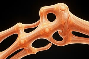
- Fractures are defined as the loss of bone integrity
Fracture Classification
- Simple fracture: Overlying skin remains intact
- Compound fracture: Bone communicates with the skin surface
- Comminuted fracture: The bone is fragmented
- Displaced fracture: The ends of the bone at the fracture site are not aligned
- Stress fracture: Bone is damaged due to increased physical activity and repetitive loads
- Greenstick fracture: Occurs in infants with soft bones and extends only partially through the bone
- Pathologic fracture: Results from a disease process such as a tumor
Fracture Repair
- Bone repair is genetically mediated
- Immediately post-fracture, hematoma formation provides a fibrin mesh for fracture sealing
- Inflammatory cell influx, fibroblast ingrowth, and capillary proliferation lead to granulation tissue formation
- Degranulated platelets and inflammatory cells release PDGF, TGF-β, and FGF
- Osteoprogenitor cells are activated in the periosteum, medullary cavity, and surrounding soft tissues, leading to osteoclastic and osteoblastic activity.
- Soft Tissue Internal Callus or Procallus: Provides anchorage but lacks structural rigidity for weight bearing
- Within 2 weeks of injury, activated osteoprogenitor cells form subperiosteal trabeculae of woven bone in the medullary cavity
- Procallus transforms into bony callus and enables fx stabilization
- Endochondral ossification: Bridging fx bone ends with a contiguous network of newly deposited bone trabeculae increases mineralization, stiffness, and strength of callus to bear weight
- Healing completes through restoration of the medullary cavity
- Inadequate immobilization results in delayed union or nonunion of the fracture
- If nonunion persists, the malformed callus undergoes cystic degeneration with a luminal surface lined by synovial-like cells, forming a false joint or pseudoarthrosis
Osteomyelitis
- Osteomyelitis: Inflammation of bone and marrow, secondary to bacterial, viral, fungal, or parasitic infection
- In about 50% of patients, the specific organism is unidentified
- Pyogenic osteomyelitis is a bacterial infection that spreads hematogenously, extends from a contiguous site or results from direct implantation, such as traumatic injury.
- Staphylococcus aureus accounts for 80% to 90% of culture-positive pyogenic osteomyelitis cases
- Escherichia coli, Pseudomonas, and Klebsiella are etiological agents also
- Haemophilus influenzae and group B streptococci occur in neonates
- Sickle cell disease predisposes to Salmonella infection
Acute Osteomyelitis
- Proliferation and recruitment of neutrophils within 48 hours, along with necrosis of bone cells and marrow.
- Bacterial infection leads to a subperiosteal abscess when bacteria infect the periosteum
- Periosteal lifting impairs blood supply
- Necrosis and soft tissue abscesses follow periosteal rupture, which opens to the skin as draining sinuses
- Dead bone, or sequestrum, forms, crumbles, and releases fragments into sinus tracts, leading to a chronic stage called CLOACAE in 5-25% of cases
- Chronic inflammation stimulates bone resorption, fibrosis, and reactive bone deposition
- New bone forms a living shell, called an involucrum, around devitalized, infected bone.
- X-ray may reveal a lytic focus of bone destruction surrounded by sclerosis
- Draining osteomyelitis reveals a drainage tract in the subperiosteal shell of viable new bone, or involucrum, and inner native necrotic cortex, or sequestrum
Seronegative Spondyloarthropathies
- Undefined microbial antigens cross-react with musculoskeletal system components to cause this Seronegative Spondyloarthropaty
- The triggering antigens cause T-cell responses
- The conditions are immune-mediated
- This is a heterogeneous group of disorders
- Absence of rheumatoid factor
- Pathologic changes occur in ligamentous attachments, not the synovium
- Sacroiliac joint involvement
- Association with HLA-B27
- Bony proliferation leading to ankylosis (fusion)
- Examples include ankylosing spondylitis, infectious arthritis, and reactive arthritis
Ankylosing Spondylitis
- Characterized by destruction of articular cartilage and bony ankyloses
- Sacroiliac and vertebral apophyseal joints located between tuberosities and processes are involved
- Low back pain and spinal immobility typically manifest during the second and third decades of life
- Approximately 90% of patients test positive for HLA-B27
- The role of HLA-B27 in the disease process remains unknown
- Infectious arthritis due to microorganisms is characterized by hematogenous dissemination, direct inoculation, or contiguous spread from soft tissue such as abscess or osteomyelitis
Reactive Arthritis (Reiter’s Syndrome)
- Reactive arthritis presents as a triad of symptoms
- The triad consists of arthritis, non-gonococcal urethritis or cervicitis, and conjunctivitis
- Mono or oligoarticular arthritis
- Triggered by intestinal infections, such as Salmonella, Shigella, and Campylobacter
- Occurs following sexually transmitted diseases and Chlamydia trachomatis infections
- Rarely, proceeds group A streptococcal infections
- The condition is RF negative and HLA-B27 positive
- Common in young adults
- 50% experience recurrent arthritis, tendonitis, lumbosacral pain
- Active disease lasting >6 months indicates chronic reactive arthritis
- Initial acute-onset asymmetric oligo-arthritis typically affects the knee and causes inflammatory low back pain
- Inflammation presents at insertions of the Achilles tendon and plantar fascia, heel swelling, digit inflammation, joint stiffness, and lower back pain.
- Extra-articular manifestations include conjunctivitis, uveitis, dysuria, pelvic pain, urethritis, balanitis, cervicitis, mucosal ulcers, skin rashes, and cardiac valvular disease
Lyme Arthritis
- Lyme arthritis is caused by the spirochete Borrelia burgdorferi spread by deer ticks (Ixodes)
- It is the leading arthropod-borne disease in the US, particularly in New England, the mid-Atlantic states, and the upper Midwest
- Classic Lyme disease affects multiple organ systems and has three clinical stages
- Early localized stage presents with an initial skin infection, followed by organism dissemination to other cutaneous sites, cranial nerves, heart, and meninges
- Untreated or late disseminated disease presents as migratory arthritis (Lyme arthritis) in 80% of cases, months after infection, lasting weeks to months
- Anti-Borrelia antibodies are present in serology
- Arthritis occurs in 10% of Borrelia infections, and is often treated at earlier stages, and cured
- One or two joints are affected i.e. knees, shoulders, elbows, and ankles
- Lyme rash often presents as a bull's-eye with circles around the middle
- The rash is typically round, red, and at least 2 inches across
- It grows to 10-12 inches across within days
- It is warm to the touch
- Not itchy or painful
- Occurs anywhere on the body
- Lyme arthritis is a sparse superficial and deep lymphocytic infiltrate
- It has an unremarkable epidermis
- Epidermal and or dermal necrosis
- Treatment involves doxycycline, amoxicillin, or cefuroxime axetil and yields cure rates of 90%
Fibrous Dysplasia
- Fibrous dysplasia is a benign tumor
- All components of normal bone
- Structures do not differentiate into mature structures
- Occurs during skeletal development
- Pathogenesis involves various somatic gain-of-function mutations in GNAS1.
- Results in constitutive activation of G s protein, which results in cellular proliferation and inhibition of osteoblast differentiation,
- Presence of intramedullary lytic lesions with bowing and cortical bone thinning occurs
- Histologically, curvilinear trabeculae of woven bone exhibit moderately cellular fibroblastic proliferation
- Histo: No prominent osteoblastic rimming
- Clinically exhibits: pain, swelling, pathologic fractures, and severe bone deformity.
- Early Monostotic fibrous dysplasia occurs during early adolescence and affects femur, tibia, ribs, jawbones, and calvarium.
- Asymptomatic, can cause pain, discrepancies in limb length, and pathologic fracture.
- Treated by curettage which is commonly recurrent
- Polyostotic fibrous dysplasia develops earlier and presents later in adulthood, affecting the femur, skull, and tibia
- Craniofacial involvement occurs
- It is progressive with crippling deformities, and fractures;
- Bisphosphonates can reduce bone pain
- Rare malignant transformation into sarcoma can occur
- McCune-Albright Syndrome: Syndrome involves precocious sexual development, especially in girls
- Additional features: hyperthyroidism, pituitary adenomas, growth hormone secretion, and primary adrenal hyperplasia
- A unilateral cafe-au-lait spot lesion occurs on the same side of body
Simple Bone Cyst (Unicameral Bone Cyst)
- Incidental finding
- Not a neoplasm
- Fluid-filled lesion with a fibrous lining
- Occurs during the first 20 years of life
- Located near the growth plate of long bones
- May reoccur or disappear with maturity
- Cyst enlargement may cause a fracture
Aneurysmal Bone Cyst (ABC)
- Multiloculated blood-filled spaces affect all ages, mostly in adolescence and often in femur, tibia, or vertebral body
- X-ray shows; expansive, well-circumscribed lytic lesion has well-defined margins and shows central lysis with thin sclerotic shell of reactive bone,
- Multiple blood-filled cystic spaces divided by thin, tan-white septa composed of plump spindle cells, multinucleated osteoclast-like giant cells, and reactive woven bone lined by osteoblasts 70% have chromosome 17p13 rearrangements within plump spindle cells resulting in the fusion of the USP6 coding region with promoters of genes highly expressed in osteoblasts
- Clinic Manifestations: Localized pain and swelling with resulting lesion and nerve compression
- Benign but locally aggressive
- Treatment involves curettage or excision with a 10% to 50% recurrence rate
Ganglion & Synovial Cysts
- Ganglion cysts are 1.5 cm near a joint capsule/tendon sheath, common on wrists, firm, fluctuant, pea-sized translucent nodules
- Composed of cystic or myxoid connective tissue degeneration
- The cyst wall lacks a cell lining and it may be multilocular
- Synovial cysts are herniation of synovium through a joint capsule or massive enlargement of a bursa
- Popliteal Synovial Cyst, known as Baker cyst, is associated with rheumatoid arthritis
- The cyst's lining resembles synovium
- The Cyst and synovium :hyperplastic and Cyst contains inflammatory cells and fibrin
Primary Bone Tumors
- Rare and outnumbered by metastases and affect long bones of extremities
- Asymptomatic, identified incidentally
- May be painful, slow-growing, or cause pathologic fracture
- Clinical presentation, radiographic appearance, patient age, lesion location, and microscopic appearance are important for determination
- In the first three decades, these affect skeletal growth rate
- Manifestations occur in Distal femur and proximal tibia
- Benign are more common than malignant Bone Tumors
- Age is the most important clinical factor for diagnosis
- Painful lesions indicate: malignancy or locally aggressive tumor
Benign Tumors
- Osteoid osteoma and osteoblastoma are histologically similar tumors that differ in size, site of origin, and symptoms
- Malignant transformations are rare
- Round-to-oval in shape, well-circumscribed masses, with hemorrhage and tan tissue
- Micro: trabeculae of woven bone rimmed by one layer of prominent osteoblasts
- The connective tissue exhibits dilation and congested capillaries
- Reactive bone surrounds the actual neoplasm, or nidus a small mineralized radiolucent core
- Osteoid osteoma can be treated by radiofrequency ablation
- Osteoblastoma: curettage or en bloc excision removes large pieces of tissue
Osteoid Osteoma
- Benign with no malignant potential
- It is less than 2 cm in size
- Those under 25 years old are affected
- Occurs in long bones and on the surface of facial cones or skull
- Gardner’s Syndrome can cause it: Familial Adenomatous Polyposis + osteomas and soft tissue tumors
- It is slow-growing, and has no clinical significance unless there is obstruction or cosmetic issues
- Characterized by Nocturnal bone pain relieved by Aspirin and NSAIDs due to an excess of prostaglandin E2 produced by a proliferating osteoblast
- Clinical Presentation: A thick rim of reactive cortical bone, it can be only found upon investigation
Osteoblastoma
- Greater than 2cm in size
- More common in males under 25 years old
- Arises in spine
- Pain which is not nocturnal and will not be relieved by aspirin or other NSAIDS
Osteochondroma (Exostosis)
- It is common
- Mass attached to the skeleton by a bony stalk capped by cartilage.
- 85% are solitary/sporadic
- 15% of the AD multiple hereditary exostosis syndrome
- Patients may be single or have multiple solitary osteochondromas
- Occurs in late adolescence or early adulthood
- Men affected more commonly than women
- Occurs in bones of endochondral origin
- Occurs in the metaphysis near the growth plate of long tubular bones
- Palpable to the touch
- Slow-growing and cause pain through nerve impingement
- Histology: disorganized growth plate, undergoes endochondral ossification
- The cartilage cap is lined by perichondrium, and is contiguous and mature
Chondroma (Enchondroma)
- Benign tumor of hyaline cartilage: in medullary cavity (enchondromas) and on bone surface ( juxtacortical chondromas).
- Patients aged 20-50+
- Asymptomatic but can cause painful or pathological fractures
- Well-circumscribed nodules usually 3cm
- Gray-blue, translucent cut surface.
- Heterozygous mutations in IDH1 and IDH2 genes
- Histology: hypocellular lesion with hyaline cartilage. Chondrocytes are generally normal
- If periphery exhibits endochondral ossification, a peripheral endochondral ossification must be noted
- Circumscribed lucency appears on x-ray with central irregular calcifications, a sclerotic rim, and intact cortex
- Treatment: Usually observation or curettage
- Rare for chondromas to undergo sarcomatous transformation outside of Ollier disease and Maffucci syndrome
Ollier disease and Maffucci syndrome
- Characterized by nonhereditary disorders characterized by multiple enchondromas
- Enchondromas confined to one side and limbs.
- Increased risk chondrosarcoma at about 25-30% near 40 years of age
- Increased risk gliomas, pancreatic and juvenile granulosa cell tumors of the ovary
- Syndromes can exhibit multiple spindle cell hemangiomas
- Patients are at greater risk for other developing malignancies, including brain gliomas
Giant Cell Tumor (Osteoclastoma)
- It is Rare to find benign tumors made of multinucleated, osteoclast-type giant cells which means it is locally aggressive
- 4% metastasize to lungs or regress spontaneously
- rarely fatal
- knee, involving distal femur or proximal tibia are affected
- Patients experience, arthritis-like symptoms, and pathologic fractures
- May destroy the overlaying cortex resulting in bulging soft tissue mass
- Macroscopically, they are reddish-brown masses that undergo cystic degeneration
- Histology: uniform oval mononuclear tumor cells and abundant osteoclast-type giant cells with 100 or more nuclei with necrosis and mitotic activity
- X-ray: "Soap Bubble" indication
- Therapy: curettage, and 40-60% of the locally treated cases will reoccur
Osteosarcoma
- Most common primary malignant tumor of the bone at 20%
- Bimodal distribution with those occurring before 20 years of age, and a smaller peak in older adults
- Related to Paget's disease or bone infarcts
- Caused by Prior radiation
- More prevalent in males
- Prefers the metaphyseal region of long bones.
- ~50% of near the knee in distal femur or proximal tibia
- The tumors are Bulky, gritty, with gray-white color, hemorrhage and tumors
- Destroy the cortices produces soft tissues masses and extend into the medullary canal replacing hematopoietic Marrow
- Pleomorphism or large hyperchromatic nuclei with bizarre tumor giant cells and abundant mitoses Extensive necrosis with intravascular invasion
- Composed of malignant tumor cells that make unmineralized or mineralized osteoid bone
- All cases of tumors are assumed to have occult metastases at the time of its diagnosis
- Can affect many Hematogenous metastasis, such as lungs, bones, brain and more.
- 5-year survival rate of <20 with metastatic, recurrent, and secondary osteosarcoma
Clinical and Genetic Features;
- Pain and path fractures
- It has a peak onset in fast growing individuals during Adolescents.
- in a Growth Plate and rapidly expanding bones
- Genetic Ab with Chrom aberrations are usually mutated such as: RB with 70% or sporatic OST
- Germin Mutation w TP to those with the Li Fraumell Syndrome for example cancer in breast, brain cancers
- CDAKNZA encodes w/ 2 Tumor Suppresses that Inacti Xray: destructive, mixed lytic and blastic mass infiltrative The cortex lifts and breaks resulting Reactive periosteal formations Sun burst stretched- fibers form rapidly
Ewing Sarcoma
Malignant and and primitive tumor cells which make 6- 10% primary malignant bone tuma Most occur with children and affects of those 80% are under the age of 20. Boys has greater chance with it (African and Asian) Diaphysis of the long tuma the femurs humerus and the ribs Pain, Systemic symptoms fever and and inflammation and the ESR and more. Tumos are lytic and deposit
Chondrosarcomas
- Enlarging and most are malignant Conventional clear and variant tumors. Patients older than 40 and man will twice the rate and spread as foci
Metastatic Bone Tumors
- Affects Metastatic Cancers & primary bone, direct extention Mechanisms for tumors include: lymphatic, dissemination, and/or hematogenous intraspinal seeding and Adults: skeletal metastases from Prostate, breast, kidney, and lung cancers and with Children with: neuroblastoma, Wilms Osteosacoma is in all X ray: may lysis the forms
Studying That Suits You
Use AI to generate personalized quizzes and flashcards to suit your learning preferences.