Podcast
Questions and Answers
In flow cytometry, what signals are analyzed to characterize cells?
In flow cytometry, what signals are analyzed to characterize cells?
- Only emitted light from fluorochromes attached to the cells.
- Only physical signals related to cell size and granularity.
- Both optical and physical signals emitted or scattered by the cells. (correct)
- Primarily electrical signals, with secondary analysis of optical data.
What does 'hydrodynamic focusing' achieve in flow cytometry?
What does 'hydrodynamic focusing' achieve in flow cytometry?
- It amplifies the intensity of the laser beam.
- It separates cells based on their size and granularity.
- It aligns cells into a single stream for sequential analysis. (correct)
- It enhances the fluorescent signal emitted by the cells.
In flow cytometry, forward scatter (FSC) is proportional to what cellular property?
In flow cytometry, forward scatter (FSC) is proportional to what cellular property?
- Cell size. (correct)
- Cell fluorescence intensity.
- Cell granularity.
- Cell internal complexity
What does side scatter (SSC) indicate about a cell in flow cytometry?
What does side scatter (SSC) indicate about a cell in flow cytometry?
What is the purpose of using lasers in flow cytometry?
What is the purpose of using lasers in flow cytometry?
What is the primary role of photomultiplier tubes (PMTs) in flow cytometry?
What is the primary role of photomultiplier tubes (PMTs) in flow cytometry?
Which of the following best describes the function of dichroic mirrors in flow cytometry?
Which of the following best describes the function of dichroic mirrors in flow cytometry?
What is the MOST important purpose of compensation in flow cytometry?
What is the MOST important purpose of compensation in flow cytometry?
In flow cytometry, what does a 'single-parameter histogram' represent?
In flow cytometry, what does a 'single-parameter histogram' represent?
Which type of plot is MOST useful for detecting a small number of events in flow cytometry?
Which type of plot is MOST useful for detecting a small number of events in flow cytometry?
In the context of fluorochromes used in flow cytometry, what is a 'tandem dye'?
In the context of fluorochromes used in flow cytometry, what is a 'tandem dye'?
Why is it important to protect tandem dyes from light and certain fixatives (like PFA) when preparing samples for flow cytometry?
Why is it important to protect tandem dyes from light and certain fixatives (like PFA) when preparing samples for flow cytometry?
When choosing a fluorochrome for a flow cytometry experiment, what is the MAIN factor to consider?
When choosing a fluorochrome for a flow cytometry experiment, what is the MAIN factor to consider?
What is the MOST appropriate control to use for defining positivity and negativity in flow cytometry?
What is the MOST appropriate control to use for defining positivity and negativity in flow cytometry?
What is the purpose of using 'isotype controls' in flow cytometry?
What is the purpose of using 'isotype controls' in flow cytometry?
In cell sorting, what is the MOST relevant basis for cell separation?
In cell sorting, what is the MOST relevant basis for cell separation?
In mass cytometry, what type of tags are attached to antibodies for cell analysis?
In mass cytometry, what type of tags are attached to antibodies for cell analysis?
What is a PRIMARY advantage of mass cytometry compared to traditional flow cytometry?
What is a PRIMARY advantage of mass cytometry compared to traditional flow cytometry?
Spectral flow cytometry differs from conventional flow cytometry primarily in what way?
Spectral flow cytometry differs from conventional flow cytometry primarily in what way?
Which of the following represents a common application of flow cytometry in a clinical laboratory?
Which of the following represents a common application of flow cytometry in a clinical laboratory?
If the goal is to perform cell cycle analysis using flow cytometry, which DNA dyes would be MOST appropriate to use?
If the goal is to perform cell cycle analysis using flow cytometry, which DNA dyes would be MOST appropriate to use?
Flashcards
What was Moldovan's contribution in 1934?
What was Moldovan's contribution in 1934?
Cell count by capillary/photoelectric detector.
What was Wallace Coulter's patent in 1949?
What was Wallace Coulter's patent in 1949?
Device for counting and measuring cell size.
What did Kamentsky add in 1965?
What did Kamentsky add in 1965?
Addition of cellular constituents analysis.
What did Herzenberg describe in 1969?
What did Herzenberg describe in 1969?
Signup and view all the flashcards
What happened in 1974 with Beckton-Dickinson?
What happened in 1974 with Beckton-Dickinson?
Signup and view all the flashcards
What was added in 1980?
What was added in 1980?
Signup and view all the flashcards
What was added in 1990?
What was added in 1990?
Signup and view all the flashcards
What did Sony do in 2012?
What did Sony do in 2012?
Signup and view all the flashcards
What is flow cytometry?
What is flow cytometry?
Signup and view all the flashcards
What signals are analyzed in flow cytometry?
What signals are analyzed in flow cytometry?
Signup and view all the flashcards
What is Simultaneous measurement?
What is Simultaneous measurement?
Signup and view all the flashcards
What is Measurement carried out?
What is Measurement carried out?
Signup and view all the flashcards
What does Forward Scatter (FSC) measure?
What does Forward Scatter (FSC) measure?
Signup and view all the flashcards
What does Side Scatter (SSC) measure?
What does Side Scatter (SSC) measure?
Signup and view all the flashcards
What is Forward Scatter?
What is Forward Scatter?
Signup and view all the flashcards
What is Side Scatter?
What is Side Scatter?
Signup and view all the flashcards
What is CELL SIZE?
What is CELL SIZE?
Signup and view all the flashcards
What is Fluorescence?
What is Fluorescence?
Signup and view all the flashcards
What is Fluidics?
What is Fluidics?
Signup and view all the flashcards
What is Optics?
What is Optics?
Signup and view all the flashcards
What is Electronics?
What is Electronics?
Signup and view all the flashcards
How many cells at a time is analysed?
How many cells at a time is analysed?
Signup and view all the flashcards
What is hydrodynamic focusing?
What is hydrodynamic focusing?
Signup and view all the flashcards
What is used for Emission optics?
What is used for Emission optics?
Signup and view all the flashcards
What signals are routed to detection systems?
What signals are routed to detection systems?
Signup and view all the flashcards
What happens to Light?
What happens to Light?
Signup and view all the flashcards
What are Optical filters?
What are Optical filters?
Signup and view all the flashcards
What are 5-ELECTRONICS?
What are 5-ELECTRONICS?
Signup and view all the flashcards
What is a Fluorochrome?
What is a Fluorochrome?
Signup and view all the flashcards
What is a Fluorochrome composed of?
What is a Fluorochrome composed of?
Signup and view all the flashcards
What is function of Annexin-V?
What is function of Annexin-V?
Signup and view all the flashcards
What is correct compensation?
What is correct compensation?
Signup and view all the flashcards
How Cells are separated?
How Cells are separated?
Signup and view all the flashcards
How is Separation Achieved?
How is Separation Achieved?
Signup and view all the flashcards
What is Mass Cytometry for?
What is Mass Cytometry for?
Signup and view all the flashcards
What does Biological markers are detected by?
What does Biological markers are detected by?
Signup and view all the flashcards
What does Conventional flow cytometry use?
What does Conventional flow cytometry use?
Signup and view all the flashcards
What does Spectral flow cytometry uses?
What does Spectral flow cytometry uses?
Signup and view all the flashcards
What is fluidic Column?
What is fluidic Column?
Signup and view all the flashcards
Study Notes
- Introduction to flow cytometry as of 2025
Summary of Topics
- Overview of historic and defining milestones in flow cytometry
- Flow Cytometry is built on some basic, but important principles to allow simultaneous data collection
- The Applications
- Other, special cases
Historic and Definitions
- In 1934, Moldovan created the first device to count cells by capillary/photoelectric detector
- Wallace Coulter patented a device in 1949 for counting and measuring cell size
- Kamentsky added cellular constituents analysis in 1965
- In 1969, Herzenberg wrote one of the first articles to describe mammalian cell sorting, followed by its application in 1973 and Marvin Van Dilla started to use lasers as a light source
- Becton-Dickinson commercialized the FACSII in 1974
- 3 fluorescence parameters per cell could be analyzed by 1980 for the first time
- By 1990, 7 fluorescence parameters could be analyzed
- 11 fluorescence parameters could be analyzed by 2001
- 2004 saw Robinson describe spectral approaches
- 18 fluorescence parameters could be analyzed by 2006
- Sony Biotech launched the first commercial spectral flow cytometer in 2012
- 20 fluorescence parameters could be analyzed by 2016
- 2020 saw the use of artificial intelligence to help analysis
- Precise study of isolated cells through a liquid flow defines flow cytometry
- Flow cytometry characterizes individual particles suspended in a liquid, both quantitatively and qualitatively, as well as assessing their function
- Flow cytometry analyzes optical or physical signals emitted by a particle
- This cutting happens via the light beam from either a laser or arc lamp
Basic Principles
- Simultaneous measurement of different characteristics of a cell or particle
- Measurements are carried out as cells pass one by one through an analysis chamber at varying speeds
- The data obtained for each cell includes:
- Size, via forward scatter (FSC)
- Granularity or internal complexity, via side scatter (SSC)
- Fluorescence intensity depending on fluorochromes
- Each event will diffract light as it passes in front of the laser beam
- Cell size (FSC) is proportional to diffraction
- Cell structure (SSC) is proportional to cell granularity or complexity
Forward Scatter
- Light is diffracted at small angles
- Diffracted light is collected at a small angle, between 1 and 10 degrees
- The signal is proportional to cell size
- A detector is aligned with the laser, in the cone of diffracted light
Side Scatter
- Light is diffracted at large angles
- Diffracted light is collected at 90° to the laser axis
- The signal is proportional to cell granularity and complexity
- The detectors are perpendicular to the laser beam
- They are located in the zone of widely diffracted light
- Representational Scatter plots of lysed normal whole blood
- FSC vs SSC alone can distinguish cell types
- Cell size measures particles from 100 nM to 100 μM
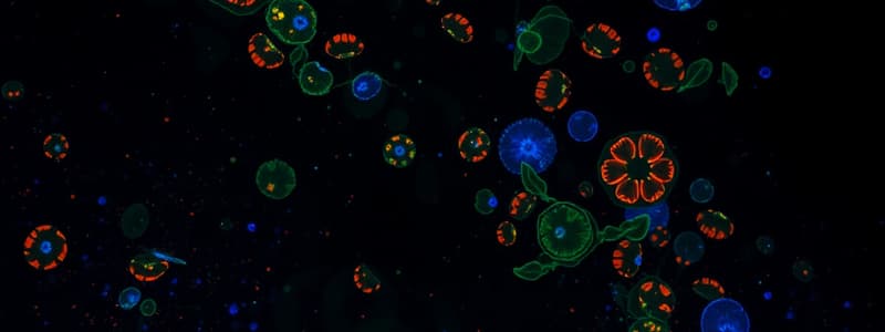
Fluorescence
- Fluorescence is measured at 90° when an event passes in front of a laser
- Photoluminescence is radiative phenomenon from the light excitation (visible or UV photons)
- Chemiluminescence is a radiative phenomenon resulting from a chemical reaction
- Many things can produce natural fluorescence including several minerals
- Adamite fluoresces green
- Hemimorphite fluoresces blue
- Calcite fluoresces red or pink
- There are also examples of imported fluorescence such as in the GFP-Rabbit, GFP-mice, and GFP-fly fruit due to animals carrying the GFP ubiquitary promoter
Equipment: Function
- Fluidics introduce and position cells
- Optics produce and collect light signals
- Electronics convert optical signals into electronic signals and digitize them for computer analysis
Equipment: Fluidics
- Cytometers analyze one cell at a time by placing the cells in suspension and driving them with a pump, one by one, in front of one or more lasers
- Centering of cells in laminar flow comes from hydrodynamic focusing
- Cell separation achieved via progressive acceleration
- Cells pass through a nozzle for channeling and centering
- Hydrodynamic focusing centers and aligns cells in laminar flow
- Sheath liquid stretches the sheath liquid to achieve cell separation via progressive acceleration
- Cells then move through a nozzle to channel and center it
Controlling the Speed of Flow
- Low Pressure uses 10μL/mn
- Medium Pressure uses 30 µL/mn
- High Pressure uses 60 µL/mn
- Sheath liquid pressure remains constant, unlike the sample
- Increase in pressure leads to an increase in flow
- The higher the pressure, the greater the freedom of movement of the cells
Equipment: Optics
- Necessary to focus the light source (laser) on the cells
- Illumination must excite dyes at a wavelength close to their excitation maximum
- Lasers are commonly used as powers source and offer stability
- Vapor lamps such as mercury and xenon, are expensive but precise
3-Emission Optics
- A thin laser beam allows focus on one cell at a time
- Background noise is reduced
- Energy concentrates on a small surface
- Monochromatic light excitation becomes very powerful
- Light coming from a laser is monochromatic and unidirectional
- Common lasers and their output include:
- UV Laser 325 nm: UV
- Laser 405 nm: violet
- Laser 488 nm: blue
- Laser 532 nm: green
- Laser 561 nm: yellow
- Laser 592 nm: orange
- Laser 635 nm: red
4-Reception Optics
- Optical signals emitted by the cells must be focused and separated
- The aforementioned signals need to be routed to detection systems: via photomultipliers or photodiodes
- Selections should be performed by different optical circuits composed of mirrors and filters
- A dichroid mirror directs each signal to the chosen detector
- The signal to pass in one direction is transmission, while being sent in another is reflection
4-Reception Optics: Optical Filters
- Optical filters are composed of materials that absorb certain wavelengths and transmit others
- The three modalities are:
- LP 500 (Longpass)
- SP 500 (Shortpass)
- BP520/20 (Bandpass)
4-Reception Optics: Light Conversion
- Light is collected and converted to an electrical signal after it passes through the series of mirrors and filters
- This can occur via:
- a Photomultiplier (PMT), which are very sensitive and used for weak signals and high gain, but can assess fluorescence/structure
- a Photodiode (PD), which has lower sensitivity, for strong, very intense signals and if detector saturation is a potential issue. These are a means to asses size
5-Electronics
- The objective is to transform optical light signals (photons) into electrical signals to be digitized
- These are PMTs (photomultipliers) or photodiodes that convert a photon into an electric current
- A pulse can be created to help collect the data with a laser
- The pulse is characterized by:
- Height, signifying pulse height which measures the intensity of the scattered light from the particle
- Area, showing pulse area that indicates the overall fluorescence of the particle
- Pulse duration to show pulse width
- Each numerical value corresponds to the channel whose counter is incremented by the number of events
- A single-parameter histogram graphically describes the number of events per channel
Electronics: Data
- The simplest method of data collection is a histogram to collect relative fluorescence in number of events on one parameter
- A Dot plot is a characteristic to see relative characteristics in multiple other parameters
- This allows one to use a good way to detect where the number of cells are, and where populations are well separated
- Density Plot is the measure of relative density
- Pseudo colors plots can be used to simulate 3D representation where the third parameter is the number of events while also highlighting the discrete populations
- Contour plots show 2D representation of populations with similar events, and will show high percentages, but some low percentages may disappear
- Three-parameter histograms generated from a two-parameter plot help provide an idea of the proportion of different cell categories.
Applications : Fluorochromes
- Fluorochromes are molecules capable of emitting fluorescent light after excitation
- The emission wavelength is longer than the excitation wavelength
Fluorochromes over Time
- Late 1800s saw the basis for development of future fluorescent probes
- Chemicals used included: Methyl violet, malachite green, safranin O, methylene blue, fluorescein, rhodamine, or acridine orange
- Early 1930s used fluorescence microscopy: first vital stains for bacteria and protozoa
- Early 1940s saw the development of techniquesto label secondary antibodies coupled with a large variety of fluorochromes
- 1992: Cloning of the GFP-coding gene enabled development of techniques for producing fusion proteins where numerous spectral variants of GFP could be used
- Far-red and near-infrared fluorophores are used for deep tissue imaging, most notably Cy7 and Alexa Fluor dyes
- Advanced super-resolution microscopy using fluorochromes -Spectral cytometry is used, including instruments with 5 lasers and 40 colors
- Advanced synthetic fluorophores are now available, with enhanced brightness and photostability for long-term imaging
- Fluorogenic probes that are only activated with a specific protein improve signal-to-noise ratios
- Machine learning optimizes flourescence signal by having improved sensitivity and resolution
- The discovery of new flourescent proteins are optimized for highly specific wavelengths.
- Fluorescent molecules can combine to form multiple aromatic rings composed of one or more π bonds
- Excitation is usually activated by a certain wavelength but emit other specific ones
Applications: Fluorescence Intensity & Tandem
- Tje amount of Fluorescence intensity emitted is proportional to number of binding sites
- Tandem describes two different molecules covalently attached to each other allowing signal amplification
- Limits to tandem molecules involve the possible degradation of one of the dyes due to environment or reagents used
- metabolic activity can also degrade them
Applications: How to best use Tandem dyes
- Keep antibody vials and labeled samples at 4°C
- protect from light
- Fix and read within a maximum of 30 minute windows of reading
- Compensate for spectral spillover using single stain controls read with with the same machine settings
- Keep in mind, using tandem configurations may force the need to compansate to achieve clean data to offset "spillover"
- Match color choice in your assay to the specific equipment used and the biological question that you require answering
- Make sure to also pay attention to brightness as well as spectral overlap and other detectors
Applications: Fluorochrome Characteristics
- The number of photons emitted over photons absorbed characterizes how well photons can be delivered
- Each fluorochrome is characterized by an excitation spectrum and an emission spectrum of fluorescence
Applications: Fluorochrome Compensation
- If there is a partial overlap of fluorochrome, it can cause fluorescence issues
- This in turn gives off artifact fluorescence picked up erroneously at the the wrong detection gates
- For this reason, a certain percentage of flourescence should be removed, and this is termed COMPENSATION
Applications: Compensations - A How To
- It is important to have proper controls during compensation setup and execution
- These controls should include Running of the tubes containing many cells such as:
- Unstained cells for threshold gates
- Cells stained with cells of the same type used in assay to eliminate background
- Use PMT settings for SSC/FSC gate determination
- Be aware of cells single stained, as they should be accounted for
- When performing FITC, it alligns with FL2, and this is referred to a fluorescence
- Remember*, correct compansation can only be done if the median of the positive to the negative population are relatively similar
Applications: Mulitcolor Considerations
- Match brightness to antigen density in a multi staining experiment
- Use HIGH STAIN for low antigen density, LOW stain for HIGH antigen.
- Minimize overstaining
- Pay attention tandem dye use when doing a multi color experiment
Application: Multi panel
- Understand the limitations of the instruments used
- Understand if antibodies used are able to activate your specific assay target (proliferation or inhibition) It is highly recommended to use pre-made assay/instrument builder websties such as the BD spectrum viewer and select compenstaition These speciffically highlight where spillover will occur
- Pro Tips*
- Isotype: Use with same type if assay is looking at single cell activity
- PMT settings: are needed to adjust data properly
- Gating: Know how your instrument works and plan accordingly
Positivity Threshholds
Is an assay positive that you were trying to stain for?
- Control autogflourescence to ensure what is true signal an what is backgroun
- Can use a control
- can use non reactive antibodies to the test sample, directly with use of antibody
- IMPORTANT*- Can use any cells that are missing fluorescence for new combinations when trying to determin threshold
Applications: Examples
- Clinical labratory Use:*
- HIV identifcation
- Absoute Counts
- Cell Enumeration
- Lemphoima Assays
- Research lab Use:*
- Hematopioteic sorting
- Cell Sorting
Examples: Viability & What things can be studied in flow
- Viability at mortatliy
- PH change
- Apotosotos
Reagents for certain viability
- DNA
- Live and dead
Studying That Suits You
Use AI to generate personalized quizzes and flashcards to suit your learning preferences.


