Podcast
Questions and Answers
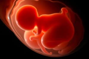
What are fetal membranes?
What are fetal membranes?
Tissue layers that cover the fetus, separating it from the maternal endometrial lining.
List the main components of the fetal membranes.
List the main components of the fetal membranes.
Amniotic membrane (Amnion), Yolk sac (umbilical vesicle), Allantois, Chorionic plate.
When and where does the Amniotic Membrane form?
When and where does the Amniotic Membrane form?
It forms within the Inner Cell Mass during the 2nd week of development.
What cells secrete amniotic fluid?
What cells secrete amniotic fluid?
What are the primary sources of amniotic fluid during the embryonic period?
What are the primary sources of amniotic fluid during the embryonic period?
What additional sources contribute to amniotic fluid during the early fetal period besides amnioblast cells and embryonic surfaces?
What additional sources contribute to amniotic fluid during the early fetal period besides amnioblast cells and embryonic surfaces?
What are the main sources of amniotic fluid during the late fetal period?
What are the main sources of amniotic fluid during the late fetal period?
List the three main pathways for the clearance of amniotic fluid.
List the three main pathways for the clearance of amniotic fluid.
How is amniotic fluid cleared via fetal swallowing?
How is amniotic fluid cleared via fetal swallowing?
What occurs in the intramembranous pathway of amniotic fluid clearance?
What occurs in the intramembranous pathway of amniotic fluid clearance?
What occurs in the transmembrane pathway of amniotic fluid clearance?
What occurs in the transmembrane pathway of amniotic fluid clearance?
List three functions of the Amniotic Sac.
List three functions of the Amniotic Sac.
What is the fate of the amniotic sac?
What is the fate of the amniotic sac?
What is Oligohydramnios?
What is Oligohydramnios?
List three causes of Oligohydramnios.
List three causes of Oligohydramnios.
What are potential complications of Oligohydramnios?
What are potential complications of Oligohydramnios?
List three common fetal causes of Polyhydramnios.
List three common fetal causes of Polyhydramnios.
Besides fetal causes, what are other potential causes of Polyhydramnios?
Besides fetal causes, what are other potential causes of Polyhydramnios?
What causes Amniotic Band Disruption Syndromes?
What causes Amniotic Band Disruption Syndromes?
When is the Yolk Sac (Umbilical Vesicle) formed?
When is the Yolk Sac (Umbilical Vesicle) formed?
What are the main functions of the Yolk Sac?
What are the main functions of the Yolk Sac?
What is the fate of the dorsal part of the Yolk Sac?
What is the fate of the dorsal part of the Yolk Sac?
What happens to the ventral part of the Yolk Sac?
What happens to the ventral part of the Yolk Sac?
What is Meckel's diverticulum?
What is Meckel's diverticulum?
What is the Allantois?
What is the Allantois?
What structures develop from the lower part of the Allantois?
What structures develop from the lower part of the Allantois?
What happens to the upper part of the Allantois?
What happens to the upper part of the Allantois?
What can persistence of the allantois cause?
What can persistence of the allantois cause?
What are the three layers composing the Chorionic Plate?
What are the three layers composing the Chorionic Plate?
What part of the chorion forms the fetal component of the placenta?
What part of the chorion forms the fetal component of the placenta?
What is the clinical utility of the chorion in early pregnancy diagnosis?
What is the clinical utility of the chorion in early pregnancy diagnosis?
What is the primary site of nutrient and gas exchange between the mother and fetus?
What is the primary site of nutrient and gas exchange between the mother and fetus?
What are the two main components of the placenta, and from where are they derived?
What are the two main components of the placenta, and from where are they derived?
What is the 'afterbirth'?
What is the 'afterbirth'?
What is the decidua?
What is the decidua?
What are the three regions of the decidua?
What are the three regions of the decidua?
Which region of the decidua forms the maternal part of the placenta?
Which region of the decidua forms the maternal part of the placenta?
What is the decidua capsularis?
What is the decidua capsularis?
What is the decidua parietalis?
What is the decidua parietalis?
What causes decidual cells to form?
What causes decidual cells to form?
What constitutes the decidual reaction?
What constitutes the decidual reaction?
What are the functions of the decidual reaction?
What are the functions of the decidual reaction?
By the end of which week are the anatomic arrangements necessary for physiologic exchanges between mother and embryo established?
By the end of which week are the anatomic arrangements necessary for physiologic exchanges between mother and embryo established?
What is the difference between the smooth chorion and the villous chorion?
What is the difference between the smooth chorion and the villous chorion?
What structure attaches the fetal part of the placenta to the maternal part (decidua basalis)?
What structure attaches the fetal part of the placenta to the maternal part (decidua basalis)?
What are placental septa?
What are placental septa?
What are cotyledons?
What are cotyledons?
What happens to the decidua capsularis by 22 to 24 weeks?
What happens to the decidua capsularis by 22 to 24 weeks?
From where is the intervillous space of the placenta derived?
From where is the intervillous space of the placenta derived?
How does maternal blood enter the intervillous space?
How does maternal blood enter the intervillous space?
How is the intervillous space drained?
How is the intervillous space drained?
What does the maternal blood in the intervillous space provide to the fetus?
What does the maternal blood in the intervillous space provide to the fetus?
What fetal waste products are transferred into the maternal blood in the intervillous space?
What fetal waste products are transferred into the maternal blood in the intervillous space?
List the three main functions of the placenta.
List the three main functions of the placenta.
What is Placenta Previa?
What is Placenta Previa?
What is Placental Abruption?
What is Placental Abruption?
Flashcards
Fetal Membranes
Fetal Membranes
Tissue layers covering the foetus and separating it from the mother.
Fetal Membrane Components
Fetal Membrane Components
Amniotic membrane or amnion, yolk sac, allantois, and chorionic plate.
Amniotic Membrane Formation
Amniotic Membrane Formation
Forms within the inner cell mass during the 2nd week of development.
Amniotic Membrane Development
Amniotic Membrane Development
Signup and view all the flashcards
Sources of Amniotic Fluid
Sources of Amniotic Fluid
Signup and view all the flashcards
Amniotic Fluid Clearance
Amniotic Fluid Clearance
Signup and view all the flashcards
Functions of the Amniotic Sac
Functions of the Amniotic Sac
Signup and view all the flashcards
Fate of Amniotic Sac
Fate of Amniotic Sac
Signup and view all the flashcards
Oligohydramnios
Oligohydramnios
Signup and view all the flashcards
Complications of Oligohydramnios
Complications of Oligohydramnios
Signup and view all the flashcards
Common Foetal Causes of Polyhydramnios
Common Foetal Causes of Polyhydramnios
Signup and view all the flashcards
Causes of Polyhydramnios
Causes of Polyhydramnios
Signup and view all the flashcards
Amniotic Band Disruption
Amniotic Band Disruption
Signup and view all the flashcards
Yolk Sac Formation
Yolk Sac Formation
Signup and view all the flashcards
Functions of the Yolk Sac
Functions of the Yolk Sac
Signup and view all the flashcards
Fate of the Yolk Sac
Fate of the Yolk Sac
Signup and view all the flashcards
Clinical Correlates of the Yolk Sac
Clinical Correlates of the Yolk Sac
Signup and view all the flashcards
The Allantois
The Allantois
Signup and view all the flashcards
Allantois Functions
Allantois Functions
Signup and view all the flashcards
Fate of Allantois
Fate of Allantois
Signup and view all the flashcards
Clinical Correlates of Allantois
Clinical Correlates of Allantois
Signup and view all the flashcards
Chorionic Plate Components
Chorionic Plate Components
Signup and view all the flashcards
Functions of the Chorion
Functions of the Chorion
Signup and view all the flashcards
Fate of Chorion
Fate of Chorion
Signup and view all the flashcards
Clinical Utility of the Chorion
Clinical Utility of the Chorion
Signup and view all the flashcards
The Placenta
The Placenta
Signup and view all the flashcards
Fetal part of the placenta
Fetal part of the placenta
Signup and view all the flashcards
Maternal part of the placenta
Maternal part of the placenta
Signup and view all the flashcards
Decidua basalis
Decidua basalis
Signup and view all the flashcards
Decidua capsularis
Decidua capsularis
Signup and view all the flashcards
Decidua parietalis
Decidua parietalis
Signup and view all the flashcards
Decidua Transformation
Decidua Transformation
Signup and view all the flashcards
Endometrial arteries pass through
Endometrial arteries pass through
Signup and view all the flashcards
Development of the placenta involves
Development of the placenta involves
Signup and view all the flashcards
The blood carries oxygen and fetal waste
The blood carries oxygen and fetal waste
Signup and view all the flashcards
Maternal blood is derived from
Maternal blood is derived from
Signup and view all the flashcards
Main Placenta Functions
Main Placenta Functions
Signup and view all the flashcards
Placenta Related abnormalities
Placenta Related abnormalities
Signup and view all the flashcards
Study Notes
Fetal Membranes & Placentations
- Fetal membranes are tissue layers that cover the fetus, separating it from the mother's endometrial lining.
- Fetal membranes originate from the zygote.
- Key functions of fetal membranes include protection, nutrition, respiration, excretion, and hormone production.
- The placenta and fetal membranes are expelled from the uterus after birth.
- The outcomes covered are formation, key roles during pregnancy, eventual fate, and related clinical aspects.
Fetal Membrane Components
- Amniotic membrane (Amnion)
- Yolk sac (umbilical vesicle)
- Allantois
- Chorionic plate
The Amniotic Membrane
- It forms within the inner cell mass during the 2nd week of development.
- Amnioblast cells migrate from the epiblast layer to form it.
- It secretes amniotic fluid into the amniotic cavity.
- The amniotic sac grows bigger as the pregnancy progresses.
Sources of Amniotic Fluid
- Embryonic period: Amnioblast cells and embryonic surfaces
- Early fetal period: Amnioblast cells, embryonic surfaces, fetal urine, and fetal lung secretions
- Late fetal period: Fetal urine, fetal lung secretions, fetal oral secretions, and fetal nasal secretions
Functions of the Amniotic Sac
- Protects the fetus from trauma to the maternal abdomen
- Cushions the umbilical cord from compression
- Has antibacterial properties
- Serves as a reservoir of fluid and nutrients for the fetus
- Provides the environment for normal development of the fetal lungs, musculoskeletal system (MSK), and gastrointestinal tract (GIT)
- Thermoregulation
- Lubricates fetal skin and the birth canal
Fate of the Amniotic Sac
- Tears around the time of delivery, causing "rupture of membranes"
- Expelled after birth along with the placenta
Oligohydramnios
- Occurs when amniotic fluid volume is less than expected for the gestational age
- Generally, the Amniotic Fluid Index (AFI) is less than 5cm
- Possible causes: fetal demise, drugs, renal abnormalities, intrauterine growth restriction (IUGR), premature rupture of membranes (PROM), placental insufficiency, chromosomal anomalies (Trisomy 13, Trisomy 18)
- Can lead to complications like pulmonary hypoplasia, fetal limb anomalies, or fetal demise
Polyhydramnios
- Common fetal causes: CNS anomalies, gastrointestinal obstruction, multiple pregnancy, cardiac anomalies, Trisomy 21 (or 18 & 13)
- Other causes: Idiopathic in more than 50% of cases, maternal causes such as diabetes mellitus (DM) or congestive heart failure (CCF)
Amniotic Band Disruption Syndromes
- Results from entrapment of fetal body parts in a disrupted fibrous amnion
- Involves a wide spectrum of abnormalities
- Multiple defects can occur in this condition
The Yolk Sac (Umbilical Vesicle)
- Forms in the 2nd week of development
- Forms by migrating cells from the hypoblast layers.
- Forms later by endodermal cells.
Functions of the Yolk Sac
- Nutrient supply
- Site of early hemopoiesis
- Gives rise to primordial germ cells (PGCs)
- Fate of the Yolk Sac
- Its dorsal part is incorporated into the embryo during embryonic folding, becoming the primordial gut
- Its ventral part degenerates, initially as the vitelline duct
The Allantois
- Extension of the yolk sac into the connecting stalk
- Has similar functions as the yolk sac, like nutrient transfer, hematopoiesis, and giving rise to primordial germ cells (PGCs)
Fate of Allantois
- Lower part gets incorporated to form the urinary bladder
- Upper part degenerates as the urachus
- The remaining structure becomes the median umbilical ligament
Clinical correlates of the Allantois
- Persistence of the allantois causes urachal anomalies
The Chorionic Plate
- COMPONENTS
- Somatic layer of the extraembryonic mesoderm
- Cytotrophoblast layer
- Syncytiotrophoblast
Functions of the Chorion
- Chorion frondosum forms the fetal component of the placenta
- Protects the embryo
- Serves as a hemopoietic center during the yolk sac phase
- Forms the fetal components of the placenta
Fate of Chorion
- The villous chorion forms the fetal components of the placenta
The Placenta
- Primary site of nutrient and gas exchange between mother and fetus
- Feto-maternal organ with 2 components:
- Fetal part: Develops from the chorionic sac
- Maternal part: Derived from the endometrium
- The placenta and umbilical cord form a transport system.
- Some functions include protection, nutrition, respiration, excretion, and hormone production.
The Decidua
- Also known as the gravid endometrium; is the functional layer of the endometrium
- Separates from the remainder of the uterus after parturition
- Consists of three regions:
- Decidua basalis: deep to the conceptus
- Decidua capsularis: superficial part overlying the conceptus
- Decidua parietalis: The remaining parts
- Due to elevated progesterone, decidua enlarge, forming pale-staining decidual cells that accumulate glycogen and lipids.
- Cellular and vascular changes in the endometrium due to blastocyst implantation constitute the decidual reaction.
- The decidual reaction protects maternal tissue against uncontrolled invasion by the syncytiotrophoblast
- Ultrasonography can be used for early detection of pregnancy
Development of the Placenta
- Involves rapid trophoblast proliferation and chorionic sac/villi development
- By the end of the third week, the anatomy enables physiological exchanges
- A complex vascular network forms by the end of the fourth week, facilitating maternal-embryonic exchanges
- Chorionic villi cover the entire chorionic sac until the eighth week
- As the sac grows, villi associated with the decidua capsularis are compressed, resulting in the smooth chorion
- Villi associated with the decidua basalis increase, branch and enlarge forming the villous chorion
- The uterus, chorionic sac, and placenta enlarge with embryo growth, with placenta growth rapid by 18 weeks.
- A fully developed placenta covers 15%-30% of the decidua and weighs 1/6 that of the fetus.
- The placenta consists of a fetal part and a maternal part, the latter replaced by the fetal part by the fourth month.
- The fetal part of the placenta attaches to the maternal part (decidua basalis) via the cytotrophoblastic shell, with the chorionic villi anchoring to the decidua basalis.
- Endometrial arteries and veins pass through cytotrophoblastic shell gaps to interface with the intervillous space.
- Chorionic villi are showered with maternal blood that circulates through the intervillous space
- Maternal blood carries both oxygen and nutrients for support
- Maternal blood also holds fetal salts, carbon dioxide, waste and products of protein metabolism
Functions of the Placenta
- Three main functions: metabolism, transport, and endocrine secretion, which ensure normal fetal development
Studying That Suits You
Use AI to generate personalized quizzes and flashcards to suit your learning preferences.