Podcast
Questions and Answers
Flashcards
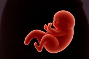
Fetal Period
Fetal Period
Period from ninth week to birth focused on rapid growth and differentiation.
Viability
Viability
Ability of a fetus to survive outside the uterus.
Ultrasonography
Ultrasonography
Used to estimate fetal age and monitor development throughout pregnancy.
Diagnostic Amniocentesis
Diagnostic Amniocentesis
Signup and view all the flashcards
Alpha-Fetoprotein Assay
Alpha-Fetoprotein Assay
Signup and view all the flashcards
Chorionic Villus Sampling
Chorionic Villus Sampling
Signup and view all the flashcards
Percutaneous umbilical cord blood sampling
Percutaneous umbilical cord blood sampling
Signup and view all the flashcards
Magnetic Resonance Imaging (MRI)
Magnetic Resonance Imaging (MRI)
Signup and view all the flashcards
Study Notes
Fetal Period Overview (Ninth Week to Birth)
- The fetal period marks a shift where major systems have already formed in the embyro
- Development now focuses on rapid body growth and tissue/organ/system differentiation.
- There is a noticeable change in head growth relative to the rest of the body, with head growth slowing down.
- Fetal weight increases a lot during the final weeks of the pregnancy.
- Continuous growth occurs but can have periods of absent growth also
Estimation of Fetal Age
- Fetal age can be estimated using ultrasound measurements of the crown-rump length (CRL).
- Measurements are used along with fetal head and femur length
- Measurement ultimately provides a predicted date of delivery.
- Gestational age is usually calculated from the onset of the last normal menstrual period (LNMP) in clinical practice.
- In embryology, gestational age based on the LNMP date is not actually accurate because fertilization occurs mid-menstrual cycle.
- It should be specified whether age is calculated from LNMP or fertilization to avoid confusion.
- The intrauterine period is divided into days, weeks or months, which can lead to confusion
- Calendar months (28-31 days) vs lunar months (28 days).
- Fetal age in the book refers to time since fertilization, unless specified.
Viability of Fetuses
- Viability is defined by a fetus' ability to survive outside of the uterus.
- Fetuses weighing less than 500 g at birth usually do not survive.
- There have been increasing reports of survival at 22-23 weeks of gestation, blurring the line of viability.
- Expert postnatal care can help fetuses less than 1000 g survive, and such infants are referred to as extremely low-birth-weight infants.
- Many full-term, low-birth-weight infants have intrauterine growth restriction (IUGR).
- Most fetuses weighing between 750 and 1500 g can usually survive, though complications may occur.
- Approximately 500,000 preterm infants (<37 weeks) are born in the US each year.
- Many preterm infants suffer from severe medical complications or early mortality.
- Both antenatal steroids and postnatal endotracheal surfactant administration have decreased acute and long-term morbidity.
- Prematurity is a common cause of morbidity and perinatal death.
Criteria for Estimating Fertilization Age
- Various criteria are used to estimate fertilization age during the fetal period based on physical characteristics such as:
- Crown-rump length (mm)
- Foot length (mm)
- Fetal weight (g)
- Main external characteristics
- Measurements may not apply in all cases because dimensional variations increase with age.
- Weights refer to fetuses fixed for about 2 weeks in 10% formalin, fresh specimens typically weigh 5% less.
- Babies under 500g or 22 weeks rarely survive
- Babies born between 26-28 weeks have difficulty surviving because of under developed respiratory/nervous systems
Trimesters of Pregnancy
- The gestational period has three trimesters, each lasting 3 months.
- Major systems have developed by the end of the first trimester.
- The fetus will grow sufficiently in the second trimester so anatomical detail can be viewed using ultrasonography allowing major birth defects to be detected by real-time ultrasonography
- The fetus can likely survive being born prematurely by the beginning of the third trimester.
- At 35 weeks the fetus weighs approximately 2500 g and has a higher chance of surviving if born prematurely
Measurements and Characteristics of Fetuses
- Various measurements + external characteristics can be used to estimate fetal age
- CRL is the best until end of first trimester
- Several structures will be measure ultrasonograpically in the second/third trimester
- Biparietal diameter
- Head circumference
- Abdominal circumference
- Femur length
- Foot length
- Weight is useful for estimating age
- There may be discrepancy between weight and age such as in mothers with diabetes
- Fetal dimensions obtained via ultrasounds approximates CRL from spontaneously aborted fetuses.
- Determining size of a fetus especially its head circumference is important for obstetricians to manage patients.
Highlights of Fetal Period: Overview
- There is no formal staging system for the fetal period.
- Changes that occur are usually described in periods of 4-5 weeks.
Nine to Twelve Weeks
- At the beginning of the fetal period the head accounts for ~1/2 of the CRL
- Body length growth accelerates rapidly subsequently
- By end of 12 weeks the CRL has almost doubled, and head growth has slowed
- Head is still disproportionately large in comparison to the body
- At 9 weeks, the face is broad, the eyes are widely separated, the ears are low set, and the eyelids are fused.
- By the end of 12 weeks, ossification centers appear in the skeleton, especially in the cranium and long bones.
- Early in the ninth week, legs are short, thighs are also small
- By the end of 12 weeks, the upper limbs have almost reached their final relative lengths, lower limbs are still not so well developed, and are slightly shorter than their final relative lengths.
- The external genitalia of males and females appear similar until the end of the ninth week
- Mature form not established until 12th week.
- Intestinal coils visible in the proximal end of the umbilical cord until the middle of the 10th week.
- Intestines have returned to the abdomen by the 11th week.
- At 9 weeks the liver is the major site of erythropoiesis
- By the end of 12 weeks erythropoiesis has decreased in the liver and has begun in the spleen.
- Urine formation begins between the 9th and 12th weeks
- Urine discharged through the urethra into the amniotic fluid in the amnionic cavity.
- The fetus will reabsorb some amniotic fluid after swallowing it.
- Fetal waste products are transferred to the maternal circulation by passage across the placental membrane.
Thirteen to Sixteen Weeks
- Growth is very rapid during this period.
- By 16 weeks, the head is smaller than in the 12-week fetus
- The lower limbs have lengthened at this point.
- Limb movements, which first occur by end of embryonic period coordinated at 14th week
- Movements are too slight to be felt by the mother but are still visible during ultrasounds.
- Ossification of the fetal skeleton is active during this period, and the developing bones are clearly visible on ultrasound images by the 16th week.
- Slow eye movements occur at 14 weeks
- Scalp hair patterning also determined during period.
- By 16 weeks, the ovaries are differentiated and contain primordial ovarian follicles, which contain oogonia (primordial germ cells).
- Genitalia of male/female fetuses can be recognized by 12-14 weeks
- By 16 weeks the eyes face anteriorly, and the external ears reach their positions on the sides of the head.
Seventeen to Twenty Weeks
- Growth slows down but CRL increases by around 50mm.
- Fetal movements (quickening) commonly felt by the mother.
- The skin becomes covered with a greasy material, the vernix caseosa
- Vernix protects the delicate fetal skin from abrasions, chapping, and hardening due to amniotic fluid.
- Fetuses become covered with fine, downy hair (lanugo)
- Lanugo helps the vernix adhere to the skin.
- Eyebrows and head hair are visible at 20 weeks.
- Brown fat forms during this period (site of heat production)
- Tissue found mostly at the root of the neck, posterior to the sternum, and in the perirenal area.
- Brown fat produces heat via oxidizing fatty acids.
- By 18 weeks, the fetal uterus is formed, and canalization of the vagina has begun.
- Many primordial ovarian follicles containing oogonia are also visible.
- By 20 weeks, the testes have started to descend, but remain located on the posterior abdominal wall, like the ovaries.
21 to 25 Weeks
- Substantial weight gain occurs as well as better proportions.
- The skin is usually wrinkled and more transparent, especially initially
- The skin appears pink to red due to capillaries that are visible in the skin.
- Rapid eye movements initiate at week 21
- Blink-startle responses reported at 22-23 weeks
- Secretory epithelial cells (type II pneumocytes) in the interalveolar walls of the lung initiate the secretion of surfactant
- Surfactant is a surface-active lipid that maintains the patency of the developing alveoli of the lungs
- Fingernails present by week 24
- Between 22-25 weeks if a fetus is born prematurely can survive if intensive care is given
- However there is risk of death because of the immaturity of the respiratory system, with risk for neurodevelopmental disability being high in fetuses born before 26 weeks
26 to 29 Weeks
- During period fetuses can survive if they're born prematurely with intensive care being given
- Lungs and pulmonary vasculature have developed sufficiently to provide gas exchange.
- The central nervous system can direct rhythmic breathing movements and controls body temperature
- The highest rate of neonatal mortality occurs in those of low birth weight (≤2500 g) and very low birth weight (≤1500 g).
- Eyelids open at 26 weeks
- Lanugo (fine downy hair) + head hair becomes well developed.
- Toenails visible, considerable subcutaneous fat is under the skin, smoothing out many of the wrinkles.
- Quantity of white fat increases to approximately 3.5% of the body weight during this period.
- In this period, fetal spleen is an important site of erythropoiesis (formation of red blood cells; ends by week 28).
- By said time bone marrow has become the major erythropoietic site.
30 to 34 Weeks
- The pupillary reflex can be elicited at this time
- The skin is pink and smooth, limbs have a chubby appearance,.
- Quantity of white fat is 8% of the body weight
- Fetuses 32 weeks and older usually survive if born prematurely.
- The papillary dilates up to full term
35 to 38 Weeks
- Fetuses born at 35 weeks have a firm grasp and orient to light spontaneously
- Integrative functions and the nervous system sufficiently mature
- Most fetuses are plump during finishing period.
- By 36 weeks, circumferences of head/abdomen essentially =
- After 36 weeks the circumference of the abdomen may be greater than head's
- The foot length of fetuses will be slightly larger than femoral length at 37th week
- Foot can be an alternative parameter for fetal age confirmation slowing of growth as birth approaches
- At full term (38 weeks) most fetuses are at CRL of 360 mm and weigh approximately 3400 g
- White fat is approximately 16% of the body
Low Birth Weight
- Not every low birth weight baby is premature
- About about one third those with 2500g or less birth weight are actually small or gestational date
- They may be underweight due to placental insufficiency
- Placentas are poorly attached and/or has degenerative changes reducing the provision of oxygen and food to fetus
- It is imporatnt to separate full term, low weight babies versus premature with low weight babies
- IUGR can be preeclampsia, smoking or illicit drugs, multiple gestation, cardiovascular defects, inadequate mineral nutriiton as well as mother and fetal hormone
- Babies with asymmetrical IUGR who have large head circumference relative to weight and length show lack of subs fatty tissue and wrinkly skin, and suggests fat loss
Impaired Uteroplacental and Fetoplacental Blood Flow
- Conditions resulting from decreasing uterine bllod flow or reduction of maternal placental circualtion
- Small chorionic vessels
- Severe maternal hypotension
- Renal disease
- Chronic reduction may cause fetal starvation leading to IUGR, possible due to placental dysfunction, or infarction
- There will be less total area for exchange of nutrients between the fetal/maternal bloodstreams
- It is difficult to separate effect of changes resulting from matneral blood flow to the placenta
- Maternal vascular changes in the uterus can be primary while placental defects can be secondary
Genetic Factors and Growth Retardation
- Genetic factors can cause IUGR and repeats could be the cause of abnormal growth
- Both structural and nummeric chromosomal aberrations have been displayed to be associated with impaired growth
- IUGR is pronounced in infants with Down syndrome with it being charactertic of fetuses with trisomy 18 syndrome
Procedures for Assessing Fetal Status
- Perinatology is the branch concerned wit hthe well-being of the fetus and the neonate
- Period is between 26 weeks following fertilization, up to 4 weeks after birth
- It is subspecialty of medication and combines aspects of obstertics and pediatrics
Ultrasound Scans
- primary for imaging for assessment of fetus/es b/c: readily available, inexpensive, have quality imaging, lack adverse affects
- chorionic sac(k) contents can be visualized
- Multiple births, as well abnormalities in placenta and presentations can be determined
- Details can give accurate measurements of biparietal diameter of fetal cranium, through which length and age can be determined
- Rapid advancements, including 3-D, have made it a large tool for prenatal imaging
- Biopsy of tissues (skin/liver/kidney/muscle) can be performed with ultrasound assistance
Diagnostic Amniocentesis
- An common prenatal diagnostic procedure.
- Sampling and be done starting on or after week fifteen.
- A 22-gauge needle is inserted and traverses the abdominal and uterine walls and proceeds by way of the chorion and amnion
- Volume close to 200ml, where 15-20 can be taken
- Procedure possesses risks, specifically, pregnancy loss, yet minimal, (0.5-1.0%), when done and supervised by an experienced physician
- Ultrasound is used to outline placement of organs to ensure placement will not hit certain areas.
Alpha-Fetoprotein Assay
- AFP is synthesized by the fetal liver/umbilical vesicle, and is a glycoprotein.
- High concentrations in fetal serum
- Concentrations are greatest at ~week fourteen of LNMP
- There exists a low amount of AFP fluid in the amnion
Alpha-Fetoprotein and Fetal Anonmalies
- When sever defects are present such as CNSs, as well as abdominal walls AFP has an elevation.
- AFP fluid in the amnion is through immunoassay .
- In these sever cases roughly 99% can be diagnosed prenatally.
- Elevated concentrations are in serum when there is something in the neural tubes.
- Serum is lower in fetuses when syndromes are occurring.
Spectrophotometric Studies
- examination of the amniotic fluid in order to assess erythroblastosis.
- Erythroblastosis is also hemolysis, where there is damage to the red cells of the fetus.
- The concentration of the bilirubin is in correlation to the amount of hemolysis
Chorionic Villus Sampling
- The biopsies are five to twenty.
- A needle is inserted and guided by an ultrasound and goes into chamber from one side walls.
- Transcervically sampling is possible, by way of poly catheter inserted under ultrasound.
- Condition of the subject can be obtained, and karyotype can be known of the offspring
- With the knowledge of the karyotype, weeks of assessment is prior to amniocentesis
Diagnostic Value of Chorionic Villus Sampling
- Used for detection of abnormalities in the chromosomes and errors, as metabolic problems and X problems
- It can be done as of 10/12 weeks and fetal loss is lower, .5/1% similar number to amniocentesis
- There are not consistent reports of upper extremities/limb malfunctions after use
- The edge sampling over amniocent is time. Analysis of chrom is over several weeks prior.
Cell Cultures and Chromosomal Analysis
- The prevalence of chromosomal disorders is approximately 1 in 120 neonates.
- Aneuploidy + sex are known when chromosomes from fetus which is amniocent + chorionic
- High resolution is displayed when there is chrom microassay.
- In cases where conceiving is done with assistive methods, cells can be found.
- Knowledge helpful for diagnosis of disease by gender.
- Subtelometric + microduplicatons are found by hybridization
Noninvasive Prenatal Diagnosis
- Down syndrome (trisomy 21) is the most commonly known chromosomal disorder.
- Children born with this condition have varying degrees of intellectual disabilities. Noninvasive screening for trisomy 21 is based on the isolation of fetal cells in maternal blood and the detection of cell-free fetal DNA and RNA.
- DNA-based prenatal diagnostic testing and sequencing of maternal plasma are reliable for the early detection of fetal aneuploidies.
- Chromosomal microarray analysis and whole-exome sequencing advancements allow for advanced prenatal diagnostic and screening.
Fetal Transfusion
- Fetuses with hemolytic disease of the neonate can be treated by intrauterine blood transfusions.
- Blood can be injected via needle into fetal peritoneal cavity.
- A fetal anemia treatment can involve blood and packed red injected with blood via umbilical transfusion with advances.
- There is reduction with mothers using -Rh immunoglobulin, which negates the need to transfuse/helps the disease
- Treatment infusion through vein in umbilical can be done when is needed.
Fetoscopy
- Exterior of the body of a fetus is examined with fiberoptics
- Fetoscope is put by way of stomach + uterus and into the amnion/cavity.
- The scope is put in during week seventeen/twenty and thinner gauges in the abdomen help.
- Risks a bit steep so other procedures are used
- Combining with laser can treat like twin.
- Also can release amnion band.
Percutaneous Umbilical Cord Blood Sampling
- Samples attained by way of the umbilical vein
- Cordocentesis for diagnosis of fetal abnormal condition: Aneuoploidy Fetal growth restriction. Infection, anemia
- Done 18 weeks under guidance via ultrasound for locating
- Danger in loss is roughly 1.3, yet can go a little more steeply if issue exist
- Procedure also has way of curing ailments. Blood injection happens in these times as well
Magnetic Resonance Imaging
- Needed for planning, provide insight to disorders
- No use radiation and uses similar ultra sound
- Provides quality soft tissues/resolution.
Fetal Monitoring
- Monitor for high risk pregnancies
- Gives information about the fetus and its oxygenated levels
- Heart rate can have issues
- In such occasions the fetus is suspect/in danger
- Simple method: with a transducer which is placed on the abdomen from outside
Summary of Fetal Period
- The fetal period begins 8 weeks after fertilization (10 weeks after the LNMP) and ends at birth.
- Rapid body growth and differentiation of tissues and organ systems occur
- Obvious change in the fetal period is the relative slowing of head growth compared with that of the rest of the body.
- At start of week 20 lanugo, and hair are visible.
- Coating on skin with vernix caseosa happens.
- Lids are closed through this time until the baby is nearly cooked.
- During end, usually capable of extrauterine function due to the respiratory systems
- Up to week thirty. Skin will be thin with no subdermal fat. The baby develop.
Studying That Suits You
Use AI to generate personalized quizzes and flashcards to suit your learning preferences.