Podcast
Questions and Answers
What is the process of fusion of the genetic material of spermatozoon with mature ovum (secondary oocyte)?
What is the process of fusion of the genetic material of spermatozoon with mature ovum (secondary oocyte)?
Fertilization
What wafts the oocyte?
What wafts the oocyte?
Cilia and peristaltic contraction of the uterine tube.
What secretes alkaline mucus to attract the spermatozoa?
What secretes alkaline mucus to attract the spermatozoa?
Cervix
What does the zona pellucida of the oocyte produce, that attracts capacitated sperms?
What does the zona pellucida of the oocyte produce, that attracts capacitated sperms?
What enzyme from the acrosome of the sperms disperses corona radiata, allowing access to zona pellucida?
What enzyme from the acrosome of the sperms disperses corona radiata, allowing access to zona pellucida?
Oocyte becomes mature when the plasma membrane of sperm and egg fuses.
Oocyte becomes mature when the plasma membrane of sperm and egg fuses.
What is formed when the nucleus of sperm and ova then fuses?
What is formed when the nucleus of sperm and ova then fuses?
What is the composition of sperm regarding sex chromosomes?
What is the composition of sperm regarding sex chromosomes?
The ovum only contains what chromosome?
The ovum only contains what chromosome?
XY is male and XX is female.
XY is male and XX is female.
What is the origin of fraternal (dizygotic) twins?
What is the origin of fraternal (dizygotic) twins?
What is the origin of monozygotic (identical) twins?
What is the origin of monozygotic (identical) twins?
What are the 3 phases of zygote development?
What are the 3 phases of zygote development?
How long is the pre-embryonic period after fertilization?
How long is the pre-embryonic period after fertilization?
How long is the fetal period?
How long is the fetal period?
What surrounds the zygote as it travels along the uterine tube to the uterus?
What surrounds the zygote as it travels along the uterine tube to the uterus?
Where does the zygote receive nutrients during the pre-embryonic period?
Where does the zygote receive nutrients during the pre-embryonic period?
What is the mitotic division of the zygote called?
What is the mitotic division of the zygote called?
What are the small cells called that undergo mitotic division?
What are the small cells called that undergo mitotic division?
What is formed by 16 cells after 3 days?
What is formed by 16 cells after 3 days?
When cells bind tightly together, what do they become?
When cells bind tightly together, what do they become?
What occurs when outermost cells secrete fluid into the morula and a fluid filled cavity appears?
What occurs when outermost cells secrete fluid into the morula and a fluid filled cavity appears?
What is the blastocyst made up of?
What is the blastocyst made up of?
What does the inner cell mass become?
What does the inner cell mass become?
What does the trophoblast become?
What does the trophoblast become?
After implantation of the cell mass, the inner cell mass now called the epiblast cells differentiates into what?
After implantation of the cell mass, the inner cell mass now called the epiblast cells differentiates into what?
The two cell types, epiblast and hypoblast differentiate and fuse into 3 layers called what?
The two cell types, epiblast and hypoblast differentiate and fuse into 3 layers called what?
The two cell types, epiblast and hypoblast differentiate and fuse into 3 layers called trilaminar embryonic disc using what process?
The two cell types, epiblast and hypoblast differentiate and fuse into 3 layers called trilaminar embryonic disc using what process?
During the gastrulation process of embryo cells differentiating and fusing, what 3 layers are known as the primitive streak and appear at day 15?
During the gastrulation process of embryo cells differentiating and fusing, what 3 layers are known as the primitive streak and appear at day 15?
What does Trophoblast cells produce that is a hormone that prevents menstruation and maintains pregnancy by sustaining the corpus luteum?
What does Trophoblast cells produce that is a hormone that prevents menstruation and maintains pregnancy by sustaining the corpus luteum?
What does the corpus luteum produce?
What does the corpus luteum produce?
What is the main function of the placenta?
What is the main function of the placenta?
Name the three regions that endometrium/decidua has
Name the three regions that endometrium/decidua has
What is the fluid surrounding the fetus from above with fluid that is aseptic and bactericidal, during labour called?
What is the fluid surrounding the fetus from above with fluid that is aseptic and bactericidal, during labour called?
Approximately what percentage of amniotic fluid is water?
Approximately what percentage of amniotic fluid is water?
At what week can the umbilical cord be formed?
At what week can the umbilical cord be formed?
What is the approximate length of the umbilical cord measured in centimetres?
What is the approximate length of the umbilical cord measured in centimetres?
Flashcards
Growth
Growth
Increase in size through increase in cell number via mitosis.
Development
Development
Includes growth and changes in life phases that accomplish physiological changes.
Fertilization
Fertilization
The fusion of genetic material from a spermatozoon and a mature ovum (secondary oocyte).
Sperm capacitation
Sperm capacitation
Signup and view all the flashcards
Hyaluronidase
Hyaluronidase
Signup and view all the flashcards
Zygote
Zygote
Signup and view all the flashcards
Dizygotic twins
Dizygotic twins
Signup and view all the flashcards
Monozygotic twins
Monozygotic twins
Signup and view all the flashcards
Pre-embryonic period
Pre-embryonic period
Signup and view all the flashcards
Cleavage
Cleavage
Signup and view all the flashcards
Morula
Morula
Signup and view all the flashcards
Cavitation
Cavitation
Signup and view all the flashcards
Blastocyst
Blastocyst
Signup and view all the flashcards
Trophoblast
Trophoblast
Signup and view all the flashcards
Embryoblast
Embryoblast
Signup and view all the flashcards
Implantation
Implantation
Signup and view all the flashcards
Villi
Villi
Signup and view all the flashcards
Lacunae
Lacunae
Signup and view all the flashcards
Inner cell mass differentiation
Inner cell mass differentiation
Signup and view all the flashcards
Two cell types
Two cell types
Signup and view all the flashcards
Gastrulation
Gastrulation
Signup and view all the flashcards
3 layers
3 layers
Signup and view all the flashcards
Ectoderm
Ectoderm
Signup and view all the flashcards
Mesoderm
Mesoderm
Signup and view all the flashcards
Endoderm
Endoderm
Signup and view all the flashcards
Trophoblast cells
Trophoblast cells
Signup and view all the flashcards
Placenta at Term
Placenta at Term
Signup and view all the flashcards
Maternal surface
Maternal surface
Signup and view all the flashcards
Fetal Surface
Fetal Surface
Signup and view all the flashcards
Chorion villi
Chorion villi
Signup and view all the flashcards
Anchoring placenta
Anchoring placenta
Signup and view all the flashcards
Placenta: Respiratory Function
Placenta: Respiratory Function
Signup and view all the flashcards
Placenta: Nutrition
Placenta: Nutrition
Signup and view all the flashcards
Placenta: Excretory Function
Placenta: Excretory Function
Signup and view all the flashcards
Placenta: Storage.
Placenta: Storage.
Signup and view all the flashcards
Placenta: Protection.
Placenta: Protection.
Signup and view all the flashcards
Placenta: Endocrine Function
Placenta: Endocrine Function
Signup and view all the flashcards
Placental Membranes
Placental Membranes
Signup and view all the flashcards
Amnion membrane
Amnion membrane
Signup and view all the flashcards
Chorion membranes
Chorion membranes
Signup and view all the flashcards
Amniotic Fluid
Amniotic Fluid
Signup and view all the flashcards
Study Notes
- Pregnancy involves fertilization and early embryonic development.
Ovum Structure
- The ovum contains follicle cells, corona radiata, nucleus, nucleolus, cytoplasm, zona pellucida, plasma membrane (vitelline membrane), and perivitelline space.
Sectional View of Ovary
- The Ovary contains:
- Primary follicle
- Secondary follicle
- Tertiary follicle
- Graafian follicle
- Blood Vessels
- Corpus Luteum
- Ovum
Sperm structure
- A Sperm contains:
- Head (with Acrosome and Nucleus)
- Acrosome – digestive enzymes
- Nucleus – 23 chromosomes
- Mid piece (with Mitochondria and Centriole)
- Collar (containing many mitochondria)
- Tail (with Flagellum)
- Flagellum – causes sperm to swim
Definition of Terms
- Growth is an increase in size through an increase in cell number by mitosis.
- Development includes growth and changes in life phases to accomplish physiologic changes.
- Fetus development can be divided into 4 stages.
- Fertilization takes 24 hours.
- Blastocytes form between the 2nd and 6th day.
- The embryonic stage is from the 7th day to the 4th week.
- The fetal stage is from the 4th week onward.
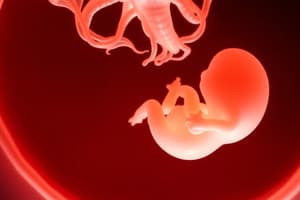
Zygote Development
- The stage of development is Zygote -> Embryo -> Fetus -> Baby.
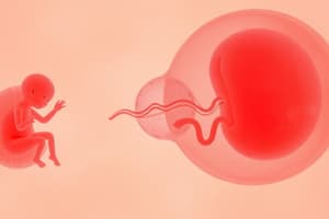
Fertilization
- Fertilization, also known as conception, is the fusion of the genetic material of a spermatozoon with a mature ovum (secondary oocyte).
- The ovum's nucleus is surrounded by cytoplasm, the zona pellucida (jelly coat), and the corona radiata (follicular cells).
- Sperm includes structures like the acrosome, sperm nucleus, cortical granules, cytoplasm, zona pellucida, and follicle cells.
- During fertilization, the oocyte is moved by cilia and peristaltic contractions of the uterine tube.
- The cervix secretes alkaline mucus to attract spermatozoa.
- After deposition in the posterior fornix of the cervix, 2 million sperms reach loose cervical mucus and uterine tubes out of 300 million sperm -Some sperm are destroyed by the acidic medium in the vagina
- Sperm undergo capacitation, which involves physiological changes such as increased flagella action and removal of glycoproteins, enabling penetration and fertilization within the uterine tube.
- The zona pellucida of the oocyte produces chemicals that attract capacitated sperms.
- Hyaluronidase, an enzyme from the acrosome of sperm, disperses the corona radiata, allowing access to the zona pellucida.
- Only one spermatozoon penetrates the zona pellucida, after which the cell membrane becomes impenetrable to other sperms.
- Oocyte maturation occurs when the plasma membranes of sperm and egg fuse.
- The sperm contains either a Y or an X chromosome, while the ovum contains only an X chromosome
- XY sperm yields a male and XX sperm yields a female
- Fraternal (dizygotic) twins form from two ooctyes released independently. Monozygotic (identical) twins develop from a single zygote that divides into two during division/cleavage.
- Dizygotic twins have genetically distinct separate placentas
- Monozygotic twins have genetically identical shared placentas during zygote splits
Development of Zygote
- The development of a zygote includes three phases: pre-embryonic, embryonic, and fetal periods.
- Pre-embryonic period (2 weeks after fertilization)
- Embryonic period (2 to 8 weeks after fertilization)
- Fetal period (weeks 8 to birth)
Pre-embryonic Period
- After fertilization, the zygote travels along the uterine tube to the uterus, surrounded by the zona pellucida.
- The zygote is nourished by nutrients from goblet cells and secretory cells in the uterine tubes and uterus.
- The zygote undergoes mitotic division called cleavage, resulting in small cells called blastomeres.
- Cell division occurs:
- 2 cells on day 1
- 4 cells on day 2
- 8 cells on day 2.5
- 16 cells on day 3 forming a morula
Pre-Embryonic period: Cavitation & Blastocyst
- Cavitation occurs when the outermost cells secrete fluid into the morula, creating a fluid-filled cavity, forming a blastula through the process of blastulation.
- During cleavage, the size of the developing structure does not initially increase, but cells divide into smaller cells bounded by the zona pellucida.
- By day 4, the blastocyst enters the uterus.
- Endometrial glands secrete fluid that nourishes the blastomeres.
- The outer cell mass, or trophoblast, and the inner cell mass, or embryoblast, makeup a blastocyst.
- Day 1: Fertilization
- Day 2: Cleavage occurs
- Day 3: Compaction begins
- Day 4: Differentiation in cells are seen
- Day 5: Cavitation
- Day 6: Zona Hatching
- Day 7: Implantation
- Day 9: Cell mass differentiation
- Day 12: Bilaminar Disc Formation
Blastocyte
- The inner cell mass (embryoblast) within the blastocyst attaches to the wall of the uterus via the endometrium.
- A blastocyst contains the inner cell mass, trophoblast, and blastocyst cavity (blastocoele).
- The inner cell mass becomes the embryo, amnion, and umbilical cord, and the trophoblast becomes the placenta and chorion.
- Implantation of the trophoblast into the decidua (endometrium) begins with the help of proteolytic enzymes.
- The trophoblast invades the decidua and forms finger-like projections called villi.
- The spaces between the villi are called lacunae which fill with maternal blood.
Embryoblast Differentiation & Gastrulation
- Embryoblast cells differentiate into two types: epiblast (closest to the trophoblast) and hypoblast (closest to the blastocyst cavity).
- Epiblast and hypoblast then differentiate and fuse into 3 layers, called the tri-laminar embryonic disc, through a process called gastrulation.
Gastrulation and Germ Layers
- At day 15, there are 3 layers in the primitive streak
- Ectoderm: forms the epidermis layer of skin, hair, nails, and nervous system
- Mesoderm: forms muscles, skeleton, dermis of skin, urogenital glands, blood vessels, blood and lymph cells, and connective tissues
- Endoderm: forms the epithelial lining of digestive, respiratory and urinary systems, and glandular cells of organs (e.g., liver, pancreas)
Early Placenta Development
- Trophoblast produces hCG, which ensures the endometrium is ready for implantation.
- Decidualization of the endometrium occurs, with increased vascularity, and structural changes.
- Decidua basalis: between the embryo and stratum basalis of uterus at implantation site.
- Decidua capsularis: covers developing embryo.
- Decidua vera/parietalis: lines remainder of uterine cavity.
- During embryonic development, the uterine glands produce nutrients the embryo requires prior to full placenta development.
- Adaptation of the mother's immune system prevents rejection of the fetus.
- Implantation follows prelancunar and lacunar stages.
- Prelacunar is the stage before villi develop
- Lacunar stage refers to when the villi have formed, and spaces between them are evident (the spaces are called lacuna, which further develops to form intervillous space)
Placental Development
- During weeks 0 - 40, the placenta develops alongside the fetus, eventually forming the uterus wall, placental vili, cytotrophoblast, syncytiotrophoblast, and the maternal blood.
Placenta at Term
- The fully formed structure is a spongy disc that is circular, 20 cm in diameter, 2.5 cm thick, dark red.
- The placenta is fully formed by the 16th week of pregnancy, but starts developing around the 14th day after fertilisation.
- The placenta weighs around 500g, and has a fetal size to placenta ratio of 7:1.
- It is mainly composed of chorionic villi, also know as Chorion frondosum.
- The maternal surface of the placenta is the one attached to the uterine wall.
- The adjacent fetal surface lies to the fetus, covered by the shiny amnion.
- It is divided into 15-20 lobes separated by deep grooves and has an umbilical cord attached near its center.
Placenta Villi
- The developing chorion villi are of two kinds:
- The ones that deeply enter the basal deciduas (anchoring the placenta)
- The ones that hang loosely in the maternal blood, absorbing nutrients through the nutritive vili.
- The mature chorionic villi are tree like, and contain the foetal capillaries network arising from one stem.
- Each villum is covered with few layers of tissue, which make it impossible for the foetal and maternal blood to mix in the placenta.
- Foetal blood is pumped by the foetal heart through umbilical arteries to capillaries in the chorionic villi
- In the chorionic villi, the blood absorbs oxygen and nutrients from the maternal blood, and returns via umbilical vein to the foetus.
Placenta Function
- Respiratory: Oxygen from the mother's hemoglobin goes to the fetus; the fetus lets off carbon dioxide by simple diffusion.
- Nutrition: The fetus gets nourishment from the mother's diet.
- Excretory: Waste products and carbon dioxide move into maternal blood by diffusion.
- Storage: Stores glycogen, iron, and fat soluble vitamins.
- Protection. The placenta acts as barrier against illnesses, but some organisms like rubella and syphilis may pass through them.
- Endocrine: Releases Human Chorionic Gonadotrophin (HCG, for maintaining pregnancy), oestrogens, progesterone, and Human Placental Lactogen (HCL).
- Human placental lactogen stimulates somatic growth, breast tissue growth, food intake and weight gain.
Fetal & Maternal circulation
Placental structure, composition and function
- The basal and chorionic plates meet to form the membrane around the amniotic fluid
- The chorion-amnion membranes are composed of:
- the amnion
- the chorion
Amnion
- It is comprised of an inner membrane derived from the inner cell mass
- Its is a tough, smooth, translucent, and continuous with outer umbilical cord
- It contains amniotic fluid, it moves atop the aiding mucous
- It produces small amounts amniotic fluid and prostaglandin in small amounts, playing role in labour initiation
Chorion
- Its is the continuous outermost membrane touching the placenta
- It is a thick, rough, fibrous, and opaque membrane connected to the decidura vernix
- It also produced platelet activating factors, oxytocin, and prostaglandins
- It can easily rupture and be retained in the uterus after birth
The amniotic fluid
- Fluid is alkaline, transparent, and yellowish inside the amniotic sac.
- It's 99% water circulating constantly.
- The placenta and amnion emit it during pregnancy.
- Later, the fetus swallows and releases it through the kidneys.
- The outer chorion and inner amnion form the fetal sac.
- Its volume changes, as at 10 weeks it can be 2oml, and at term, between 500 - 1200ml
- if lower then 500ml it termed oligohydramniouS, and above 1500 volume it termed polyhydramnious
Amniotic fluid functions
- It enables fetal movement.
- It acts as a shock absorber.
- It equalizes the pressure from uterine contractions.
- It keeps a stable intrauterine temperature and protects against heat.
- It provides the fetus with few smaller nutrients
Amniotic Fluid Composition and Significance
- Amniotic fluid contains 99% water alongside solid, and fetal skin cells.
- Vernix caseosa and lanugo can also be found in Amniotic fluid
- Vernix caseosea is a whitish, cheese substance. Long fine hair called Lanugo cover newborns.
- Meconium in amniotic fluid indicates fetal distress.
- Amniocentesis can assess fluid for condition compromises.
- During labour, when membranes break, the liquor cleans birth canal.
- Aseptic and bactericidal fluid helps labour progress
Umbilical Cord (Funis)
- The Umbilical Cord, also known as Funis, extends from the foetus to the placenta.
- It is formed by 5 weeks of pregnancy
- It is roughly 50 centimetres in lengths, and is considered shorts if it measures below 40 Centimetres
- The function of the Umbilical cord os to transport nutrients and oxygen as, and remove wastes from the foetus
- It possesses no nerves, and therefore cutting does not cause it pain
- Structure of the Umbilical Cord (Funis)
- Two arteries carry blood from fetus to placenta. One umbilical vein transports blood to fetus
- Blood vessels are covered and protected by the Wharton's Jelly, and by a layer of Amnion
Problems of the Umbilical cord
- It features true and false knots (Wharton's Jelly)
- A long umbilical cord( above 100 cms ) may wrap around the body or knotted, preventing normal circulation.
Anatomical Variation of Placenta
- Succenturiate Lobe
- separated lobe joined by vessels, which is also referred to as the significant variations. It can cause hemorrhage and infections
- Circumvallate placenta
- A ring forms from form from opaque membranes. The placenta is nearer to centre and in 2 sections
- Bipartite Placenta
- Here 2 lobes from the placenta exists. There are 2 cords, adjoined to the placenta
- Tripartite placenta
- Here there are 3 sections of the placenta exists. It is similar to the Bipartite
Variations of Cord
- Velamentous
- Cord inserted in membranes
- Vasa praevia
- Vessels cross in the os of the uterus
- Battledore insertion
- In the Battledore the Cord exist on edges
- Malformation of structure can causes structural issues, issues with complete incomplete separate of the septum and malformation of tissues.
Studying That Suits You
Use AI to generate personalized quizzes and flashcards to suit your learning preferences.