Podcast
Questions and Answers
What is the retina?
What is the retina?
The light-sensitive inner surface of the eye, containing the receptor rods and cones plus layers of neurons that begin the processing of visual information.
What is the eyeball?
What is the eyeball?
Organ of vision.
What is the sclera?
What is the sclera?
White of the eye.
What are retinal blood vessels?
What are retinal blood vessels?
What is the lens?
What is the lens?
What is the anterior compartment?
What is the anterior compartment?
What is the optic nerve?
What is the optic nerve?
What is the iris?
What is the iris?
What is the cornea?
What is the cornea?
What is the posterior compartment?
What is the posterior compartment?
What is the pupil?
What is the pupil?
What is the suspensory ligament?
What is the suspensory ligament?
Flashcards are hidden until you start studying
Study Notes
Eye Anatomy and Functions
-
Retina: The eye's inner surface that is sensitive to light, containing specialized receptor cells (rods and cones) along with neurons that start the visual processing.
-
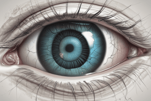
Eyeball: The anatomical structure responsible for vision; it contains various components essential for capturing and processing visual information.
-
Sclera: The tough, white outer layer of the eye that provides structure and protection.
-
Retinal Blood Vessels: Blood vessels supplying oxygen and nutrients to the retina's rods and cones, essential for their function and health.
-
Lens: A transparent structure located behind the pupil that adjusts its shape to focus light onto the retina for clear vision.
-
Anterior Compartment: The front part of the eye filled with aqueous humor, which maintains intraocular pressure and provides nutrients to the eye structures.
-
Optic Nerve: A critical nerve that transmits visual information from the retina to the brain for interpretation.
-
Iris: A muscular, colored ring that surrounds the pupil, regulating the amount of light entering the eye by adjusting the pupil's size.
-
Cornea: The clear, dome-shaped tissue that covers the front of the eye, acting as the primary lens that refracts light.
-
Posterior Compartment: The section of the eye located behind the lens, filled with vitreous humor, which helps maintain the eye's shape and supports the retina.
-
Pupil: The central opening in the iris, allowing light to pass into the eye. Its size changes based on light conditions.
-
Suspensory Ligament: Connects the lens to the ciliary body, facilitating movement and adjustment of the lens to focus on near and distant objects.
Studying That Suits You
Use AI to generate personalized quizzes and flashcards to suit your learning preferences.


