Podcast
Questions and Answers
What structure in the muscle fiber is primarily responsible for the transmission of action potentials?
What structure in the muscle fiber is primarily responsible for the transmission of action potentials?
- Sarcoplasmic reticulum
- Myofibrils
- Sarcolemma
- Transverse tubules (correct)
How does the action potential travel along the muscle fiber?
How does the action potential travel along the muscle fiber?
- Across the sarcolemma only
- Via diffusion in the interstitial fluid
- Through the cytoplasm
- Along the transverse tubules (correct)
What is the primary role of the transverse tubules in muscle fibers?
What is the primary role of the transverse tubules in muscle fibers?
- Generate ATP
- Conduct action potentials (correct)
- Facilitate oxygen transport
- Store calcium ions
In which part of the muscle fiber does the action potential primarily propagate?
In which part of the muscle fiber does the action potential primarily propagate?
Which of the following statements is true regarding the action potential in muscle fibers?
Which of the following statements is true regarding the action potential in muscle fibers?
Who is the writer associated with the lecture in Block 1.2?
Who is the writer associated with the lecture in Block 1.2?
What role does Zainab Adel Alali serve in the context of the lecture?
What role does Zainab Adel Alali serve in the context of the lecture?
What does the abbreviation 'Doctor explanation' refer to in the context of the lecture?
What does the abbreviation 'Doctor explanation' refer to in the context of the lecture?
Which of the following is indicated as deleted in the notes for the lecture?
Which of the following is indicated as deleted in the notes for the lecture?
What information can be found on pages 221-223 of the notes?
What information can be found on pages 221-223 of the notes?
How much greater is the heart blood flow during exercise compared to resting conditions?
How much greater is the heart blood flow during exercise compared to resting conditions?
Which part of the heart is indicated as receiving blood supply from the coronary arteries?
Which part of the heart is indicated as receiving blood supply from the coronary arteries?
What are the primary arteries responsible for supplying blood to the heart's inner surface?
What are the primary arteries responsible for supplying blood to the heart's inner surface?
During exercise, which of the following physiological changes occurs concerning blood flow?
During exercise, which of the following physiological changes occurs concerning blood flow?
Where are the branches of the coronary arteries primarily found?
Where are the branches of the coronary arteries primarily found?
What are the end products of glycogen breakdown in glycolysis?
What are the end products of glycogen breakdown in glycolysis?
What percentage of energy used by muscles for sustained, long-term contraction comes from fats?
What percentage of energy used by muscles for sustained, long-term contraction comes from fats?
Which of the following correctly represents the process of reconstituting ATP and phosphocreatine?
Which of the following correctly represents the process of reconstituting ATP and phosphocreatine?
Which method is an excellent measure of the chemical energy liberated by the heart?
Which method is an excellent measure of the chemical energy liberated by the heart?
What reactions occur during glycolysis to energize muscle contractions?
What reactions occur during glycolysis to energize muscle contractions?
What is the primary role of ATP in muscle contraction?
What is the primary role of ATP in muscle contraction?
What happens to the myosin head when an ATP molecule binds to it?
What happens to the myosin head when an ATP molecule binds to it?
Which of the following statements is true regarding the effect of ATP on myosin during muscle contraction?
Which of the following statements is true regarding the effect of ATP on myosin during muscle contraction?
What is the consequence of ATP not being present for the myosin head?
What is the consequence of ATP not being present for the myosin head?
In the context of muscle contraction, what is the significance of the ATP re-binding process?
In the context of muscle contraction, what is the significance of the ATP re-binding process?
What is the primary ion responsible for muscle activation during phase 2 of the action potential?
What is the primary ion responsible for muscle activation during phase 2 of the action potential?
How long does the membrane remain depolarized during the plateau phase?
How long does the membrane remain depolarized during the plateau phase?
What occurs at the beginning of phase 1 of the action potential?
What occurs at the beginning of phase 1 of the action potential?
What causes the plateau phase of the action potential?
What causes the plateau phase of the action potential?
What happens to Ca⁺⁺ and Na⁺ channels during the repolarization phase?
What happens to Ca⁺⁺ and Na⁺ channels during the repolarization phase?
Flashcards
Myosin Detachment
Myosin Detachment
The binding of a new ATP molecule to the myosin head breaks the connection between myosin and actin, allowing the myosin head to detach.
ATP's Role in Muscle Contraction
ATP's Role in Muscle Contraction
ATP binding to myosin signals the completion of the muscle contraction cycle, allowing for the process to restart and continue for sustained contraction.
Muscle Relaxation
Muscle Relaxation
The myosin head detaches from the actin filament, allowing the filament to move back to its resting position.
The Importance of Myosin Detachment
The Importance of Myosin Detachment
Signup and view all the flashcards
Restarting the Cycle
Restarting the Cycle
Signup and view all the flashcards
Action Potential
Action Potential
Signup and view all the flashcards
Transverse Tubules (TT)
Transverse Tubules (TT)
Signup and view all the flashcards
Action Potential Transmission
Action Potential Transmission
Signup and view all the flashcards
Action Potential and Sarcoplasmic Reticulum
Action Potential and Sarcoplasmic Reticulum
Signup and view all the flashcards
Calcium Release and Muscle Contraction
Calcium Release and Muscle Contraction
Signup and view all the flashcards
Glycolysis
Glycolysis
Signup and view all the flashcards
Reconstitution of ATP and Phosphocreatine
Reconstitution of ATP and Phosphocreatine
Signup and view all the flashcards
Glycogen Breakdown
Glycogen Breakdown
Signup and view all the flashcards
Oxidative Metabolism
Oxidative Metabolism
Signup and view all the flashcards
Heart's Oxygen Consumption
Heart's Oxygen Consumption
Signup and view all the flashcards
Heart Blood Flow During Exercise
Heart Blood Flow During Exercise
Signup and view all the flashcards
Subendocardial Portion
Subendocardial Portion
Signup and view all the flashcards
Left Ventricle
Left Ventricle
Signup and view all the flashcards
Coronary Arteries
Coronary Arteries
Signup and view all the flashcards
Coronary Artery Location
Coronary Artery Location
Signup and view all the flashcards
Excitation-Contraction Coupling
Excitation-Contraction Coupling
Signup and view all the flashcards
Neuromuscular Junction
Neuromuscular Junction
Signup and view all the flashcards
Acetylcholine (ACh)
Acetylcholine (ACh)
Signup and view all the flashcards
Acetylcholinesterase (AChE)
Acetylcholinesterase (AChE)
Signup and view all the flashcards
Muscle Contraction Mechanism
Muscle Contraction Mechanism
Signup and view all the flashcards
Phase 1: Early Repolarization
Phase 1: Early Repolarization
Signup and view all the flashcards
Phase 2: Plateau Phase
Phase 2: Plateau Phase
Signup and view all the flashcards
Role of L-type Ca⁺⁺ Channels in Muscle Contraction
Role of L-type Ca⁺⁺ Channels in Muscle Contraction
Signup and view all the flashcards
Duration of L-type Ca⁺⁺ Channel Opening
Duration of L-type Ca⁺⁺ Channel Opening
Signup and view all the flashcards
Repolarization after Plateau Phase
Repolarization after Plateau Phase
Signup and view all the flashcards
Study Notes
Excitation-Contraction Lecture Notes
- The lecture covered excitation-contraction coupling in cardiac muscle.
- Steps in excitation-contraction were outlined, including events between action potential initiation and final contraction/relaxation.
- The role of calcium ions in regulating cardiac muscle fiber contraction and relaxation was discussed.
- Extracellular calcium ions also play a role in the contraction strength of the heart.
- Other calcium sources affecting excitation-contraction coupling were explained, along with how intracellular calcium concentration affects cardiac contraction strength.
- The lecture addressed how cardiac and skeletal muscles gain strength, and how the heart is supplied with adequate blood.
Learning Objectives
- Students should be able to describe the steps in excitation-contraction coupling processes.
- They should understand the events occurring between action potential initiation and muscle fiber contraction/relaxation.
- Students should know the role of calcium ions in regulating cardiac muscle contraction and relaxation.
- The effect of extracellular calcium ions on contraction strength was also discussed.
- Other calcium sources involved in these processes were detailed.
- Students should also describe how cardiac and skeletal muscles gain strength and the blood supply to the heart.
Action Potential (AP) in Cardiac Muscle
- The AP in cardiac muscle fibers is 105 mV.
- AP is due to voltage-activated fast Na+ channels (phase 0), K+ Channels (phase 1), L-type Ca++ channels (phase 2), and closing of Calcium and sodium channels (phase 3), and K+ channel efflux (phase 4).
- Various channels play different roles.
- The plateau phase of the AP is important.
Refractory Period (RP)
- The refractory period is the interval during which cardiac muscles cannot transmit impulses.
- Absolute RP lasts 0.25-0.30 seconds for ventricles and 0.15 seconds for atria.
- Relative refractory period is approximately 0.05 seconds, during which the muscle is harder to excite than normal.
Advantages of Long Plateau
- The long plateau in cardiac muscle contraction is important for maintaining long contractions, in contrast to skeletal muscle contractions.
- The prolonged period prevents excessively fast re-excitation in the ventricles, allowing for proper filling of the heart
Heart Muscle and Tetanus
- Heart muscle can't exhibit tetanus.
- Contraction proceeds into relaxation before generating the next one, allowing uninterrupted flow of blood
Syncytial Nature of Cardiac Muscle Fibers
- Cardiac muscle fibers are interconnected.
- Gap junctions facilitate rapid transmission of action potentials.
Contractile Proteins
- Thick filaments (myosin)
- Thin filaments (actin, tropomyosin, troponin)
Calcium Role in Cardiac Muscle Contraction
- Calcium ions play a crucial role in cardiac muscle contraction by inhibiting the inhibitory effect of troponin and tropomyosin on actin filaments.
- The mechanism involves calcium ions binding to troponin and moving tropomyosin, exposing actin binding sites.
- This leads to actin binding to myosin and contraction.
The Walk-Along Theory of Contraction
- Myosine ATPase hydrolyzes ATP to energize contraction.
- Binding to actin, ADP release, and power stroke lead to myosin filaments sliding over actin filaments.
- New ATP binding causes detachment of myosin from actin.
ATP Hydrolysis
- ATP hydrolyzes into ADP and phosphate, energizing myosin heads.
- Myosin-actin binding, ADP dissociation, and bending of myosin head leads to actin filament pulling.
- ATP attaching to myosin detaches it from actin, allowing the cycle to repeat.
Excitation-Contraction Coupling
- Mechanism by which action potentials lead to muscle contraction.
- Action potential travels into transverse tubules (TTs).
- Voltage-dependent calcium channels open, allowing calcium influx.
- Calcium release channels open, releasing calcium into sarcoplasm.
- Calcium initiated contraction of muscles.
Extracellular Ca++ Role in Heart Contraction Strength
- Extracellular Ca++ significantly contributes to cardiac muscle contraction strength.
- Cardiac muscle's sarcoplasmic reticulum (SR) is less well-developed compared to skeletal muscle.
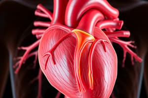
Coronary Blood Supply
- Coronary arteries supply blood to the heart.
- Small arteries penetrate deeper to supply the nutritive mass within the muscle.
Venous Blood
- Most venous blood from the left ventricle passes through the coronary sinus.
- Small anterior cardiac veins also drain venous blood.
- Venous blood is pumped into the right atrium of the heart.
Normal Coronary Blood Flow
- Resting coronary blood flow averages 225 ml/min.
- Increased blood flow occurs during exercise to meet increased nutrient demand.
Subendocardial Portion of the Left Ventricle
- Pressure in this area is slightly greater than the aorta during systole. Blood flow primarily occurs during diastole.
- Coronary blood flow in the subendocardium will not be drastically influenced by systole.
Studying That Suits You
Use AI to generate personalized quizzes and flashcards to suit your learning preferences.