Podcast
Questions and Answers
Which of the following best describes the primary mechanism by which the gastrointestinal tract moves food?
Which of the following best describes the primary mechanism by which the gastrointestinal tract moves food?
- Segmentation
- Diffusion
- Osmosis
- Peristalsis (correct)
The esophagus connects which two anatomical structures within the digestive system?
The esophagus connects which two anatomical structures within the digestive system?
- The mouth and the stomach
- The larynx and the trachea
- The pharynx and the small intestine
- The pharynx and the stomach (correct)
At what vertebral level does the esophagus typically pass through the diaphragmatic hiatus?
At what vertebral level does the esophagus typically pass through the diaphragmatic hiatus?
- T11 (correct)
- C6
- T6
- L1
What is the primary cause of Barrett’s esophagus?
What is the primary cause of Barrett’s esophagus?
Which cellular change characterizes Barrett's esophagus?
Which cellular change characterizes Barrett's esophagus?
Dysphagia, a common clinical feature of esophageal carcinoma, is best defined as:
Dysphagia, a common clinical feature of esophageal carcinoma, is best defined as:
A patient with a long-term burning sensation and indigestion is diagnosed with Barrett's esophagus. What is the most critical next step in managing this patient?
A patient with a long-term burning sensation and indigestion is diagnosed with Barrett's esophagus. What is the most critical next step in managing this patient?
A patient presents with progressive dysphagia and unintentional weight loss. Which condition should be of primary concern?
A patient presents with progressive dysphagia and unintentional weight loss. Which condition should be of primary concern?
A patient presents with haematemesis and is diagnosed with esophageal varices. Which of the following is the MOST likely underlying cause?
A patient presents with haematemesis and is diagnosed with esophageal varices. Which of the following is the MOST likely underlying cause?
Why are alcoholics at a particularly high risk of developing oesophageal varices?
Why are alcoholics at a particularly high risk of developing oesophageal varices?
The oesophagus begins at the level of the cricoid cartilage. At which vertebral level is this located?
The oesophagus begins at the level of the cricoid cartilage. At which vertebral level is this located?
Which part of the stomach surrounds the superior opening of the stomach at the T11 level?
Which part of the stomach surrounds the superior opening of the stomach at the T11 level?
Which of the following describes the location of the stomach in relation to other digestive organs?
Which of the following describes the location of the stomach in relation to other digestive organs?
A surgeon is performing a procedure involving the pylorus of the stomach. At which vertebral level would they expect to find the pyloric sphincter?
A surgeon is performing a procedure involving the pylorus of the stomach. At which vertebral level would they expect to find the pyloric sphincter?
Which of the following structures attaches to the lesser curvature of the stomach?
Which of the following structures attaches to the lesser curvature of the stomach?
During a surgical procedure on the lesser curvature of the stomach, which arterial supply would the surgeon need to be MOST aware of?
During a surgical procedure on the lesser curvature of the stomach, which arterial supply would the surgeon need to be MOST aware of?
A patient reports a sharp, precisely localized abdominal pain that worsens with movement. Which type of pain is the patient most likely experiencing?
A patient reports a sharp, precisely localized abdominal pain that worsens with movement. Which type of pain is the patient most likely experiencing?
A patient experiencing dull, poorly localized abdominal pain describes it as a constant ache. Which mechanism is least likely to be the cause of their pain?
A patient experiencing dull, poorly localized abdominal pain describes it as a constant ache. Which mechanism is least likely to be the cause of their pain?
What anatomical structure marks the transition from the ascending colon to the transverse colon?
What anatomical structure marks the transition from the ascending colon to the transverse colon?
Which scenario best describes the concept of referred pain?
Which scenario best describes the concept of referred pain?
Which of the following is a characteristic feature of the large intestine that distinguishes it from the small intestine?
Which of the following is a characteristic feature of the large intestine that distinguishes it from the small intestine?
A patient who underwent a colonoscopy complains about lower abdominal pain. The doctor suspects bowel distention during the procedure. Which type of pain is this patient most likely experiencing?
A patient who underwent a colonoscopy complains about lower abdominal pain. The doctor suspects bowel distention during the procedure. Which type of pain is this patient most likely experiencing?
The teniae coli are three strips of muscle that contract to shorten the wall of the bowel, producing sacculations. What are these sacculations called?
The teniae coli are three strips of muscle that contract to shorten the wall of the bowel, producing sacculations. What are these sacculations called?
A physician is evaluating a patient with hemorrhoids and recommends conservative management. What symptoms indicate this treatment approach is appropriate?
A physician is evaluating a patient with hemorrhoids and recommends conservative management. What symptoms indicate this treatment approach is appropriate?
The rectum begins at the level of which sacral vertebra as a continuation of the sigmoid colon?
The rectum begins at the level of which sacral vertebra as a continuation of the sigmoid colon?
Which part of the large intestine is approximately 40cm long and located in the left lower quadrant?
Which part of the large intestine is approximately 40cm long and located in the left lower quadrant?
What is the primary role of the internal and external anal sphincters?
What is the primary role of the internal and external anal sphincters?
Which of the following features is NOT characteristic of the rectum when distinguishing it macroscopically from the colon?
Which of the following features is NOT characteristic of the rectum when distinguishing it macroscopically from the colon?
What type of muscle primarily forms the internal anal sphincter, and is its function voluntary or involuntary?
What type of muscle primarily forms the internal anal sphincter, and is its function voluntary or involuntary?
What is the primary role of segmentation contractions in the small intestine?
What is the primary role of segmentation contractions in the small intestine?
Which of the following best describes the function of the hormone Ghrelin?
Which of the following best describes the function of the hormone Ghrelin?
A patient is experiencing difficulty digesting fats. Which of the following secretions is most likely deficient?
A patient is experiencing difficulty digesting fats. Which of the following secretions is most likely deficient?
What is the primary function of the colon?
What is the primary function of the colon?
Where does the majority of fat, protein, carbohydrates, sodium, and chloride absorption take place within the small intestine?
Where does the majority of fat, protein, carbohydrates, sodium, and chloride absorption take place within the small intestine?
A patient's stool sample is pale in color. This suggests a potential issue with which of the following?
A patient's stool sample is pale in color. This suggests a potential issue with which of the following?
Which of the following components of gastric juice is responsible for neutralizing the acidity of chyme as it enters the small intestine?
Which of the following components of gastric juice is responsible for neutralizing the acidity of chyme as it enters the small intestine?
Which of the following correctly lists the order in which chyme travels after leaving the stomach?
Which of the following correctly lists the order in which chyme travels after leaving the stomach?
Which of the following is a known factor that may alter the balance of an individual's gut microbiome?
Which of the following is a known factor that may alter the balance of an individual's gut microbiome?
What is the role of Peyer's patches in the small intestine?
What is the role of Peyer's patches in the small intestine?
A patient reports experiencing early satiety, bloating, and belching. Which category of conditions is MOST likely indicated by these symptoms?
A patient reports experiencing early satiety, bloating, and belching. Which category of conditions is MOST likely indicated by these symptoms?
A patient with GERD is complaining of dyspepsia. How does the presentation of dyspepsia typically change in older adults with GERD?
A patient with GERD is complaining of dyspepsia. How does the presentation of dyspepsia typically change in older adults with GERD?
During an abdominal assessment, a healthcare provider is using the 'IAPP' sequence. What does 'IAPP' stand for?
During an abdominal assessment, a healthcare provider is using the 'IAPP' sequence. What does 'IAPP' stand for?
When assessing a patient's oral cavity, what finding during the 'ah' test would indicate an intact vagus nerve (10th cranial nerve)?
When assessing a patient's oral cavity, what finding during the 'ah' test would indicate an intact vagus nerve (10th cranial nerve)?
A patient is experiencing excessive flatulence. Where do gases in the small intestine typically proceed before being expelled?
A patient is experiencing excessive flatulence. Where do gases in the small intestine typically proceed before being expelled?
A patient is diagnosed with a pilonidal cyst. This condition is characterized by:
A patient is diagnosed with a pilonidal cyst. This condition is characterized by:
When inspecting a patient's oral cavity, what instructions should be given to optimize visualization of the tonsils, uvula, and posterior pharynx?
When inspecting a patient's oral cavity, what instructions should be given to optimize visualization of the tonsils, uvula, and posterior pharynx?
Which of the following factors is LEAST relevant when taking a comprehensive patient history for gastrointestinal complaints?
Which of the following factors is LEAST relevant when taking a comprehensive patient history for gastrointestinal complaints?
Flashcards
Squamous Cell Carcinoma
Squamous Cell Carcinoma
Most common type of esophageal cancer.
Adenocarcinoma
Adenocarcinoma
Esophageal cancer type in the inferior third of the esophagus.
Esophageal Varices
Esophageal Varices
Abnormally dilated sub-mucosal veins in esophagus.
Portal Hypertension
Portal Hypertension
Signup and view all the flashcards
Hematemesis
Hematemesis
Signup and view all the flashcards
Stomach
Stomach
Signup and view all the flashcards
Cardia
Cardia
Signup and view all the flashcards
Fundus
Fundus
Signup and view all the flashcards
GI function disruptors?
GI function disruptors?
Signup and view all the flashcards
GI tract anatomy?
GI tract anatomy?
Signup and view all the flashcards
GI tract function?
GI tract function?
Signup and view all the flashcards
Esophagus?
Esophagus?
Signup and view all the flashcards
Esophagus location?
Esophagus location?
Signup and view all the flashcards
Esophagus and diaphragm?
Esophagus and diaphragm?
Signup and view all the flashcards
Barrett’s Esophagus?
Barrett’s Esophagus?
Signup and view all the flashcards
Esophageal carcinoma types?
Esophageal carcinoma types?
Signup and view all the flashcards
Haemorrhoids
Haemorrhoids
Signup and view all the flashcards
Somatic Abdominal Pain
Somatic Abdominal Pain
Signup and view all the flashcards
Visceral Abdominal Pain
Visceral Abdominal Pain
Signup and view all the flashcards
Referred Pain
Referred Pain
Signup and view all the flashcards
Ptyalin (Salivary Amylase)
Ptyalin (Salivary Amylase)
Signup and view all the flashcards
Right Colic Flexure
Right Colic Flexure
Signup and view all the flashcards
Transverse Colon
Transverse Colon
Signup and view all the flashcards
Transverse Mesocolon
Transverse Mesocolon
Signup and view all the flashcards
Sigmoid Colon
Sigmoid Colon
Signup and view all the flashcards
Rectum
Rectum
Signup and view all the flashcards
Omental Appendices
Omental Appendices
Signup and view all the flashcards
Teniae Coli
Teniae Coli
Signup and view all the flashcards
Anal Sphincters
Anal Sphincters
Signup and view all the flashcards
Ghrelin
Ghrelin
Signup and view all the flashcards
Leptin
Leptin
Signup and view all the flashcards
Chyme
Chyme
Signup and view all the flashcards
Pancreatic Secretions (Alkaline)
Pancreatic Secretions (Alkaline)
Signup and view all the flashcards
Bile
Bile
Signup and view all the flashcards
Sphincter of Oddi
Sphincter of Oddi
Signup and view all the flashcards
Villi
Villi
Signup and view all the flashcards
Main function of the colon
Main function of the colon
Signup and view all the flashcards
Feces Composition
Feces Composition
Signup and view all the flashcards
Dyspepsia
Dyspepsia
Signup and view all the flashcards
Dyspepsia Symptoms
Dyspepsia Symptoms
Signup and view all the flashcards
Belching
Belching
Signup and view all the flashcards
Flatulence
Flatulence
Signup and view all the flashcards
Pilonidal Disease
Pilonidal Disease
Signup and view all the flashcards
IAPP Sequence
IAPP Sequence
Signup and view all the flashcards
Oral Inspection Criteria
Oral Inspection Criteria
Signup and view all the flashcards
Tongue Depression Technique
Tongue Depression Technique
Signup and view all the flashcards
Normal Uvula Movement
Normal Uvula Movement
Signup and view all the flashcards
Study Notes
Assessment of Digestive and Gastrointestinal Function
- Bleeding, perforation, obstruction, inflammation, and cancer indicate digestive and gastrointestinal dysfunction.
- The GI tract is susceptible to many organic diseases.
- Extrinsic factors such as stress, anxiety, fatigue, and sudden diet changes can affect digestive and gastrointestinal function.
Anatomic and Physiologic Overview
- The GI tract is a long, folded tube from the mouth to the anus.
- The GI tract includes the mouth, esophagus, stomach, small intestine, cecum, colon (large intestine), rectum, and anal canal.
- Food moves through the tract via peristalsis for digestion and absorption.
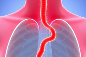
Esophagus
- It is a fibromuscular tube, approximately 25cm in length, that transports food from the pharynx to the stomach.
- It originates at the inferior border of the cricoid cartilage (C6) and extends to the cardiac orifice of the stomach (T11).
- Its passes through the diaphragm at an opening called diaphragmatic hiatus.
Important Sphincters in Digestion
- The Upper Esophageal Sphincter (UES) is an anatomical sphincter at the top of the esophagus that connects to the pharynx.
- The UES controls the entry of food and prevents air from entering; it is made of skeletal muscle (cricopharyngeus and pharyngeal constrictor muscles).
- The Lower Esophageal Sphincter (LES) is a functional sphincter comprised of smooth muscle that connects to the stomach.
- The LES prevents backflow of stomach contents and maintains a high-pressure zone.
Barrett's Esophagus
- Metaplasia involves the change of lower esophageal squamous epithelium to gastric columnar epithelium.
- The cause is chronic acid exposure from a malfunctioning lower esophageal sphincter.
- Acid irritates the esophageal epithelium, leading to a metaplastic change.
- A common symptom is a long-term burning sensation of indigestion.
- It can be detected via endoscopy and monitored for cancerous changes.
Esophageal Carcinoma
- Around 2% of malignancies in the UK are esophageal carcinomas.
- Clinical features include dysphagia, which worsens over time as the tumor grows, and weight loss.
- Two major types: squamous cell carcinoma and adenocarcinoma.
- Squamous cell carcinoma is the most common.
- Adenocarcinoma occurs in the inferior third of the esophagus and is associated with Barrett's esophagus.
Esophageal Varices
- Abnormally dilated sub-mucosal veins occur in this anastomosis.
- These are caused by an increased pressure in the portal system (portal hypertension).
- Portal hypertension often results from chronic liver disease (e.g., cirrhosis) or portal vein obstruction.
- Esophageal Varices are prone to bleeding, and patients often present with haematemesis [vomiting blood].
- Alcoholics are particularly at risk of developing esophageal varices.
Esophagus Location
- The esophagus begins at the level of the cricoid cartilage at vertebral level C6.
- The esophagus opens into the cardia of the stomach.
Stomach Divisions
- The stomach, an intraperitoneal digestive organ, sits between the esophagus and the duodenum.
- The cardia surrounds the superior opening of the stomach at the T11 level.
- The Fundus is the rounded, often gas-filled portion superior to and left of the cardia.
- The Body is the large, central portion inferior to the fundus.
- The Pylorus connects the stomach to the duodenum and is divided into the pyloric antrum, pyloric canal, and pyloric sphincter.
- The pyloric sphincter demarcates the transpyloric plane at the level of L1.
Stomach Curvatures and Attachments
- The greater curvature forms the long, convex, lateral border of the stomach.
- The lesser curvature forms the shorter, concave, medial surface of the stomach.
- The hepatogastric ligament attaches to the lesser curvature and is supplied by the left gastric artery and the right gastric branch of the hepatic artery.
Stomach Sphincters
- The pyloric sphincter is between the pylorus and the duodenum.
- This sphincter controls the exit of chyme from the stomach.
- It is an anatomical sphincter with smooth muscle that constricts to regulate stomach content discharge.
Stomach Peritoneum and Omenta
- The peritoneum is a double-layered membrane within the abdominal cavity.
- The peritoneum supports most abdominal viscera and assists with attachment to the abdominal wall.
- Omenta are structures consisting of peritoneum folded over itself (four membrane layers in total).
- Both the greater and lesser omenta attach to the stomach and important anatomical landmarks.
Gastro-Esophageal Reflux Disease (GERD)
- GERD is a digestive disorder affecting the lower esophageal sphincter, leading to the movement of gastric acid and food into the esophagus.
- GERD affects approximately 5-7% of the population and includes symptoms such as dyspepsia, dysphagia, and a sour taste in the mouth.
- The three main causes of reflux disease are dysfunction of the lower esophageal sphincter, delayed gastric emptying, and hiatal hernia.
- Treatment includes lifestyle changes/medication such as a PPI to reduce stomach acid, and as a last resort, surgery.
Hiatal Hernia
- A hiatal hernia occurs when part of the stomach protrudes into the chest through the esophageal hiatus in the diaphragm
Small Intestine
- The Small Intestine is a 6.5m long organ in the gastrointestinal tract; aiding in digestion and absorption.
- It extends from the stomach's pylorus to the ileocecal valve, meeting the large intestine.
- Three parts: duodenum, jejunum, and ileum.
- All major digestive events and most of the absorption takes place in the small intestine
Duodenum
- The duodenum is the most proximal part of the small intestine, forming a 'C' shape around the pancreas.
- It has four parts: superior, descending, transverse and ascending
- Only the beginning of the first part is an intraperitoneal region (connected to the liver by the hepatoduodenal ligament)
Jejunum and Ileum
- They are intraperitoneal and cannot be clearly distinguished from one another.
- The Jejunum is long and leaf-like with villi, more plicae circulares, and an intermediate number of goblet cells and lacks peyer's patches.
- The Ileum has fewer shorter villi.
- The Ileum is abundant, occupies the RLQ and appears pale pink and has is longest in lengh.
Cecum
- It is the most proximal part of the large intestine, located in the right iliac fossa of the abdomen.
- It lies inferiorly to the ileocecal junction.
- It can be palpated if enlarged due to feces, inflammation, or malignancy.
- The name 'Cecum' originates from the Latin word 'caecus', which means 'blind' (referring to its inferior bind-end)
Appendix
- Is a narrow, blind-ended tube attached to the posteromedial end of the cecum (large intestine).
- It contains a large amount of lymphoid tissue but is thought to have no vital functions.
Appendicitis
- Inflammation of the appendix causes acute severe abdominal pain.
- Maximum tenderness occurs at McBurney's point (located one-third of the way from the right anterior superior iliac spine to the umbilicus).
- In young individuals, it is commonly caused by an increase in lymphoid tissue size, occluding the lumen.
- In adults over 30 years, it’s more likely to be caused by a faecolith blockage.
- Pain starts in the umbilical region and then localizes to the right lower quadrant as inflammation increases.
- If untreated, the appendix can necrotize and rupture, leading to peritonitis (inflammation of the peritoneum).
Colon
- It is the distal part of the gastrointestinal tract that receives digested food from the small intestine.
- It's main functions involves absorption of water and electrolytes to form feces.
- The colon averages 150 cm in length.
- The Colon has 4 parts: Ascending, transverse, descending, and sigmoid.
- The colon forms an arch around the small intestine.
Ascending Colon
- The ascending colon is a retroperitoneal structure & begins as the colon.
- It ascends superiorly from the cecum.
- The colon turns 90 degrees horizontally when meeting the right lobe of the liver, and this is known as the right colic flexure (or hepatic flexure).
Transverse Colon
- The transverse colon extends from the right colic flexure to the spleen.
- Other factors for the transverse colon are; it’s left colic flexure (or splenic flexure), phrenicocolic ligament and transverse mesocolon
Descending Colon
- Descending colon has features and is located and is located inferiorly towards the pelvis
Sigmoid Colon
- A 40cm-long sigmoid colon is located in the left lower quadrant
- It is located in an "S" shape and uses sigmoid mesocolon
Rectum
- The rectum is the most distal segment of the large intestine.
- It has an important role as a temporary store of feces
- The rectum begins at the level of the S3 (as a continuation of the sigmoid colon)
- The rectum is macroscopically distinct from the colon with an absence of tenia coli, haustra, and omental appendices.
Characteristic Features of large intestine
- The large intestine has a number of characteristic features that distinguishes it from the small intestine.
- Attached to the surface of the large intestine are omental appendices which - small pouches of peritoneum, filled with fat.
- Running longitudinally along the surface of the large bowel are three strips of muscle; known as the teniae coli; they are called the mesocolic, free, and omental coli.
- The teniae coli contract to shorten the wall of the bowel, producing sacculations known as haustra.
- The large intestine has a much wider diameter compared to the small intestine
Anal Canal
- The anal canal is the final segment of the gastrointestinal tract.
- The anal canal plays an important role in defecation and maintaining faecal continence.
- Except during defecation, the anal canal is collapsed by the internal and external anal sphincters to prevent passage of faecal material.
Anal Sphincters
- The anal canal is surrounded by internal and external anal sphincters.
- The internal anal sphincter surrounds the upper 2/3 of the anal canal and is formed from a thickening of the involuntary circular smooth muscle in the bowel wall.
- The external anal sphincter is a voluntary muscle that surrounds the lower 2/3 of the anal canal (and so overlaps with the internal sphincter).
- The external anal sphincter blends superiorly with the puborectalis muscle of the pelvic floor.
Haemorrhoids
- They are vascular cushions found within the anal canal of healthy individuals; which help with the maintenance of faecal continence .
- They can cause bleeding and itchiness, and can be managed conservatively or surgically depending on the severity.
Abdominal Pain
- Abdominal pain can be broadly classified into three groups: somatic, visceral and referred.
- Somatic pain can arise from the skin, fascia, muscles and parietal peritoneum; lateralizing if the origin is on one side and is severe /precisely localised.
- Visceral pain arises in abdominal organs, visceral peritoneum and mesenteries; it's dull and poorly localized (colic) but the stretching of a viscus or mesentery, distention of a hollow viscus, impaired blood supply (ischemia) to a viscus, and chemical damage can act as causes
- Referred pain is a feeling of pain at a location other than the site of origin of the stimulus, however are supplied by the same/adjacent segments of the spinal cord.
Portacaval Circulation
- Anastomosis between "PORTAL VEIN" and "SYSTEMIC BLOOD"
- the "PORTAL VEIN" supplies bold from GUT liver then the "Systemic Venous system'
- It is able to perform "detoxification" , "Nutrient storage and/ release"
Portal Hypertension
- An increase in pressure in the portal vein due to liver diseases (ascities)
- Portacaval Shunt is caused as esophageal varices , Caput Medusae , Hemorrhoids.
- It bypasses liver , drains to systemic circulation (alternative direction)
Function of the Digestive System
- Digestion processes food to break it down.
- Absorption: is used mainly for nutrient
- Elimination happens after nutrients are extracted.
Saliva Function
- Reflex salivation is triggered through eating, sight, smell, or taste of food.
- Daily saliva production is approximately 1.5 liters of saliva secreted daily.
- Important salivary glands - parotid, submaxillary, and sublingual glands - contribute to secretion.
- Ptyalin (salivary amylase) is an enzyme that begins starch digestion.
- Water and Mucus lubricate food, facilitating chewing and swallowing.
SWALLOWING Reflex
- Swallowing is a voluntary act regulated by medulla
- The epiglottis covers the tracheal opening > peristalsis propels the bolus of food > LES Relaxes> Food enters Stomach> ES closes",
Gastric Function Cells involved in digestion
- Parietal Cells produces "HCL acid " , "intrinsic factor" . They are eosinophilic + ditt mitochondria to produce H+ to power H+ pump(
- Chief cells secrete the enzyme pepsinogen, are basophilic and are largely found body + fundus
- Mucous cells mucus as protective layer on stomach lining
- G cells - Produce GASTRIN (goes to > parietal cells (HCL) + chief cells (pepsinogen).
- ENTEROENDOCRINE CELLS - "SOMATOSTATIN""
Stomach Hormones
- Ghrelin (hunger hormone) regulates appetite, energy balance, and fat storage.
- It aids in stimulating a patient’s release of growth hormone, promotes calorie intake and reduced energy expenditure + hormone produced in Stomach" "Small Intestine" " pancreas" " brain"
- Leptin contributes to the signaling satiety and inhibiting hunger,
Gastric Actions
Peristalsis moves food from pylorus + body, this is mainly due to the pyloric sphincter (mechanical reaction
- It takes 30minutes- several hours before Chyme (partially digested foods) can mix with gastric secretions
- Hormones/ Regulators controls secretions & mobility (speed)
Small Intestine Function
- Duodenum mainly continues + finishes the digestive process
- Enzymes included here are amylase , lipase (from bile duct and "trpsin"
- Digestive secretions enter through pancreatic duct - Common bile duct + enters ampulla van walter (bile acids)
Secretin
- Secreted by s cells in duodenum",Stimulus needed, mainly "PH<",Stimulate increase output HCL fluid", bile and pancreatic enzymes"
Contractions
- 2 Types of intestinal contraction are "Segmentation" " Intestinal Perisatslis" these cause food to stay between 3-6 hours
- Villi "fingerlike" absorb nutrients unchanged, main absorber in Jejunum
Types of Small Intestine cells
- Enterocytes - they line the surface and has microvilli", increase surface"
- Goblet - secretes lubricant and protection (mucus)
- Paneth
- Enteroendocrine
Colonic Functions
- Peristalsis pushes residue (terminal Ileum > cecum after 4 hrs with food
- Gut / Microboes" can breakdown waste and make it smaller
Colic Secretions
- Bicarbonate : counter actions of bacteria
- Mucus protects the colic mucosa and helps fecal mass adherence
- The Slow and weak peristalsis (absorbs water + electrolytes)
- Intermittent and strong (push contents)
- Transit time is 12 hrs as rectum gets expanded.
Waste Products of Digestion
- Undigested foods ,Inorganic material, water + Bacteria
- 75%) Fluid / 25%) solid
- "Bile > bacteria (Sterc obilum
- Odour chemical " Methane " hydrogen sulfide and ammonia "Gas"(150mL within GI")
- flatus
Gut/ micro biome: in digestion and absorption
Colonic Functions
- Colonization short period + starts shortly after birth and is 2yrs / assistin waste ,Vit synthesis"
- Genetics [ "bacteroides"] or Diet [Lactobacillus, Bifidobacterium, Bacteroides"]
- Protection : against pathogens , increase anti inflammatory , prevents pathogens.
Clinical Tests
- Gag Reflex Test: stimulate the back of neck , this checks for symmetry and checks "vagus nerve" function
- Uvula Movement , This checks for nerves when AHHH Swallowing , check muscles that are used with swallowing"
Abdominal Inspection, Auscultation, Percussion, and Palpation
- Inspection:
- Note skin changes, nodules, lesions, scarring, discolorations, inflammation, bruising, or striae.
- Lesion (diseases and changes. Observe contour and symmetry of the abdomen.
- Identify localized bulging, distention, or peristaltic waves
Abdominal contour/structure: for check up.
- structure of abdomen and see if it feels flat, rounded, or scaphoid
-
Auscultation Before Percussion and Palpation:
- the "Reason",is to see how the" bowel sounds are"
- sounds "Clicks " 5-30 times per minute / hypoactive
- Diaphragm = normal
- bell (Bruits )
Studying That Suits You
Use AI to generate personalized quizzes and flashcards to suit your learning preferences.