Podcast
Questions and Answers
During an equine neurologic exam, which of the following is the MOST likely reason for assessing mentation?
During an equine neurologic exam, which of the following is the MOST likely reason for assessing mentation?
- To assess the horse's ability to respond to touch and proprioceptive stimuli.
- To determine the presence of gait abnormalities such as ataxia or spasticity.
- To identify potential cortical dysfunction or altered brain activity that may indicate neurological compromise. (correct)
- To evaluate the function of the peripheral nervous system's response to pain stimuli.
When evaluating a horse with suspected Horner's syndrome, which combination of clinical signs would MOST strongly suggest an interruption of sympathetic input?
When evaluating a horse with suspected Horner's syndrome, which combination of clinical signs would MOST strongly suggest an interruption of sympathetic input?
- Mydriasis, anhidrosis, and ptosis.
- Mydriasis, hyperhidrosis, and protrusion of the third eyelid.
- Miosis, anhidrosis, and enophthalmos. (correct)
- Ptosis, hyperhidrosis, and exophthalmos.
When performing a neurological examination on a horse, how does evaluating cranial nerve function contribute to lesion localization?
When performing a neurological examination on a horse, how does evaluating cranial nerve function contribute to lesion localization?
- Cranial nerve deficits always suggest multifocal lesions involving both the brainstem and spinal cord simultaneously.
- Cranial nerve function is not useful for lesion localization, as cranial nerves are only affected in diffuse diseases.
- Isolated cranial nerve deficits assist in localizing lesions in the brainstem or along the specific nerve pathway, distinct from more generalized central or peripheral lesions. (correct)
- Cranial nerve deficits can only indicate the presence of a central lesion within the cerebral cortex.
A horse exhibits jerky, bobbing head movements that worsen when it attempts to eat. This presentation is MOST indicative of which condition?
A horse exhibits jerky, bobbing head movements that worsen when it attempts to eat. This presentation is MOST indicative of which condition?
During a neurological exam, diminished muscle tone and atrophy of the tongue are detected. Which cranial nerve is MOST likely affected?
During a neurological exam, diminished muscle tone and atrophy of the tongue are detected. Which cranial nerve is MOST likely affected?
A horse shows feed particles in its nasal discharge and clinical signs of dysphagia following guttural pouch disease. What is the MOST likely explanation?
A horse shows feed particles in its nasal discharge and clinical signs of dysphagia following guttural pouch disease. What is the MOST likely explanation?
What is the MOST critical initial step in performing an equine neurologic examination?
What is the MOST critical initial step in performing an equine neurologic examination?
When evaluating a horse with suspected cerebellar disease, what gait characteristic is MOST likely to be observed?
When evaluating a horse with suspected cerebellar disease, what gait characteristic is MOST likely to be observed?
During a neurological examination of a horse, you observe a ventrolateral ocular position that is unopposed by cranial nerve VI. This finding indicates a lesion of which cranial nerve?
During a neurological examination of a horse, you observe a ventrolateral ocular position that is unopposed by cranial nerve VI. This finding indicates a lesion of which cranial nerve?
If a horse exhibits a consistently delayed or absent palpebral response, which combination of cranial nerves should be evaluated to determine the lesion's location?
If a horse exhibits a consistently delayed or absent palpebral response, which combination of cranial nerves should be evaluated to determine the lesion's location?
During evaluation of the pupillary light reflex (PLR) in a horse, which nerves are assessed and what are their respective roles?
During evaluation of the pupillary light reflex (PLR) in a horse, which nerves are assessed and what are their respective roles?
Following a head trauma, a horse exhibits asymmetry of the muscles of mastication with marked atrophy of the right temporalis muscle. Which nerve is MOST likely affected?
Following a head trauma, a horse exhibits asymmetry of the muscles of mastication with marked atrophy of the right temporalis muscle. Which nerve is MOST likely affected?
When performing a gait analysis on a horse, what does ataxia indicate, and how should it influence your diagnostic approach?
When performing a gait analysis on a horse, what does ataxia indicate, and how should it influence your diagnostic approach?
How does the assessment of a horse's gait contribute to distinguishing between vestibular and cerebellar etiologies causing ataxia?
How does the assessment of a horse's gait contribute to distinguishing between vestibular and cerebellar etiologies causing ataxia?
After observing a horse exhibit altered mentation, seizures, and head pressing, where is the MOST likely location of the lesion?
After observing a horse exhibit altered mentation, seizures, and head pressing, where is the MOST likely location of the lesion?
When evaluating a horse with suspected peripheral vestibular disease, which clinical sign would MOST likely be observed, and how does it relate to the lesion?
When evaluating a horse with suspected peripheral vestibular disease, which clinical sign would MOST likely be observed, and how does it relate to the lesion?
During a neurological examination, a horse exhibits a grade 4/5 spinal ataxia. How would you interpret this grading, and what clinical signs would you expect to observe?
During a neurological examination, a horse exhibits a grade 4/5 spinal ataxia. How would you interpret this grading, and what clinical signs would you expect to observe?
Which of the following is a critical consideration when assessing the menace response in a horse during a cranial nerve examination?
Which of the following is a critical consideration when assessing the menace response in a horse during a cranial nerve examination?
When performing a neurological exam, which of the following is the MOST important reason for including gait analysis?
When performing a neurological exam, which of the following is the MOST important reason for including gait analysis?
Which statement MOST accurately describes why diagnostic testing is vital after lesion localization in an equine neurologic examination?
Which statement MOST accurately describes why diagnostic testing is vital after lesion localization in an equine neurologic examination?
A practitioner notes that a horse has diminished sensation to its face and presents with difficulty chewing. Which cranial nerve should the practitioner examine more closely?
A practitioner notes that a horse has diminished sensation to its face and presents with difficulty chewing. Which cranial nerve should the practitioner examine more closely?
After examination of the fundus, a veterinarian determines that a horse is blind. Which cranial nerve is most likely affected?
After examination of the fundus, a veterinarian determines that a horse is blind. Which cranial nerve is most likely affected?
What is the BEST way to challenge a horse's coordination and proprioception during a neurological examination?
What is the BEST way to challenge a horse's coordination and proprioception during a neurological examination?
Cranial nerve VIII is affected in horse that has a head tilt to the right and pulls to the left when prompted to walk. Where is the lesion located?
Cranial nerve VIII is affected in horse that has a head tilt to the right and pulls to the left when prompted to walk. Where is the lesion located?
A horse is unable to retract its tongue. What cranial nerve is affected?
A horse is unable to retract its tongue. What cranial nerve is affected?
What are the afferent and efferent components of the corneal reflex, respectively?
What are the afferent and efferent components of the corneal reflex, respectively?
Which of the following is the BEST way to assess cranial nerve I in horses?
Which of the following is the BEST way to assess cranial nerve I in horses?
What cranial nerves should be assessed when looking at the corneal and palpebral reflexes, respectively?
What cranial nerves should be assessed when looking at the corneal and palpebral reflexes, respectively?
Which location should be suspected when a horse is showing gait deficits, altered consciousness, and cranial nerve deficits?
Which location should be suspected when a horse is showing gait deficits, altered consciousness, and cranial nerve deficits?
Which term BEST describes the lack of coordination of motor movements?
Which term BEST describes the lack of coordination of motor movements?
How do you BEST describe the gait of a horse graded with a 2 on the gait deficit scale?
How do you BEST describe the gait of a horse graded with a 2 on the gait deficit scale?
Upon evaluation of the cranial nerves, a right-sided facial nerve disease is detected. Which of the following correctly describes this affliction?
Upon evaluation of the cranial nerves, a right-sided facial nerve disease is detected. Which of the following correctly describes this affliction?
Which cranial nerve(s) are involved in prehension, chewing, and swallowing?
Which cranial nerve(s) are involved in prehension, chewing, and swallowing?
After performing a complete neurologic exam, you suspect that the patient has equine protozoal myeloencephalitis (EPM). Where do you suspect the lesion originates? Choose the BEST answer.
After performing a complete neurologic exam, you suspect that the patient has equine protozoal myeloencephalitis (EPM). Where do you suspect the lesion originates? Choose the BEST answer.
During an equine neurologic examination, if a horse exhibits signs of blindness and an absent menace response, but has normal pupillary light reflexes (PLRs), which is the MOST likely location of the lesion?
During an equine neurologic examination, if a horse exhibits signs of blindness and an absent menace response, but has normal pupillary light reflexes (PLRs), which is the MOST likely location of the lesion?
A horse presents with muscle atrophy along its face, particularly the masseter muscle. Which cranial nerve is MOST likely affected in this case?
A horse presents with muscle atrophy along its face, particularly the masseter muscle. Which cranial nerve is MOST likely affected in this case?
What clinical signs would strongly indicate a possible infection or lesion of the vestibulocochlear nerve (CN VIII) in a horse?
What clinical signs would strongly indicate a possible infection or lesion of the vestibulocochlear nerve (CN VIII) in a horse?
During a neurological examination, a horse demonstrates normal strength and proprioception but exhibits exaggerated, floating movements of the limbs, especially during ambulation. Which of the following lesion localizations is MOST consistent with these gait abnormalities?
During a neurological examination, a horse demonstrates normal strength and proprioception but exhibits exaggerated, floating movements of the limbs, especially during ambulation. Which of the following lesion localizations is MOST consistent with these gait abnormalities?
A horse presents with dysphagia, feed material in its nasal passages, and a head tilt. Given these clinical signs, which combination of cranial nerve deficits and corresponding lesion location is MOST likely?
A horse presents with dysphagia, feed material in its nasal passages, and a head tilt. Given these clinical signs, which combination of cranial nerve deficits and corresponding lesion location is MOST likely?
A horse exhibits signs of altered mentation, aimless wandering, and compulsive circling. Assuming a singular lesion is responsible, where is the MOST likely location?
A horse exhibits signs of altered mentation, aimless wandering, and compulsive circling. Assuming a singular lesion is responsible, where is the MOST likely location?
When evaluating a horse for cranial nerve deficits, a veterinarian observes that the horse has a weakened ability to prehend food, decreased sensation around the nose, and atrophy of the masseter muscle. Which of the following diagnostic steps would BEST help confirm the location and nature of the underlying neurological issue?
When evaluating a horse for cranial nerve deficits, a veterinarian observes that the horse has a weakened ability to prehend food, decreased sensation around the nose, and atrophy of the masseter muscle. Which of the following diagnostic steps would BEST help confirm the location and nature of the underlying neurological issue?
During a neurological examination, a horse displays a menace response in the right eye but not in the left. However, pupillary light reflexes are normal in both eyes. Where is the MOST likely location of the lesion causing these specific deficits?
During a neurological examination, a horse displays a menace response in the right eye but not in the left. However, pupillary light reflexes are normal in both eyes. Where is the MOST likely location of the lesion causing these specific deficits?
Flashcards
Goals of Equine Neurologic Exam?
Goals of Equine Neurologic Exam?
Determine neurological normality, lesion location, differential diagnosis, and treatment plan.
What are the key components?
What are the key components?
It involves assessing mentation, cranial nerves, limb responses, reflexes, and gait.
What does altered mentation indicate?
What does altered mentation indicate?
It can indicate forebrain or cortical dysfunction.
What is the role of the cerebellum?
What is the role of the cerebellum?
Signup and view all the flashcards
List Cranial Nerve Functions
List Cranial Nerve Functions
Signup and view all the flashcards
What does CN I - Olfactory control?
What does CN I - Olfactory control?
Signup and view all the flashcards
What does CN II - Optic control?
What does CN II - Optic control?
Signup and view all the flashcards
What actions help assess function?
What actions help assess function?
Signup and view all the flashcards
What nerves facilitate Palpebral Response?
What nerves facilitate Palpebral Response?
Signup and view all the flashcards
What nerves facilitate Pupillary Light Reflex?
What nerves facilitate Pupillary Light Reflex?
Signup and view all the flashcards
Ocular position nerve lesions
Ocular position nerve lesions
Signup and view all the flashcards
What are the clinical signs of Equine Horner's?
What are the clinical signs of Equine Horner's?
Signup and view all the flashcards
CN V's Role
CN V's Role
Signup and view all the flashcards
CN VII & VIII Role
CN VII & VIII Role
Signup and view all the flashcards
Fast phase nystagmus shows?
Fast phase nystagmus shows?
Signup and view all the flashcards
Guttural pouch issues affect?
Guttural pouch issues affect?
Signup and view all the flashcards
Checking CN XII Function
Checking CN XII Function
Signup and view all the flashcards
What is Ataxia?
What is Ataxia?
Signup and view all the flashcards
Assessing gait involves?
Assessing gait involves?
Signup and view all the flashcards
Gait deficits are graded?
Gait deficits are graded?
Signup and view all the flashcards
Equine Neuro Exam Steps
Equine Neuro Exam Steps
Signup and view all the flashcards
Study Notes
- A complete equine neurologic examination is key for evaluating horses with suspected neurologic disease.
- Examinations should be systematic and complete.
- Lesion localization helps determine likely differentials.
- Diagnostic testing helps confirm diagnoses and establish prognoses.
Learning Outcomes
- Students should be able to describe components of a complete equine neurologic examination.
- Students should be able to describe methods for performing components of the exam.
- The components of the exam include mentation, cranial nerve assessment, panniculus reflex and hoof placement, and basic gait analysis.
- Students should be able to interpret findings and distinguish normal from abnormal responses.
Goals of a Neurologic Examination
- The goals of a neurologic examinclude determining whether the patient is neurologically normal or abnormal.
- It is important to know what is normal versus abnormal.
- Lesion localization is important and can be central, peripheral, focal, diffuse, or multifocal.
- Differential diagnosis, diagnostic protocols, and treatment for treatable conditions are also goals.
Examination Components
- Key examination components include mentation, cranial nerve examination, limb and placing response, tail, sensory/panniculus reflexes, and gait analysis.
- Determining neurological deficits will be based on the exam.
- It is important to know what is normal and what is lame.
Central Lesions
- Central lesions affect the cerebral cortex.
Equine Neurologic Examination Specifics
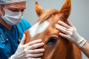
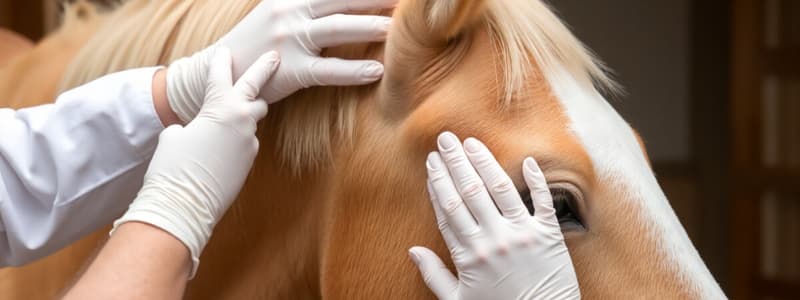
- Assess the mentation of the horse to determine if it is normal or abnormal.
- Abnormal mentation can manifest as changes in behavior, seizures, and altered mentation.
- Evaluation of cranial nerves to determine normals
- Assess foot placement to check if the horse "knows" where its feet are positioned.
- Sensory/proprioceptive deficits indicate abnormal foot placement.
- Assess gait to analyze normality
- Ataxia, spasticity, and weakness all indicate abnormal gait.
Cortical Dysfunction
- Altered mentation, seizures, head pressing, and blindness are all indicators of cortical dysfunction.
Head Pressing
- Head pressing indicates altered mentation.
Cerebellum
- The cerebellum and cranial nerve VIII work together to control smooth head movements.
- Cerebellar disease can cause jerky, bobbing head movements that worsen when the horse eats.
- Jerky head movements because of cerebellar disease are referred to as intention tremors.
Brainstem Dysfunction
- Cranial nerve deficits, altered consciousness or gait deficits are all signs related to brainstem dysfunction.
Cranial Nerves
- Cranial nerve I (Olfactory) handles olfaction and appetite.
- Cranial nerve II (Optic) controls vision and menace.
- Cranial nerve III (Oculomotor) manages ocular position and pupillary size.
- Cranial nerve IV (Trochlear) manages ocular position and strabismus.
- Cranial nerve V (Trigeminal) handles facial sensation and mastication.
- Cranial nerve VI (Abducent) controls ocular position and strabismus.
- Cranial nerve VII (Facial) handles facial expression and PS lacrimation.
- Cranial nerve VIII (Vestibulocochlear) manages balance and auditory control.
- Cranial nerve IX (Glossopharyngeal) is responsible for swallowing.
- Cranial nerve X (Vagus) manages laryngeal function and swallowing.
- Cranial nerve XI (Spinal Accessory) controls motor muscles of the neck and withers.
- Cranial nerve XII (Hypoglossal) manages movement of the tongue.
Olfactory Nerve (CN I)
- Horses need the olfactory nerve to smell, which is important for proper eating.
CN I, II, V, VII, IX, X, XII
- Prehension, chewing, looking, moving ears, and swallowing all help determine/establish cranial nerve function and identify abnormalities.
Menace Response
- Cranial nerves II and VII are evaluated during the Menace Response.
- Cranial nerves V and VII are evaluated when performing the Palpebral response.
- Corneal responses evaluate cranial nerves V and VI.
Palpebral Response
- The afferent nerve is the Trigeminal n. (CN V).
- The efferent nerve is the Facial n. (CN VII).
Pupillary Light Reflex
- Afferent nerve is the Optic n. (CN II).
- Efferent nerve is the Oculomotor n. (CN III).
Ocular Position & Strabismus
- A lesion in CN 3 results in ventrolateral position unopposed by VI.
- CN 3 innervates several components including dorsal, ventral and medial recti, ventral oblique and levator palpebre and PS pupil = constriction.
- A lesion in CN 4 means the dorsal deviation of the medial angle
- CN 4 innervates the dorsal oblique
- A lesion in CN 6 results in medial deviation.
- CN 6 innervates the lateral rectus and retractor bulbi
Equine Horner's Syndrome
- Interruption of sympathetic input results in ptosis, miosis, enophthalmos, and retraction of the 3rd eyelid.
- Sweating of the face and neck may be noted cranially to T3.
Trigeminal Nerve (CN V)
- Facial sensation, mastication
- Disease with atrophy of muscles
CN VII & VIII
- Guttural pouch disease can affect the facial and vestibulocochlearnerves.
Mandibular Fractures
- Stylohyoid fractures can happen
Vestibular Nerve (CN VIII)
- Disease of CN VIII peripheral vestibular causes fast phase away from the side with the lesion
Guttural Pouch Disease
- Guttural pouch disease involving branches of IX and X can cause clinical signs of dysphagia.
- An example of damage caused by guttural pouch disease is guttural pouch mycosis.
- Feed particles in the nasal discharge from lack of swallowing.
- Mycosis commonly damages cranial nerves IX and X.
- Fungal plaque in the guttural pouch causes marked inflammation and hemorrhage secondary to the infection.
Hypoglossal Nerve (CN XII)
- Test tone/retraction to examine the hypoglossal nerve
Gait Analysis
- Gait analysis includes walking and trotting the animal.
- Specific tasks can challenge the animal, like obstacles, hills, backing, circles, and head elevation.
- Ataxia is not a diagnosis, but rather a description of clinical signs.
- Ataxia frequently associates to weakness due to lack of coordination of motor movements.
- Vestibular ataxia is head tilt and asymmetric ataxia.
- Cerebellar ataxia shows up in foals with cerebellar abiotrophy.
- Cerebellar ataxia shows symmetric ataxia with intention tremors and retention of strength.
- Sensory ataxia is most common caused by interruption of ascending input to the cerebellum.
Grading Gait Deficits
- Grade 0 signifies no deficits detected.
- Grade 1 signifies mild deficits.
- Grade 2 signifies easily detected deficits.
- Grade 3 signifies prominent deficits.
- Grade 4 signifies marked deficits.
- Grade 5 signifies an inability to rise.
Studying That Suits You
Use AI to generate personalized quizzes and flashcards to suit your learning preferences.