Podcast
Questions and Answers
In a horse exhibiting relative erythrocytosis due to dehydration, what physiological mechanism contributes most significantly to the increased red blood cell concentration?
In a horse exhibiting relative erythrocytosis due to dehydration, what physiological mechanism contributes most significantly to the increased red blood cell concentration?
- Sequestration of plasma volume, leading to hemoconcentration. (correct)
- Decreased destruction of red blood cells in the spleen.
- Stimulation of the bone marrow to produce more red blood cells.
- Increased erythropoietin production by the kidneys.
How does the presence of Howell-Jolly bodies in equine erythrocytes relate to splenic function and erythropoiesis?
How does the presence of Howell-Jolly bodies in equine erythrocytes relate to splenic function and erythropoiesis?
- Is a typical finding and related to the unique maturation process of equine red blood cells.
- Points towards an iron deficiency impacting the terminal stages of erythropoiesis.
- Indicates accelerated erythropoiesis due to increased demand.
- Suggests splenic dysfunction, especially if present in high numbers. (correct)
Why is the absence of circulating reticulocytes in horses with anemia a key diagnostic challenge?
Why is the absence of circulating reticulocytes in horses with anemia a key diagnostic challenge?
- Reticulocytes are indistinguishable from mature erythrocytes in horses.
- Reticulocytes are only present in bone marrow samples, not in peripheral blood.
- Horses do not produce reticulocytes under any circumstances.
- It complicates the differentiation between regenerative and non-regenerative anemia. (correct)
Which condition would most likely lead to pseudothrombocytopenia in an equine blood sample?
Which condition would most likely lead to pseudothrombocytopenia in an equine blood sample?
What is the most critical implication of identifying Rouleaux formation in an equine blood sample?
What is the most critical implication of identifying Rouleaux formation in an equine blood sample?
In a horse with chronic anemia and consistently low hematocrit, what adaptive physiological response would be most expected?
In a horse with chronic anemia and consistently low hematocrit, what adaptive physiological response would be most expected?
What clinical sign would suggest acute blood loss rather than chronic blood loss in a horse?
What clinical sign would suggest acute blood loss rather than chronic blood loss in a horse?
When assessing blood loss in horses, why might splenic contraction lead to an initially misleading packed cell volume (PCV) reading?
When assessing blood loss in horses, why might splenic contraction lead to an initially misleading packed cell volume (PCV) reading?
In equine blood transfusions, what is the key consideration when selecting a universal donor, and why is it essential?
In equine blood transfusions, what is the key consideration when selecting a universal donor, and why is it essential?
In treating a horse with immune-mediated hemolytic anemia (IMHA), why is discontinuing certain medications a crucial first step?
In treating a horse with immune-mediated hemolytic anemia (IMHA), why is discontinuing certain medications a crucial first step?
How does the pathophysiology of intravascular hemolysis differ significantly from extravascular hemolysis in horses, particularly in terms of clinical presentation?
How does the pathophysiology of intravascular hemolysis differ significantly from extravascular hemolysis in horses, particularly in terms of clinical presentation?
How does the presence of neoplasia, particularly lymphoma, complicate the diagnosis and management of immune-mediated hemolytic anemia (IMHA) in horses?
How does the presence of neoplasia, particularly lymphoma, complicate the diagnosis and management of immune-mediated hemolytic anemia (IMHA) in horses?
Why might a horse with anemia of chronic disease (ACD) not respond to iron supplementation, even if iron stores appear to be low?
Why might a horse with anemia of chronic disease (ACD) not respond to iron supplementation, even if iron stores appear to be low?
What is the rationale behind performing a bone marrow aspirate or biopsy in a horse with non-regenerative anemia?
What is the rationale behind performing a bone marrow aspirate or biopsy in a horse with non-regenerative anemia?
In the context of equine bloodwork interpretation, which hematological finding would be most indicative of recent, severe intravascular hemolysis?
In the context of equine bloodwork interpretation, which hematological finding would be most indicative of recent, severe intravascular hemolysis?
How can you differentiate between true iron deficiency anemia and anemia of chronic disease (ACD) in horses based on clinical and laboratory findings?
How can you differentiate between true iron deficiency anemia and anemia of chronic disease (ACD) in horses based on clinical and laboratory findings?
What is the best approach to manage a severe case of IMHA that is unresponsive to initial corticosteroid therapy?
What is the best approach to manage a severe case of IMHA that is unresponsive to initial corticosteroid therapy?
Why is cross-matching blood samples particularly critical for horses requiring repeated transfusions?
Why is cross-matching blood samples particularly critical for horses requiring repeated transfusions?
What is the primary reason for the development of pigment-associated nephropathy in horses undergoing acute intravascular hemolysis, and how is it best prevented?
What is the primary reason for the development of pigment-associated nephropathy in horses undergoing acute intravascular hemolysis, and how is it best prevented?
How does the underlying pathophysiology of equine infectious anemia (EIA) contribute to the development of anemia in infected horses?
How does the underlying pathophysiology of equine infectious anemia (EIA) contribute to the development of anemia in infected horses?
In a horse diagnosed with guttural pouch mycosis experiencing epistaxis and subsequent anemia, what is the most likely mechanism of blood loss?
In a horse diagnosed with guttural pouch mycosis experiencing epistaxis and subsequent anemia, what is the most likely mechanism of blood loss?
A horse presents with non-regenerative anemia secondary to chronic renal failure. What is the most likely underlying mechanism contributing to the anemia?
A horse presents with non-regenerative anemia secondary to chronic renal failure. What is the most likely underlying mechanism contributing to the anemia?
A horse experiencing acute blood loss has a normal PCV upon initial presentation but develops a decreased PCV several hours later. What is the most likely explanation for this delayed drop in PCV?
A horse experiencing acute blood loss has a normal PCV upon initial presentation but develops a decreased PCV several hours later. What is the most likely explanation for this delayed drop in PCV?
During the assessment of a horse with blood loss, if the total protein is falling, besides blood loss what other condition could cause this?
During the assessment of a horse with blood loss, if the total protein is falling, besides blood loss what other condition could cause this?
What is the most important factor to consider for a horse undergoing a one-time transfusion?
What is the most important factor to consider for a horse undergoing a one-time transfusion?
Flashcards
Polycythemia
Polycythemia
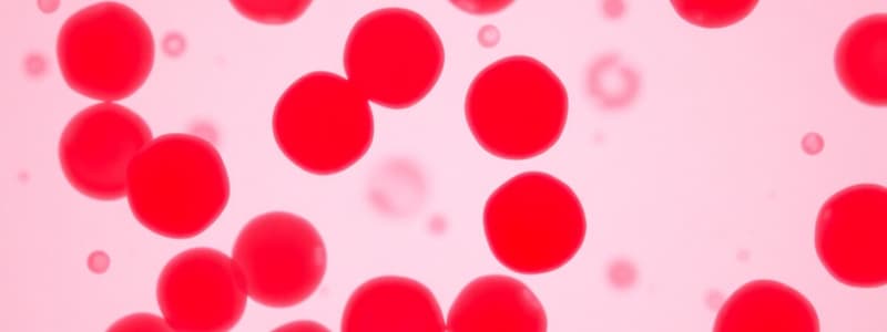
Elevated red blood cell count; can be relative (due to dehydration) or absolute (due to altitude or disease).
Equine Erythrocytes
Equine Erythrocytes
Red blood cells in horses retain marrow until maturation is complete; Rouleaux formation is common.
Reticulocytes (Equine)
Reticulocytes (Equine)
Immature red blood cells; reference range is zero in horses as they stay mature in the marrow.
Thrombocytes
Thrombocytes
Signup and view all the flashcards
Equine Anemia
Equine Anemia
Signup and view all the flashcards
Regenerative vs. Non-regenerative Anemia
Regenerative vs. Non-regenerative Anemia
Signup and view all the flashcards
Causes of Anemia
Causes of Anemia
Signup and view all the flashcards
Hemorrhage
Hemorrhage
Signup and view all the flashcards
Clinical Signs of Hemorrhage
Clinical Signs of Hemorrhage
Signup and view all the flashcards
Equine Anemia Treatment
Equine Anemia Treatment
Signup and view all the flashcards
Erythrocyte Destruction
Erythrocyte Destruction
Signup and view all the flashcards
Intravascular Hemolysis
Intravascular Hemolysis
Signup and view all the flashcards
Extravascular Hemolysis
Extravascular Hemolysis
Signup and view all the flashcards
IMHA
IMHA
Signup and view all the flashcards
Diagnosis of IMHA
Diagnosis of IMHA
Signup and view all the flashcards
Clinical Management of IMHA
Clinical Management of IMHA
Signup and view all the flashcards
Non-regenerative Anemia
Non-regenerative Anemia
Signup and view all the flashcards
Chronic Blood Loss Point
Chronic Blood Loss Point
Signup and view all the flashcards
Study Notes
- Equine bloodwork interpretation and anemia are important topics in veterinary medicine
Polycythemia
- Polycythemia refers to an increase in red blood cell mass
- Relative erythrocytosis is a type of polycythemia caused by dehydration or splenic contraction
- Absolute erythrocytosis is a type of polycythemia caused by hypoxia (high-altitude), chronic pulmonary disease or inappropriate RBC production, like hepatocellular carcinoma.
Equine Erythrocytes
- Equine erythrocytes have marrow retention until maturation is complete
- Polychromasia and macrocytosis can be observed in equine erythrocytes
- Howell-Jolly bodies can be found in low numbers in healthy horses
- Rouleaux formation is common in equine erythrocytes
- Agglutination can be differentiated from rouleaux formation using a 1:4 saline solution
Reticulocytes
- Reticulocytes, immature RBCs, normally have a reference range of 0 in horses
- They have a fine reticulum of nucleotide material that stains positive with methylene blue
- In other species, increased presence indicates regeneration
- In horses, they stay in the marrow until mature
Thrombocytes
- Thrombocytes reference range is 117-256 X 103/µl
- Decreased thrombocytes may indicate DIC, IMT, endotoxemia, EIA, equine ehrlichiosis, or lymphoma
- Pseudothrombocytopenia, clumping in EDTA, or use of citrate or heparin can lead to decreased thrombocytes
- Increased thrombocytes may indicate chronic inflammation or Rhodococcus equi
Summary
- Clinical pathology is useful in case management
- Serum chemistry and CBC tests are commonly used for evaluating equine patients
- Serial monitoring can determine the course of disease
- A repeat analysis is indicated if there are any unexpected results
- Repeating analysis can help in ruling out laboratory errors and artifactual results
Equine Anemia
- Equine Anemia involves a decrease in circulating RBC mass, decreased packed cell volume (PCV), and decreased hemoglobin (except with intravascular hemolysis)
- Determining if anemia is regenerative or non-regenerative is important
- Polychromasia can indicate regenerative anemia
- Horses do not release reticulocytes into circulation so bone marrow aspirate/biopsy from the rib or sternum must be performed to determine the cause of anemia
- Monitoring RBC increase over time can help determine the response to anemia, 3 days is needed for response
Causes of Anemia
- Anemia is caused by blood loss, destruction or reduced production
Hemorrhage
- Acute blood loss results in clinical signs earlier
- Acute blood loss will cause a transfusion PCV of 15-20%
- Chronic blood loss causes a transfusion PCV of 10-12%
- External blood loss occurs through wounds or from other external sources, like Guttural pouch mycosis or middle uterine artery rupture
- Internal blood loss occurs into a cavity such as the thoracic or peritoneal cavity due to splenic trauma or hemangiosarcoma
Clinical Signs
- Clinical signs are determined by the magnitude of blood loss
- Pale mucous membranes (pallor) can indicate clinical signs
- Tachycardia (> 60 bpm), and tachypnea (> 24 bpm) are vital signs of anemia
- Weakness and syncope is an additional indication of anemia
Assessment of Blood Loss in Horses
- Splenic contraction during blood loss may result in normal PCV despite marked loss, decline may be seen in 4-6 hours
- Total solids/total protein generally falls with blood loss
- Heart rate increases (>60 bpm), increased lactate circulation, depressed demeanor, and reduced appetite indicate other clinical findings impacted by reduced oxygen carrying capacity
Treatments for Equine Anemia
- Treatment includes hemorrhage control (hemostasis)
- Volume depletion should be treated by intravenous fluids
- Blood loss should be treated with blood transfusions in cases of anemia, tachycardia, tachypnea, reduced appetite, and/or depressed demeanor
- A gelding or maiden mare can receive one-time transfusion due to no previous RBC antigen exposure
- A Universal donor is Aa and Qa blood cell antigen and antibody negative
- A broodmare is a candidate for cross matching for repeated transfusions
Equine Blood Transfusion
- The amount of blood to administer for a 500-600 kg horse is approximately 5-10 L of whole blood
- The volume to administer is given by the formula: VOLUME = (BW KG) (0.08) (PCV desired – PCV patient)/PCV donor
- Example calculation: 500 KG (0.08) (20% - 12%) / 32% = 10 L
Important Points
- Patients with chronic blood loss will tolerate lower red blood cell mass
- Vital parameters and overall clinical demeanor help determine the need for transfusion
- PCV is important, but it can be lower in a patient with chronic RBC loss
Erythrocyte Destruction
- Immune-mediated destruction is also known as IMHA (Immune-Mediated Hemolytic Anemia)
- Immune-mediated destruction can be extravascular (antibody target RBC destruction) or intravascular (complement mediated destruction)
- Toxins such as red maple toxicity can cause oxidative injury
- Infectious agents can cause RBC fragility
- Hypotonic solutions such as water, and hypertonic solutions such as DMSO can cause erythrocyte destruction
Intravascular Hemolysis
- Intravascular hemolysis is the acute and severe destruction of RBC
- Signs of intravascular hemolysis are pigmenturia (hemoglobinuria), pink plasma and icterus
Extravascular Hemolysis
- Extravascular hemolysis involves erythrocyte removal from an extravascular site through opsonization and splenic removal
- Icterus occurs with yellow plasma
- No hemoglobinemia is present
IMHA
- Antibodies coat erythrocytes in IMHA
- Autoantibody is rare
- Secondary immune-mediated is most common
- Foreign RBC antigens or neonatal isoerythrolysis
- Medications, especially beta lactam antibiotics
- Anemia viruses
- Neoplasia (Lymphoma and paraneoplastic syndrome) cause
- Anemia is caused by foreign RBC antigens, anaemia viruses, beta lactam antibiotics and neoplasia
Diagnosis of IMHA
- Diagnosis involves Identification of surface-associated erythrocyte by testing for autoagglutination, Coomb's testing, direct immunofluorescence/flow cytometry, and IgG, IgA, IgM
- False negatives occur with corticosteroid administration or massive hemolysis
Clinical Management of IMHA
- Discontinue medications
- Administer Corticosteroid therapy for immune suppression
- Whole blood transfusion if indicated
- Treatment should avoid pigment associated nephropathy using diuresis
Non-regenerative Anemia
- Anemia of chronic disease (ACD) is a common cause of mild anemia in the mid 20% range
- Resolution of primary disorder is needed by controlling a small protein called hepcidin that limits the availability of iron
- Equine example of ACD is chronic pneumonia where anemia will resolve once pneumonia is resolved
- Iron deficiency is an uncommon cause of anemia due to true loss of iron stores
- External hemorrhage, caused by a severe wound with blood loss, may result in iron loss and supplementation is indicated
- Bone marrow suppression involves medications that cause a direct suppression on bone marrow function
- Myelophthisis - bone marrow destruction
- Chronic renal failure leads to reduced erythropoietin secretion
Summary
- Anemia can result from hemorrhage, destruction, or chronic disease
- Determine severity with whole blood transfusion, PCV and careful and acute patient evaluation that will decompensate more rapidly
- Immune-mediated anemia requires determining the inciting cause
- External blood loss may require iron supplementation with whole blood transfusion, low PCV, and in critical patient conditions with high HR and/or worsening condition
- Anemia associated with ACD requires treating the primary disease to resolve it
Studying That Suits You
Use AI to generate personalized quizzes and flashcards to suit your learning preferences.