Podcast
Questions and Answers
Which gait abnormality is most likely related to pain as opposed to a mechanical or neurological issue?
Which gait abnormality is most likely related to pain as opposed to a mechanical or neurological issue?
- Neurologic gait abnormality
- Gait alterations due to conformation
- Mechanical lameness involving non-painful gait
- Pain-induced lameness (correct)
When evaluating lameness, what is the primary reason for performing a static examination (hands on and hands off) before exercise?
When evaluating lameness, what is the primary reason for performing a static examination (hands on and hands off) before exercise?
- To desensitize the horse to handling
- To save time during the lameness evaluation
- To avoid diagnostic anesthesia
- To identify the location of pain (correct)
In horses with chronic lameness, how do they typically adapt their weight distribution, and what physical change might be observed in their hooves?
In horses with chronic lameness, how do they typically adapt their weight distribution, and what physical change might be observed in their hooves?
- They distribute weight evenly across all limbs, maintaining hoof symmetry
- They increase weight on the lame limb, leading to a steeper hoof angle
- They decrease weight on the non-lame limb, resulting in a smaller hoof
- They shift weight to the non-lame limb, which will have a flatter foot (correct)
What is the most common vertical compensatory movement observed in a horse with forelimb lameness?
What is the most common vertical compensatory movement observed in a horse with forelimb lameness?
What gait abnormality is defined as evident both when the limb is moving (swing phase) and when it is supporting weight (stance phase)?
What gait abnormality is defined as evident both when the limb is moving (swing phase) and when it is supporting weight (stance phase)?
When assessing lameness in multiple limbs, why is it essential to identify and evaluate the primary lameness first?
When assessing lameness in multiple limbs, why is it essential to identify and evaluate the primary lameness first?
A horse exhibits forelimb lameness on the right limb, leading to uneven weight distribution. Which compensatory lameness pattern is most likely to develop?
A horse exhibits forelimb lameness on the right limb, leading to uneven weight distribution. Which compensatory lameness pattern is most likely to develop?
Given the typical distribution of weight in horses, where do most lameness issues tend to originate, and why?
Given the typical distribution of weight in horses, where do most lameness issues tend to originate, and why?
When evaluating a horse for lameness, what percentage of hindlimb lameness cases typically involve the hock or stifle region?
When evaluating a horse for lameness, what percentage of hindlimb lameness cases typically involve the hock or stifle region?
Why are young horses considered more prone to lameness compared to mature horses?
Why are young horses considered more prone to lameness compared to mature horses?
When evaluating lameness in an older crossbred horse used for pleasure riding, where are problems most likely to originate?
When evaluating lameness in an older crossbred horse used for pleasure riding, where are problems most likely to originate?
A young racehorse is being evaluated for lameness. Based on the common injuries in this specific discipline, where is the lameness most likely to originate?
A young racehorse is being evaluated for lameness. Based on the common injuries in this specific discipline, where is the lameness most likely to originate?
What question is MOST relevant when acquiring a horse's medical history during a lameness exam?
What question is MOST relevant when acquiring a horse's medical history during a lameness exam?
When performing a hands-off examination, what observation from a distance is LEAST useful in assessing potential lameness?
When performing a hands-off examination, what observation from a distance is LEAST useful in assessing potential lameness?
During a hands-on examination, what observation in the forelimbs is most indicative of chronic lameness?
During a hands-on examination, what observation in the forelimbs is most indicative of chronic lameness?
When assessing a horse's conformation, what is the MOST important consideration regardless of breed variations?
When assessing a horse's conformation, what is the MOST important consideration regardless of breed variations?
How does the length of the neck typically influence a horse's balance and movement?
How does the length of the neck typically influence a horse's balance and movement?
What does the term 'substance of bone' refer to, and why is it important in assessing a horse's conformation?
What does the term 'substance of bone' refer to, and why is it important in assessing a horse's conformation?
What is the consequence of deviations in limb alignment that can cause strain on the collateral support structures and lead to uneven loading of the hinge joints?
What is the consequence of deviations in limb alignment that can cause strain on the collateral support structures and lead to uneven loading of the hinge joints?
How does a longer humerus positively influence a horse's movement?
How does a longer humerus positively influence a horse's movement?
When evaluating the symmetry and weight-bearing of a horse's hindlimbs from the rear, what bony landmark should be carefully assessed for equal height?
When evaluating the symmetry and weight-bearing of a horse's hindlimbs from the rear, what bony landmark should be carefully assessed for equal height?
When assessing hindlimb conformation from a lateral view (side), what anatomical landmark should a line from the point of the buttock touch to indicate proper alignment?
When assessing hindlimb conformation from a lateral view (side), what anatomical landmark should a line from the point of the buttock touch to indicate proper alignment?
In a forelimb hoof, what anatomical aspect is MOST important when evaluating foot conformation?
In a forelimb hoof, what anatomical aspect is MOST important when evaluating foot conformation?
During a hands-on lameness exam, what does asymmetrical foot size typically indicate?
During a hands-on lameness exam, what does asymmetrical foot size typically indicate?
If superficial cleaning and examination of the sole reveals a convexity at the apex of the frog, what might this indicate?
If superficial cleaning and examination of the sole reveals a convexity at the apex of the frog, what might this indicate?
During a hoof tester exam, what would diffuse sole sensitivity suggest?
During a hoof tester exam, what would diffuse sole sensitivity suggest?
When palpating the pastern during a lameness exam, how can the collateral ligaments be stressed to identify potential injuries?
When palpating the pastern during a lameness exam, how can the collateral ligaments be stressed to identify potential injuries?
During palpation of the metacarpus/metatarsus, what condition does firm fingertip pressure help to elicit, particularly in racehorses?
During palpation of the metacarpus/metatarsus, what condition does firm fingertip pressure help to elicit, particularly in racehorses?
During palpation of the tarsus to assess lameness, what condition might be indicated by bone proliferation in the distal tarsal joints?
During palpation of the tarsus to assess lameness, what condition might be indicated by bone proliferation in the distal tarsal joints?
During a lameness exam, what is the purpose of the Churchill pressure test, and which anatomical structure is targeted?
During a lameness exam, what is the purpose of the Churchill pressure test, and which anatomical structure is targeted?
During the hoof should land flat or the heel shouldhit just before the toe. When should the foot land that way?
During the hoof should land flat or the heel shouldhit just before the toe. When should the foot land that way?
During dynamic examination of the shoulder musculature, shoulder of lame limb fixes or "props" just
before it hits the ground, what would that demonstrate?
During dynamic examination of the shoulder musculature, shoulder of lame limb fixes or "props" just before it hits the ground, what would that demonstrate?
Flashcards
Equine Lameness
Equine Lameness
Any condition impairing limb or axial skeleton function/structure, visible at rest or in motion.
Lameness Differentiation
Lameness Differentiation
Distinguishing pain-related lameness from mechanical or neurological issues is key.
Objectives of Lameness Examination
Objectives of Lameness Examination
To determine if the horse is lame, and identify the limb(s), site(s), and specific cause of the problem.
Adaptive Strategies of Lame Horses
Adaptive Strategies of Lame Horses
Signup and view all the flashcards
Compensatory Movements
Compensatory Movements
Signup and view all the flashcards
Supporting Limb Lameness
Supporting Limb Lameness
Signup and view all the flashcards
Swinging Limb Lameness
Swinging Limb Lameness
Signup and view all the flashcards
Mixed Lameness
Mixed Lameness
Signup and view all the flashcards
Primary Lameness
Primary Lameness
Signup and view all the flashcards
Compensatory Lameness
Compensatory Lameness
Signup and view all the flashcards
Forelimb Stress
Forelimb Stress
Signup and view all the flashcards
Forelimb Lameness Origin
Forelimb Lameness Origin
Signup and view all the flashcards
Hindlimb Lameness Origin
Hindlimb Lameness Origin
Signup and view all the flashcards
Other Factors Contributing to Lameness
Other Factors Contributing to Lameness
Signup and view all the flashcards
History and Hands Off Examination
History and Hands Off Examination
Signup and view all the flashcards
TB racehorse injuries
TB racehorse injuries
Signup and view all the flashcards
Endurance horse injuries
Endurance horse injuries
Signup and view all the flashcards
Show/pleasure horse injuries
Show/pleasure horse injuries
Signup and view all the flashcards
Western performance horse injuries
Western performance horse injuries
Signup and view all the flashcards
Jumping horse injuries
Jumping horse injuries
Signup and view all the flashcards
Draft horse injuries
Draft horse injuries
Signup and view all the flashcards
Medical History for Lameness
Medical History for Lameness
Signup and view all the flashcards
Visual Assessment of the Horse
Visual Assessment of the Horse
Signup and view all the flashcards
Close-Up Inspection
Close-Up Inspection
Signup and view all the flashcards
Conformation
Conformation
Signup and view all the flashcards
Key Functional Areas
Key Functional Areas
Signup and view all the flashcards
"Substance of bone"
"Substance of bone"
Signup and view all the flashcards
Ideal Forelimbs
Ideal Forelimbs
Signup and view all the flashcards
Humerus Slope Assessment
Humerus Slope Assessment
Signup and view all the flashcards
Hindlimbs
Hindlimbs
Signup and view all the flashcards
Coxal Angle
Coxal Angle
Signup and view all the flashcards
Caudal View - Sound Hindlimbs
Caudal View - Sound Hindlimbs
Signup and view all the flashcards
Ideal Forefoot
Ideal Forefoot
Signup and view all the flashcards
Hoof Balance
Hoof Balance
Signup and view all the flashcards
Hands-On Examination
Hands-On Examination
Signup and view all the flashcards
Study Notes
- Study notes for Lameness Evaluation, EQM400, 2025 edition by Dr. E Hollenbach
Learning Outcomes
- Demonstrate knowledge of distal limb anatomy
- Perform a basic lameness exam and understand investigation tests
- Recognize and define lameness origins
- Develop a diagnostic approach
- Recognize gait deficits like stringhalt, Sweeney, radial nerve paralysis, upward patellar fixation, and flexural deformities
- Perform diagnostic nerve blocks (palmar digital, abaxial sesamoid, low/high 4/6-point, wheat nerve block, DBLPN) to identify lameness
- Demonstrate arthrocentesis knowledge for common sites (coffin, fetlock, carpus, tarsus)
Introduction
- Lameness is any condition impairing limb or axial skeleton function/structure, visible at rest or in motion
- Causes include trauma, congenital/acquired abnormalities, developmental issues, infections, metabolic conditions, circulatory/nerve disorders, or combinations
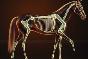
- Accurate diagnosis requires thorough understanding of equine anatomy, biomechanics, and body forces
- Differentiating pain-related, mechanical, and neurologic gait abnormalities is crucial
- Pain-induced lameness is the most common
- Lameness examination objectives:
- Determine if the horse is lame
- Identify involved limb(s)
- Locate problem site(s)
- Determine the specific cause
- Select appropriate treatment
- Predict recovery prognosis
- Routine lameness examination steps:
- Complete history (signalment/use)
- Visual exam at rest (hands off)
- Musculoskeletal system palpation (hands on)
- Motion observation
- Observation under work/saddle (if needed)
- Manipulation tests (flexion tests)
- Diagnostic anesthesia
- Diagnostic imaging
- Static examination (hands on/off) precedes exercise
- Diagnostic anesthesia/imaging follow to identify pain location, specific cause, injury extent, and recovery prognosis
Adaptive Strategies
- Horses adapt to lameness with compensatory body movements, with most attempting to "unload" the lame limb
- Chronic lameness causes higher vertical loads on the non-lame limb, resulting in a flatter foot
- Consistent compensatory movements include head displacement (forelimb lameness) and sacrum/tuber coxae displacement (hindlimb lameness)
- Overlap can occur but is typical in moderate to severe lameness
Classification
- Primary lameness contributes most to gait abnormalities
- Compensatory/secondary lameness results from overloading other limbs
- Lameness classified by occurrence within the stride:
- Supporting limb lameness occurs during weight-bearing (stance phase), caused by bone, joint, or soft tissue injuries
- Swinging limb lameness is noticeable in motion, affecting upper limbs or axial skeleton
- Mixed lameness is evident in both swing and stance phases, involving any structure combination
- Primary lameness is the most noticeable before tests, with the most severe being typically assessed first
- Compensatory lameness occurs due to uneven weight distribution from pain, frequently observed as forelimb lameness causing opposite forelimb issues
- Hindlimb lameness can mimic forelimb lameness on the same side and/or cause compensatory on opposite side
- Even minor weight-bearing shifts trigger compensatory lameness, especially at high speeds or long distances, commonly affecting feet, suspensory ligaments, sesamoid bones, hocks, and flexor tendons
- Compensatory within the same limb can occur when healthy structures protect a painful region, such as a horse with navicular toe-first landing
Other Factors
- Most lameness is in the forelimbs: bearing 60-65% of the horse's weight (70% w/rider) and absorbing more shock
- Hindlimbs propel but carry less weight/experience less concussion
- Activity matters: dressage, cutting, and reining strain hindquarters, and will cause more hindlimb lameness
- ~95% of forelimb lameness originates distal to the carpus, so examine the distal limb before assuming issues are in the upper limb
- ~80% of hindlimb lameness involves the hock/stifle; assess the foot/lower limb before these regions
- Other contributing factors: improper shoeing, unsuitable footing, and performance-related muscle fatigue
- Young horses are prone to lameness because their musculoskeletal systems are still developing
- Young horses may be raced or shown, therefore experience injuries that would typically be less likely in older horses
History and Hands-Off Examination:
- Patient age and use are important considerations
Signalment and Use
- Aged pleasure riding horses are prone to problems associated with the forefeet, low-motion joints and ligaments
- Young racehorses have lameness problems with high-motion joints, sprain/strain of flexor support structures, and stress-related fractures
- Competitive trail or endurance riding horses often sustain sprain/strain injuries, tendonitis, and phalange fractures
- Young horses starting traing can become lame from developmental orthopaedic-related problems.
- Common injuries in specific disciplines:¹
- TB racehorse (Fatigue-associated repetitive overuse)
- foot bruising
- quarter cracks
- heel pain
- Forelimb fetlock synovitis and fractures
- Carpal synovitis and fractures
- Bucked shins in young horses
- Fatigue/stress fractures (MC/MT, P1, humerus, tibia, pelvis)
- SDFT tendonitis
- Suspensory injuries
- Catastrophic fractures (PSB, P1, MC/MT, and humerus)
- Palmar/plantar osteochondral disease (POD)
- Endurance horse
- Muscle disorders including tying-up and muscle spasm, cramps, and strains
- Forelimb and hindlimb suspensory desmitis
- Foot bruising, P3 osteitis, and laminitis
- Fetlock synovitis/OA
- SDFT tendonitis
- Show/pleasure horse
- Navicular syndrome/disease
- Coffin joint synovitis/OA
- Forelimb and hindlimb PSD
- Distal tarsitis or bone spavin
- Lumbar and sacroiliac problems
- Western performance horses
- Navicular syndrome/disease including DDFT injuries
- Phalangeal fractures (primarily P2)
- Pastern ringbone
- Fetlock and carpal synovitis/OA
- Distal tarsitis or bone spavin Stifle synovitis/OA
- Forelimb and hindlimb PSD
- Thoracolumbar myositis
- Hindlimb muscle strains
- Jumping/dressage/eventing
- Navicular syndrome/disease including DDFT injuries
- Suspensory branch injuries
- Forelimb and hindlimb PSD
- Fetlock synovitis/OA
- SDFT tendonitis
- Distal tarsitis or bone spavin
- Stifle injuries
- Thoracolumbar myositis—back problems
- Sacroiliac problems
- Draft horses
- Hoof cracks and laminitis
- Subsolar abscesses, canker, and thrush Ringbone
- Sweeny
- Bone and bog spavin
- Stifle synovitis/OA
- PSSM
- Gaited horses (Hindlimb>Forelimb)
- Distal tarsitis or bone spavin
- Stifle synovitis/OA
- Thoracolumbar myositis/back problems
- DSLD
- TB racehorse (Fatigue-associated repetitive overuse)
History
- Records including the lameness's duration/intensity, specific symptoms, preceding activity, and previous treatments/therapies
- What activity does the horse perform and did the lameness occur during this activity?
- How long has the horse been lame? (Acute vs. chronic injury.)
- Has the horse been rested or exercised during the lameness period? (The horse may not exhibit the same lameness if it has been rested.)
- Has the lameness worsened, stayed the same, or improved? (May indicate severity of the problem.)
- Was the cause of the lameness observed? (This should include the character of the lameness at the onset.)
- Does the horse warm up out of the lameness?
- When do you notice the lameness most consistently?
- What treatments have been given, and have they helped? (Response to treatment may help predict the prognosis or may mask the severity of the current lameness)
- When was the horse last shod or trimmed?
- What are the abnormalities that the owner or trainer observe or feel when they ride the horse or watch the horse go?
Hands Off Examination
- Visual assessment while the horse stands squarely on a level surface
- Begin from a distance, viewing from multiple angles, and move in for closer inspection
- Assess body type, conformation, body condition, posture changes, weight distribution, and limb positioning
- Any asymmetry or alterations in limb contours need noting
- Forelimbs should bear weight evenly and be opposite each other
- Weight shifting between limbs or extending forelimbs forward may indicate bilateral forelimb discomfort
- Resting one hindlimb consistently or being unable to bear weight indicates lameness
- Any minor stance changes are a potential issue and should be investigated further
- Close inspection for symmetry comparison
- Hooves should be checked for wear, cracks, imbalance, size differences, or contraction
- Joints, tendons, and sheaths should be checked for swelling
- Muscles of limbs, back, and pelvis assessed for atrophy or enlargement
- Comparing left and right sides can detect subtle abnormalities
- Narrower hoof with higher heel and extensor muscle atrophy indicate lameness (especially chronic/bilateral)
- Hindlimb atrophy of middle gluteal/gracilis suggests an underlying problem
- An uneven tuber sacrale/pelvic tilt often corresponds to asymmetric gait pattern, to be evaluated further
Conformation
- Conformation is how a horse is shaped by bones and muscles
- Often assessed subjectively based on breed standards/discipline-specific ideals; "normal" conformation varies
- Some conformational traits increase lameness risk
- Distinguishing which abnormalities contribute to lameness improves understanding of the causes
- Structured conformation evaluation is systematic, focusing on:
- Head/neck
- Forelimbs
- Trunk
- Hindlimbs
- Evaluate each equally; examine specific conformational traits in detail after initial assessment
- Begin with a side profile, assessing balance between forehand and hindquarters
- Observe topline curvature/proportions from poll to gaskin
- Pay attention to how limbs attach, long bone lengths, and wither heights correlate to maintain proportionality across sizes
- Assess limb/hoof alignment, straightness/symmetry, and forearm/chest muscle depth/length from the front
- A side view from the right further confirms balance, topline, and limb angles
- The evaluation from behind insights into back, croup, hips, buttocks, hindquarter, and hindlimb symmetry
- The perspective is used for assessing back muscling, vertebral column alignment, and left-to-right symmetry only if the horse is standing square.
Body
- Head size/shape are breed-specific and negligibly impact performance/lameness
- The neck acts as a lever and should be long, flexible, and convex
- Longer neck aids balance over jumping fences
- Distal forelimb bears more weight than distal hindlimb
- Forelimbs tend to "bounce", while hindlimbs "slide”.
- Well-muscled forelimbs support 60-65% of the horse's body weight
- Balance helps horses move, improve efficiency, and reduce musculoskeletal system stress
Proportions and Curvature of the Top Line
- Influenced by topline proportions/curvature, loin strength, wither sharpness, croup slope, underline length to back
- Neck is measure from teh poll to the withers
- Back is measure from teh withers to teh caudal end of the loin
- Hip is measure from the caudal loin to point of the buttock
- If shorter, neck limits flexibility/balance
- A too-long back results in a hollow (concave) back
- A short hip leads to reduced propulsion/downhill body configuration
- General guideline: neck length should be equal or greater thna teh back length; hip hould measure at least ⅔ the back length
- Shape depends on the S-curve of cervical vertebrae
- Ideally, the nec rises gracefully without dipping ventrally in front
- The thickest part should be at the ventral limits
- Ewe necks display a long, downward curve, with the neck appearing to attach low on chest
- Shoulder attachment is also important
- A steep one leads to less ideal dorsal to ventral neck ratio.
- Withers blend gently into the back, ending at the midpoint, and are suitable for saddle placement
- Neck/forearm muscles and ligamentum nuchae attach at the highest point of the withers without a dip
- Low withers hinder raising of the back when lowering/extending neck
- Well-sloped shoulders typically indicate properly positioned withers
- Longissimus muscles along the spine should be flat and strong and not sloped since the horse's help counteract gravity and support rider's weight
- Loin, from the last thoracic vertebra to teh lumbosacral junction, which should be mulcusled and short; "Long-backed" horses often have a long, weak loin, putting them at risk of chronic lumbar pain or vertebral damage
- Loin and coupling transfer motion from hindquarters trhough back to forehand
- Strong/well connected lions promote power, which can lack flexibility and may be short
- The Croup from th lumbrosacral to tail, should fairly long to ensure hip and articulation
- A topline that is shorter than the underline creates a balanced combination thta indicats good stride length.
Substance
- The thickness, depth and breadth of tissue
- Evaluating muscle substance considers its type, thickness, length + attachment position
- Important elements are weight, sizes, height and the rib (the degree of curvature; best observed from the rear), and the spring
- Well sprang ribs provide ample space for the heart, lungs, digestive
- Shalllow girth/slab sides = uncomfortable
- Size relates to weight
- Use cannon size, below, to gauge size; 4 cm *100 kg for riding horses -> 500 kg needs 20 cm
- Alignment is essential for movement; the shoulder, arm, forearm, should be proportional
- Straight bones -> Smooth movement, should align with the joints
- High-quality hooves with build
Forelimbs
- Equal length and size; when standing, even length should be seen by dropping a line between limb on the shouulder to ground in a straight line
- Toes and feet should point
- Humus slope - Optimal, humerus is optimal
- Muscle taper at elbow to provide flex
- carpals should be balances
- Devations -> strain -> hinge joints
Lateral View
- Moderate angles for Shock absorption
- Measuring shoulder (Scapular and withers) - angle becomes more upright as horse matures, the shorter angle causes increase of stride
- High performance wants horizontal
- Angle facilitates elbow to ground max extension
- Humerous Length
- Improvments to stirde / Quality
- elbow to fettloc -> straight; misalignment can result in force concentration (Carpus-function)
- Fetlocks
- Straighter hind
- The hoves should be well shaped
- Extreme can raise strain
Conformation Faults
- Can lead cause musculoskelatal injureise
Hindlimbs
- Engine
- .The horses and muscles determine preformacne
Lateral Vieww - hinds
- line from buttocks -> hocks ends behind heel -> standin hindlegs, and camper out
- gluteal msucles and connect back to limb
- hip and length, stride etc
- A foreleg is faster than the hind
- Gaskin to stilthe is the one that should be shorter
From the rear - Hinds - symmetrical/length and equality
Medial / lateral
- relationship in the hoof, with a band
- asymmetry + insight
- Level from front to back
- Straighter inside
Static examination
- Visual assessment of the horse’s back begins with
- observing the muscle contour from
- the side and checking the axial alignment from the rear. The dorsal spinous processes should be palpated
- to evaluate their alignment, as well as to detect any protrusion, depression, or abnormal interspinous distances.
_ Static/Hands on exam
- A systematic approach to avoid missing abnornmalilites
- A foor exam must be done after exam of the mussocletal
- The shape, and see if normal relative size - check for abnormalities - size, cracks, imbalance, etc
- Check after clean
Hands on exam
- Palpate for problems
- With the limb off the ground, pain may be elicited from the distal sesamoidean ligaments and phalanx if a fracture is present.
- Examine for problems/fractures
Carpus
Effective when carpus if flexed
Forelimbs
- check swelling
Tarsus
palpated -> synovitis
Dynamic examination
- is best carried out on a hard, level surface
- Surface (impact)
- A jogger is very important
- Head centered, a constant speed and line, head and neck can be an indicator of health issues
- handler should focus on forelimbs
- Gait changes indicate limb concerns
Character change
- important diagnosis consideration
- when to watch the stride
- head and pelvic
EQM400 2025 is finished
Studying That Suits You
Use AI to generate personalized quizzes and flashcards to suit your learning preferences.