Podcast
Questions and Answers
Which of the following are adverse effects of PTH on the cardiovascular system?
Which of the following are adverse effects of PTH on the cardiovascular system?
- Atherosclerosis (correct)
- Myocardial infarction
- Hypertension (correct)
- Calcification (correct)
Loss of function mutations in parathyroid hormone can lead to Bartter syndrome type 5.
Loss of function mutations in parathyroid hormone can lead to Bartter syndrome type 5.
False (B)
What is the primary regulator of PTH secretion?
What is the primary regulator of PTH secretion?
Ionised calcium (Ca2+)
In senile osteoporosis, mesenchymal stem cells differentiate into __________ instead of osteoblasts.
In senile osteoporosis, mesenchymal stem cells differentiate into __________ instead of osteoblasts.
Match the receptors or agents with their respective roles:
Match the receptors or agents with their respective roles:
Which bone cells are responsible for bone resorption?
Which bone cells are responsible for bone resorption?
Sclerostin is secreted by osteoblasts to regulate bone formation.
Sclerostin is secreted by osteoblasts to regulate bone formation.
What is the primary function of osteocytes?
What is the primary function of osteocytes?
Osteocytes produce __________ in response to unloading forces or estrogen deficiency.
Osteocytes produce __________ in response to unloading forces or estrogen deficiency.
Match the following bone cells to their origin:
Match the following bone cells to their origin:
What is the main function of calcitriol in the body?
What is the main function of calcitriol in the body?
Cholecalciferol is derived only from plant sources.
Cholecalciferol is derived only from plant sources.
What enzyme in the liver converts vitamin D₃ into calcidiol?
What enzyme in the liver converts vitamin D₃ into calcidiol?
The _____ is the primary organ responsible for activating vitamin D.
The _____ is the primary organ responsible for activating vitamin D.
Match the following vitamin D metabolism components:
Match the following vitamin D metabolism components:
What effect does Vitamin D have on phosphate excretion in the kidneys?
What effect does Vitamin D have on phosphate excretion in the kidneys?
PTH leads to increased bone formation.
PTH leads to increased bone formation.
What is the suggested management for Vitamin D deficiency in terms of IU per week?
What is the suggested management for Vitamin D deficiency in terms of IU per week?
Vitamin D levels below _____ ng/ml are considered sufficient.
Vitamin D levels below _____ ng/ml are considered sufficient.
Match the following actions with the hormone responsible:
Match the following actions with the hormone responsible:
Which of the following is a bone resorption marker?
Which of the following is a bone resorption marker?
Vitamin D acts through a heterodimer receptor complex in the nucleus.
Vitamin D acts through a heterodimer receptor complex in the nucleus.
What is the action of the VDR-RXR complex when it reaches the nucleus?
What is the action of the VDR-RXR complex when it reaches the nucleus?
The marker __________ is specific to bone formation and is known as a measure of bone turnover.
The marker __________ is specific to bone formation and is known as a measure of bone turnover.
Match the following bone markers with their categories:
Match the following bone markers with their categories:
What percentage of calcium stored in the body is found in bones?
What percentage of calcium stored in the body is found in bones?
Normal corrected calcium levels are between 8.6 and 10.3 mg/dL.
Normal corrected calcium levels are between 8.6 and 10.3 mg/dL.
What is the primary site of calcium absorption in the digestive system?
What is the primary site of calcium absorption in the digestive system?
The corrected Ca²⁺ level is calculated using the formula: Corrected Ca²⁺ = Total Ca²⁺ + 0.8 (4 - ______).
The corrected Ca²⁺ level is calculated using the formula: Corrected Ca²⁺ = Total Ca²⁺ + 0.8 (4 - ______).
Which receptor is involved in calcium sensing in the body?
Which receptor is involved in calcium sensing in the body?
Acidosis causes an increase in free calcium concentration by decreasing its availability.
Acidosis causes an increase in free calcium concentration by decreasing its availability.
During calcium absorption, which protein is responsible for binding and transporting calcium within the cell?
During calcium absorption, which protein is responsible for binding and transporting calcium within the cell?
Match the following components with their roles in calcium absorption:
Match the following components with their roles in calcium absorption:
What is the primary origin of the parathyroid gland?
What is the primary origin of the parathyroid gland?
The transcription factor GCMB defect is associated with hypoparathyroidism.
The transcription factor GCMB defect is associated with hypoparathyroidism.
What type of cells in the parathyroid gland have an affinity for 99mTc sestamibi?
What type of cells in the parathyroid gland have an affinity for 99mTc sestamibi?
The development of the inferior parathyroid gland is more commonly associated with ______ location.
The development of the inferior parathyroid gland is more commonly associated with ______ location.
Match the transcription factor with its associated syndrome or defect:
Match the transcription factor with its associated syndrome or defect:
What is one of the primary sources of alkaline phosphatase (ALP)?
What is one of the primary sources of alkaline phosphatase (ALP)?
In the new hypothesis, osteoblasts are responsible for laying down the matrix.
In the new hypothesis, osteoblasts are responsible for laying down the matrix.
What enzyme is hydrolyzed by alkaline phosphatase to promote mineralization?
What enzyme is hydrolyzed by alkaline phosphatase to promote mineralization?
Osteocytes are responsible for the __________ process during bone formation.
Osteocytes are responsible for the __________ process during bone formation.
Match the following enzymes with their respective characteristics:
Match the following enzymes with their respective characteristics:
What is the primary function of parathyroid hormone (PTH)?
What is the primary function of parathyroid hormone (PTH)?
The bioactive form of PTH is made up of the first 34 amino acids.
The bioactive form of PTH is made up of the first 34 amino acids.
What effect does vitamin D have on skin, breast, and prostate malignancies?
What effect does vitamin D have on skin, breast, and prostate malignancies?
PTH predominantly affects calcium absorption in the __________.
PTH predominantly affects calcium absorption in the __________.
Match the following terms with their descriptions:
Match the following terms with their descriptions:
What is the primary role of osteoclasts in the bone remodeling process?
What is the primary role of osteoclasts in the bone remodeling process?
Osteoprotegerin (OPG) stimulates osteoclast activation.
Osteoprotegerin (OPG) stimulates osteoclast activation.
What is the role of RANKL in the bone remodeling process?
What is the role of RANKL in the bone remodeling process?
The mineralization process occurs over a period of __________ months.
The mineralization process occurs over a period of __________ months.
Which hormone inhibits osteoclast activity and stimulates renal calcium excretion?
Which hormone inhibits osteoclast activity and stimulates renal calcium excretion?
Match the following components with their functions:
Match the following components with their functions:
Osteocytes are immature osteoblasts that reside within the bone matrix.
Osteocytes are immature osteoblasts that reside within the bone matrix.
What occurs during the reversal phase of the bone remodeling cycle?
What occurs during the reversal phase of the bone remodeling cycle?
Flashcards are hidden until you start studying
Study Notes
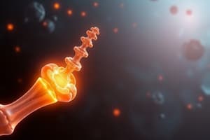
Basics of Bone & Mineral Metabolism
- Parathyroid hormone (PTH) has adverse effects on the cardiovascular system, including calcification, atherosclerosis, and hypertension
- PTH receptors are G protein-coupled receptors (GPCRs) that activate the cAMP/PKA pathway.
- The sigmoid rule describes the relationship between calcium levels and PTH secretion, highlighting the different types of PTH receptors.
- Type 1 receptors are also activated by PTH-related protein (PTHrp), which can play a paraneoplastic role in cancers like squamous cell carcinoma of the head and neck, or lung.
- Type 2 receptors are involved in calcium sensing. An increase in calcium activates the calcium-sensing receptor (CaSR) in the parathyroid gland, leading to a decrease in PTH. Conversely, a decrease in calcium inhibits the CaSR, resulting in increased PTH secretion.
- PTH secretion is always present at a basal level. Ionized calcium (Ca2+) is the most important regulator of PTH secretion.
- A loss-of-function mutation in the CaSR leads to hypoparathyroidism.
- A gain-of-function mutation in the CaSR causes Bartter syndrome type 5.
- Senile osteoporosis is characterized by mesenchymal stem cells differentiating into adipocytes instead of osteoblasts.
- Claudin 16 is a protein that promotes calcium reabsorption in the kidneys.
- The graph depicts the relationship between PTH secretion and calcium levels. PTH secretion increases exponentially with increasing calcium levels until a plateau is reached.
Vitamin D Metabolism
- Vitamin D is synthesized in the skin from cholecalciferol (vitamin D3) and can also be obtained from dietary sources.
- Vitamin D metabolism involves several steps in the liver and kidneys.
- In the liver, Vitamin D3 is hydroxylated by the enzyme 25-hydroxylase to form calcidiol (25-hydroxycholecalciferol), a form with a long half-life and the major circulating storage form.
- In the kidneys, calcidiol is further hydroxylated by 24-α hydroxylase, producing the inactive metabolite 24,25(OH)2D3.
- Calcitriol, the active form of vitamin D (1,25-dihydroxycholecalciferol), is produced by 1-α hydroxylase.
- Vitamin D is essential for intestinal calcium absorption.
- The diagram showcases the flow of vitamin D metabolism, starting from sources and progressing through the liver and kidneys to become activated vitamin D in the intestine.
Effects of PTH vs Vit D
- The Fibroblast Growth Factor 23 (FGF 23) can have both beneficial (phosphate excretion) and harmful effects (decrease in vitamin D) on the body.
- FGF 23 exerts various actions:
- Inhibition of sodium-phosphate cotransporter in the proximal convoluted tubule (PCT), resulting in decreased phosphate reabsorption.
- Inhibition of 1-alpha hydroxylase activity in the kidneys, leading to reduced vitamin D synthesis.
- Suppression of PTH via the Klotho receptor.
- The table summarizes the effects of PTH and vitamin D on different organs.
- PTH increases calcium and phosphate reabsorption in the intestine, increases phosphate excretion in the kidney, and stimulates bone resorption, leading to increased calcium mobilization from bone. Vitamin D enhances calcium and phosphate reabsorption in the intestine, reduces calcium and phosphate excretion in the kidney, and increases bone resorption and formation.
Calcium Metabolism
- The body stores most of its calcium in bones, with a small amount in soft tissues and extracellular fluid.
- Normal corrected calcium levels in the blood are between 8.6 and 10.3 mg/dL.
- Hypercalcemia is defined as a corrected calcium level greater than 10.5 mg/dL.
- Free, ionized calcium plays vital roles in the body, including sensitization of the CaSR, myocyte contractility, neuromuscular activity, and bone mineralization.
- Calcium competes with hydrogen ions for albumin binding.
- Acidosis increases free calcium levels by displacing calcium bound to albumin. A decrease in pH by 1 unit raises calcium levels by approximately 1 meq/L.
- Plasma calcium comprises both free ions and calcium complexed with anions.
Endochondral Bone Formation
- Endochondral bone formation involves the interaction of different cells:
- Osteoblasts, which are mesenchyme-derived cells responsible for bone formation.
- Chondroblasts, which are involved in cartilage formation.
- Osteocytes, which are mature osteoblasts embedded in the bone matrix and are considered the key regulators of bone formation and resorption.
- They function as mechanoreceptors, responding to loading and unloading forces.
- They are sensitive to estrogen, which inhibits osteoclast activity.
- They secrete sclerostin, a protein regulating bone remodeling.
- Osteoclasts, macrophage-derived cells that are responsible for bone resorption.
Bone Markers of Bone Formation and Resorption
- Bone resorption markers are used to monitor the breakdown of bone tissue:
- Tartrate-resistant acid phosphatase (TRAP)
- C-terminal telopeptide fragment of type I collagen (CTP)
- N-terminal telopeptide fragment of type I collagen (NTX)
- Bone formation markers reflect the formation of new bone tissue:
- Total alkaline phosphatase (ALP)
- Bone alkaline phosphatase (bsALP)
- Osteocalcin (OC)
- C-terminal propeptide of procollagen type 1 (PICP)
- N-terminal propeptide of procollagen type 1 (PINP)
Vitamin D Mechanism of Action
- Vitamin D, a group I hormone, acts through an intracellular receptor in the nucleus.
- The diagram showcases the mechanism of action of vitamin D in the cell. Vitamin D, after undergoing hydroxylation, activates its receptor, Vitamin D Receptor (VDR). The activated VDR forms a Heterodimer complex with the Retinoid X Receptor (RXR). This complex moves to the nucleus and binds to Vitamin D Response Element (VDRE) sequences on the DNA, triggering the expression of target genes and ultimately promoting calcium absorption and regulating other functions.
Bone Remodeling
- The image demonstrates the process of bone remodeling, highlighting the interplay between osteoclasts, osteoblasts, and other factors.
- The diagram illustrates a simplified version of the bone cycle.
- Osteoclast activation, which involves the recruitment and activation of osteoclasts that are responsible for bone resorption.
- Bone resorption, where active osteoclasts break down existing bone tissue, creating space for new bone formation.
- Reversal, the transition period between bone resorption and bone formation.
- Bone formation, where osteoblasts synthesize new bone matrix.
- Mineralization, which involves the deposition of calcium and other minerals onto the newly formed bone matrix.
PTH
- PTH is an 84 amino acid peptide hormone. The first 34 amino acids are the bioactive portion.
- PTH plays a crucial role in calcium homeostasis, primarily promoting calcium absorption in the jejunum.
- Its mechanism of action involves a complex interplay of proteins and transporters within the jejunal cells.
- PTH also exhibits various effects on other tissues, including the skin, breast, and prostate.
- PTH increases calcium levels and decreases phosphate levels in the blood.
- The measurement of PTH is performed using 2nd generation assays, including double antibody immunoradiometric/2-site binding assays.
- The image outlines PTH's mechanism of action for calcium absorption and provides a schematic of the involvement of proteins and transporters.
Bone Metabolism
- The image depicts the process of bone formation, including its molecular components and the cells involved.
- The traditional hypothesis of bone formation describes osteoblasts laying down the matrix and being converted into osteocytes, with bone addition from the outside to the inside.
- The new hypothesis posits that osteoblasts transform into osteocytes, which then synthesize the matrix and subsequently mineralize the bone. This mechanism also involves bone addition from the outside to the inside but involves osteocytes as the primary players.
- Alkaline phosphatase (ALP) activity plays a crucial role in mineralization, hydrolyzing inhibitors of mineralization, thus promoting bone mineralization.
Anatomy of Parathyroid Gland
- The parathyroid glands are derived from the endoderm.
- The superior parathyroid glands develop from the 4th pharyngeal pouch, while the inferior glands develop from the 3rd pouch.
- Ectopic locations, especially in the mediastinum (mainly the thymus) are common for the inferior glands.
- Several transcription factors are crucial for parathyroid gland development. Defects in these factors can lead to developmental abnormalities:
- Tbx1 defects result in DiGeorge syndrome (22q microdeletion) which affects parathyroid development.
- GATA 3, GCMB, and SOX 3 defects have been associated with hypoparathyroidism.
- Parathyroid tissue contains two main cell types:
- Chief cells, which are the primary PTH-secreting cells.
- Oxyntic cells, characterized by abundant mitochondria and an affinity for the radioisotope 99mTc sestamibi.
- The sestamibi scan is a diagnostic tool used in parathyroid imaging using the radioactive tracer 99mTc sestamibi.
- The increased uptake of 99mTc sestamibi by parathyroid adenomas is due to the higher number of oxytinic cells in these adenomas, which have a greater affinity for the tracer.
Relationship between PTH, Vitamin D & FGF 23
- The graphic illustrates the complex interactions between PTH, Vitamin D, and FGF 23.
- FGF 23 directly inhibits the production of vitamin D.
- PTH stimulates vitamin D production and indirectly inhibits the production of FGF 23 through its effects on vitamin D.
- Vitamin D promotes parathyroid proliferation and inhibits PTH.
- FGF 23 directly inhibits parathyroid proliferation and stimulates vitamin D production.
Parathyroid Gland Histology
- The image shows a microscopic view of parathyroid tissue, depicting the different cell types that constitute the gland.
Studying That Suits You
Use AI to generate personalized quizzes and flashcards to suit your learning preferences.