Podcast
Questions and Answers
What is the basic structural unit of enamel?
What is the basic structural unit of enamel?
- Enamel tubules
- Enamel prisms (correct)
- Dentin layers
- Ameloblasts
Where are the heads of the enamel rods generally oriented?
Where are the heads of the enamel rods generally oriented?
- Towards the cervical region
- Towards the occlusal/incisal surface (correct)
- Towards the dento-enamel junction
- Vertically toward the tooth axis
What is the shape of the cross-section of an enamel rod?
What is the shape of the cross-section of an enamel rod?
- Triangular shape
- Keyhole shape (correct)
- Square shape
- Round shape
What orientation do the crystallites in the head of each enamel rod have?
What orientation do the crystallites in the head of each enamel rod have?
How does the thickness of enamel vary across the tooth?
How does the thickness of enamel vary across the tooth?
What direction do enamel rods generally take at the cervical margin?
What direction do enamel rods generally take at the cervical margin?
What is the purpose of the twisted structure of enamel rods at the cusps?
What is the purpose of the twisted structure of enamel rods at the cusps?
Why is the direction of enamel rods important in cavity preparation?
Why is the direction of enamel rods important in cavity preparation?
What is the main purpose of understanding enamel structure in clinical practice?
What is the main purpose of understanding enamel structure in clinical practice?
What are enamel rods primarily associated with?
What are enamel rods primarily associated with?
Which of the following processes are discussed in relation to enamel and dentin?
Which of the following processes are discussed in relation to enamel and dentin?
How does the structure of enamel relate to dental abnormalities?
How does the structure of enamel relate to dental abnormalities?
What has been a key area of research focus regarding enamel?
What has been a key area of research focus regarding enamel?
What are enamel spindles primarily a result of?
What are enamel spindles primarily a result of?
What are Hunter-Schreger bands indicative of when observed under a microscope?
What are Hunter-Schreger bands indicative of when observed under a microscope?
What do lamellae in enamel appear as when viewed clinically?
What do lamellae in enamel appear as when viewed clinically?
How does enamel primarily protect the tooth?
How does enamel primarily protect the tooth?
Which of the following best describes the function of enamel's ability to remineralise?
Which of the following best describes the function of enamel's ability to remineralise?
What type of stress causes changes in enamel throughout a person’s life?
What type of stress causes changes in enamel throughout a person’s life?
Which part of the enamel is typically thickest?
Which part of the enamel is typically thickest?
What are the primary components of enamel contributing to its hardness?
What are the primary components of enamel contributing to its hardness?
Which of these statements is true regarding enamel's ability to perceive damage?
Which of these statements is true regarding enamel's ability to perceive damage?
What characteristic of enamel contributes to its aesthetic appeal?
What characteristic of enamel contributes to its aesthetic appeal?
What is a characteristic feature of the surface enamel compared to the body enamel?
What is a characteristic feature of the surface enamel compared to the body enamel?
What is the average width of surface enamel?
What is the average width of surface enamel?
What is the main role of fluoride in enamel?
What is the main role of fluoride in enamel?
How does the dentine-enamel junction (DEJ) appear under a microscope?
How does the dentine-enamel junction (DEJ) appear under a microscope?
What impacts the optimal etching process in dental treatments?
What impacts the optimal etching process in dental treatments?
What is the significance of the scalloped boundary at the DEJ?
What is the significance of the scalloped boundary at the DEJ?
In which areas of the dentition is body enamel most prevalent?
In which areas of the dentition is body enamel most prevalent?
Enamel tufts are thought to result from changes in what aspect of enamel structure?
Enamel tufts are thought to result from changes in what aspect of enamel structure?
Which type of dentition shows the presence of surface enamel?
Which type of dentition shows the presence of surface enamel?
What is a key characteristic of body enamel compared to surface enamel?
What is a key characteristic of body enamel compared to surface enamel?
What causes the enamel to appear yellower as a person ages?
What causes the enamel to appear yellower as a person ages?
Which condition favors the process of demineralisation in enamel?
Which condition favors the process of demineralisation in enamel?
What is the critical pH of enamel above which demineralisation is unlikely to occur?
What is the critical pH of enamel above which demineralisation is unlikely to occur?
What substance in the mouth is known to be alkaline and favours remineralisation?
What substance in the mouth is known to be alkaline and favours remineralisation?
Fluoride enhances enamel by forming which mineral structure?
Fluoride enhances enamel by forming which mineral structure?
What is a primary reason why enamel cannot repair itself?
What is a primary reason why enamel cannot repair itself?
What is formed when acid etches enamel, aiding in the bonding of composite materials?
What is formed when acid etches enamel, aiding in the bonding of composite materials?
How does the size of a carious lesion in enamel compare to that in dentine at the DEJ?
How does the size of a carious lesion in enamel compare to that in dentine at the DEJ?
The transition from enamel to dentine at the DEJ is clinically significant for what reason?
The transition from enamel to dentine at the DEJ is clinically significant for what reason?
What does a radiopaque structure in dental radiography indicate about its mineralization?
What does a radiopaque structure in dental radiography indicate about its mineralization?
Flashcards are hidden until you start studying
Study Notes
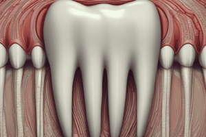
Enamel Rods
- Millions of enamel rods (prisms) make up the structure of enamel.
- They are tightly packed and organized in a keyhole shape in cross-section.
- Each rod contains millions of calcium hydroxyapatite crystallites.
- The keyhole shape reflects different parts of the enamel rod – a head and a tail.
- The head of each rod is usually oriented towards the occlusal/incisal surface relative to the tail which is oriented towards the cervical region.
- The tail is also known as an interrod.
- Each rod and interrod is surrounded by a sheath (organic material).
- There are millions of crystallites (hydroxyapatite) in each rod tightly packed in keyhole shapes.
- These are extremely long, thin and ribbon-like, they may run the thickness of enamel.
- The crystallites in the head are parallel with the long axis of the rod, and the tail diverge slightly.
- The pattern of the crystallites within the rod adds to the strength of enamel.
Enamel Rods – Orientation
- The direction of the rods varies to account for the shape of the tooth.
- At the cervical margin, they are directed more horizontally-apically, and at the cusp tips, they are almost vertical.
- The overall thickness of enamel also varies with it being thickest at the cusp tips and incisal edges, and thinnest at the cervical margins.
Enamel Rods - Clinical Significance
- The direction of the rods is a key consideration in cavity preparation to avoid unsupported enamel that will fracture, leading to failure.
Surface Enamel
- The very first and last formed enamel shows no usual prism structure where the crystals are parallel with the surface.
Body Enamel
- It is 30 microns wide at the surface, highly radio-opaque, harder and less soluble – includes more fluoride and carbon.
Clinical Significance of Surface Enamel
- It is seen in the primary dentition and 70% of the permanent dentition, greatest in the cervical regions.
- It may interfere with optimal etching.
Dentine-Enamel Junction (DEJ)
- The junction between enamel and dentine that forms once dentinogenesis and amelogenesis have started.
- Scalloped appearance under a microscope thought to strengthen the bond between the two materials almost locking them together.
Structural Features at the DEJ
- Enamel Tuft: Thought to result from abrupt changes in the direction of the enamel rods due to the scalloped boundary of enamel at the DEJ, possibly supports the bond between dentine and enamel, no known clinical significance.
Enamel Spindles
- Extension of dentine tubules into enamel.
- May result from odontoblast processes extending into the ameloblast layer becoming trapped.
- Contribute to minor sensitivity
Hunter Schreger Bands
- Light and dark bands under a light microscope.
- Longitudinal section runs upwards from dentine.
- Cross-section appears as growth rings.
Lamella
- Appear as cracks in enamel - developmental defects.
- Appear as jagged lines in the surface of the crown clinically.
- Extend inwards maybe as far as the dento-enamel junction.
- Result of ameloblast ceasing production of enamel.
- Can be mistaken for cracks in enamel and vice versa.
Functions of Enamel
- Protection of the tooth/pulp
- Eating
- Inability to repair or feel injury
- Able to remineralise and demineralise – ion exchange
- Smile - Aesthetically appealing ‘pearly whites’
How Enamel Structure Links to Function
- Protection: Thickest at cusp tips, occlusal and incisal surfaces.
- Eating: Covers the entire tooth crown.
- Inability to repair or feel injury: Inert tissue (no living cell due to ameloblasts’ limited lifecycle).
- Remineralization/Demineralization: Hardest biological tissue, Highly mineralized tissue, White translucent crystallite, Permeable 'micropores'.
Changes in Enamel Over the Life-Course
- Over time enamel is subject to tooth wear including:
- Attrition
- Abrasion
- Erosion
- For example, perkymata are worn away, scratches and cracks develop.
- Colour changes – reduced translucency and an increase in underlying dentine makes enamel appear yellower – normal aging process.
- Over time enamel is subject to reduced 'permeability' – exchange of ions such as Ca, PO, F-.
- Clinical significance for exposure to topical fluoride as a younger tooth and in the progression of early enamel lesions.
Demineralization-Remineralisation Cycle
- As a mineralised structure, enamel is subject to demineralisation (loss of mineral) and remineralisation (uptake of mineral).
- In acidic conditions, the balance favours demineralisation.
- In alkaline conditions, the balance favours remineralisation enabling uptake of fluoride and calcium phosphate.
- The critical pH of enamel is 5.5.
Clinical Application – Preventive and Restorative
- Fluoride: Enamel that has fluoride incorporated (fluorapatite) has a critical pH of 4.5, lower than hydroxyapatite, thus more resistant to acids and demin.
- Acid etch: Acid removes minerals from the enamel surface creating ‘tags’ that enable the bond to fill in and stick the composite to.
The Significance of the DEJ and Caries
- Take a close look at:
- The breakdown of enamel
- The progression into dentine and to the pulp
- Note the difference in size of the lesion in enamel compared to the lesion in dentine at the DEJ.
Key Areas of Research Focus
- Understanding how enamel can be regenerated.
Studying That Suits You
Use AI to generate personalized quizzes and flashcards to suit your learning preferences.