Podcast
Questions and Answers
During which stage of early embryonic development does the blastocoel form?
During which stage of early embryonic development does the blastocoel form?
- Gastrula
- Zygote
- Morula
- Blastocyst (correct)
Which of the following is the primary role of the trophoblast in early embryonic development?
Which of the following is the primary role of the trophoblast in early embryonic development?
- Development into the embryo itself
- Formation of the inner cell mass
- Formation of the blastocoel
- Development into extraembryonic tissues like the placenta (correct)
What key event must occur for development to proceed beyond the blastocyst stage?
What key event must occur for development to proceed beyond the blastocyst stage?
- Formation of the morula
- Secretion of estrogen
- Differentiation of the inner cell mass
- Implantation into the uterine lining (correct)
What hormonal change in the mother directly facilitates the implantation of the blastocyst?
What hormonal change in the mother directly facilitates the implantation of the blastocyst?
If implantation is successful, what hormone is secreted by the developing trophoblastic cells, and what is its primary effect?
If implantation is successful, what hormone is secreted by the developing trophoblastic cells, and what is its primary effect?
How does the endometrium support the developing embryo before the placenta is fully formed?
How does the endometrium support the developing embryo before the placenta is fully formed?
What is the correct sequence of pre-implantation development?
What is the correct sequence of pre-implantation development?
A woman is tested for pregnancy and the results show very low levels of human chorionic gonadotropin (hCG). What might this indicate?
A woman is tested for pregnancy and the results show very low levels of human chorionic gonadotropin (hCG). What might this indicate?
At what stage of meiosis is the secondary oocyte arrested until fertilization?
At what stage of meiosis is the secondary oocyte arrested until fertilization?
Which structures surround the secondary oocyte at the time of ovulation?
Which structures surround the secondary oocyte at the time of ovulation?
What event triggers the secondary oocyte to complete meiosis II?
What event triggers the secondary oocyte to complete meiosis II?
What is the immediate result of the fusion of male and female pronuclei?
What is the immediate result of the fusion of male and female pronuclei?
How does oogenesis differ from spermatogenesis in terms of gamete production from each original germ cell?
How does oogenesis differ from spermatogenesis in terms of gamete production from each original germ cell?
Why does the development of female reproductive structures occur in XX embryos?
Why does the development of female reproductive structures occur in XX embryos?
Which of the following statements regarding oogenesis is correct?
Which of the following statements regarding oogenesis is correct?
What is the role of anti-Müllerian hormone (AMH) in the development of reproductive systems during embryogenesis?
What is the role of anti-Müllerian hormone (AMH) in the development of reproductive systems during embryogenesis?
During the luteal phase of the ovarian cycle, what is the primary effect of the elevated levels of progesterone and estrogens?
During the luteal phase of the ovarian cycle, what is the primary effect of the elevated levels of progesterone and estrogens?
What event marks the beginning of the menstrual phase of the uterine cycle?
What event marks the beginning of the menstrual phase of the uterine cycle?
During the secretory phase of the uterine cycle, what is the main effect of increased progesterone secretion from the corpus luteum?
During the secretory phase of the uterine cycle, what is the main effect of increased progesterone secretion from the corpus luteum?
If fertilization of a secondary oocyte occurs, what happens to the corpus luteum, and what is the consequence of this?
If fertilization of a secondary oocyte occurs, what happens to the corpus luteum, and what is the consequence of this?
A woman is experiencing a shortened luteal phase. Which of the following hormonal imbalances is most likely contributing to this issue?
A woman is experiencing a shortened luteal phase. Which of the following hormonal imbalances is most likely contributing to this issue?
A woman on day 2 of her menstrual cycle typically experiences which of the following hormonal and physiological conditions?
A woman on day 2 of her menstrual cycle typically experiences which of the following hormonal and physiological conditions?
How would a significant decrease in estrogen levels affect the HPG axis in a menopausal individual?
How would a significant decrease in estrogen levels affect the HPG axis in a menopausal individual?
A drug inhibits the production of prostaglandins in the uterus. How would this drug most likely affect the menstrual cycle?
A drug inhibits the production of prostaglandins in the uterus. How would this drug most likely affect the menstrual cycle?
Which of the following events occurs first during fertilization?
Which of the following events occurs first during fertilization?
What is the primary role of the fimbriae in the context of fertilization?
What is the primary role of the fimbriae in the context of fertilization?
How does the acrosome reaction contribute to the process of fertilization?
How does the acrosome reaction contribute to the process of fertilization?
What cellular components of the sperm enter the oocyte during fertilization?
What cellular components of the sperm enter the oocyte during fertilization?
Why is capacitation essential for successful fertilization?
Why is capacitation essential for successful fertilization?
Which structure directly mediates the initial binding of the sperm to the outer layers of the oocyte?
Which structure directly mediates the initial binding of the sperm to the outer layers of the oocyte?
What is the zona pellucida?
What is the zona pellucida?
What is the role of the increased motility that occurs during capacitation?
What is the role of the increased motility that occurs during capacitation?
What is the primary function of gastrulation in embryonic development?
What is the primary function of gastrulation in embryonic development?
Which of the following structures is responsible for establishing the anterior-posterior axis during gastrulation?
Which of the following structures is responsible for establishing the anterior-posterior axis during gastrulation?
If the migration of mesodermal cells was inhibited during gastrulation, which of the following structures would be most affected?
If the migration of mesodermal cells was inhibited during gastrulation, which of the following structures would be most affected?
Which of the following adult structures is derived from the blastopore?
Which of the following adult structures is derived from the blastopore?
Which germ layer primarily contributes to the development of the nervous system and epidermis?
Which germ layer primarily contributes to the development of the nervous system and epidermis?
Which of the following represents the correct order of events during early embryonic development, leading up to gastrulation?
Which of the following represents the correct order of events during early embryonic development, leading up to gastrulation?
What will happen if the hypoblast fails to develop properly during the bilaminar stage?
What will happen if the hypoblast fails to develop properly during the bilaminar stage?
During gastrulation, cells migrate through the primitive streak to form different germ layers. What determines the eventual fate of these migrating cells?
During gastrulation, cells migrate through the primitive streak to form different germ layers. What determines the eventual fate of these migrating cells?
During neurulation, what process directly leads to the formation of the neural tube?
During neurulation, what process directly leads to the formation of the neural tube?
Neural crest cells are crucial for the development of various cell types. Which of the following is NOT a derivative of neural crest cells?
Neural crest cells are crucial for the development of various cell types. Which of the following is NOT a derivative of neural crest cells?
Somites, formed during organogenesis, are precursor cells for which of the following structures?
Somites, formed during organogenesis, are precursor cells for which of the following structures?
Which event occurs concurrently with the segmentation of mesodermal tissues into somites?
Which event occurs concurrently with the segmentation of mesodermal tissues into somites?
If the process of neural crest cell delamination was disrupted during neurulation, which of the following developmental defects would most likely occur?
If the process of neural crest cell delamination was disrupted during neurulation, which of the following developmental defects would most likely occur?
Ectoderm, mesoderm, and endoderm give rise to specific tissues and organs. The adrenal medulla originates from which of these germ layers, via neural crest cells?
Ectoderm, mesoderm, and endoderm give rise to specific tissues and organs. The adrenal medulla originates from which of these germ layers, via neural crest cells?
Consider a scenario where somite development is impaired during embryogenesis. Which of the following structures would be most directly affected?
Consider a scenario where somite development is impaired during embryogenesis. Which of the following structures would be most directly affected?
During a developmental study, researchers identify a signaling molecule crucial for the proper migration of neural crest cells. If this signaling pathway is blocked, predict which of the following cell types would be most affected?
During a developmental study, researchers identify a signaling molecule crucial for the proper migration of neural crest cells. If this signaling pathway is blocked, predict which of the following cell types would be most affected?
Flashcards
Meiosis II arrest
Meiosis II arrest
Meiosis II starts in the secondary oocyte but pauses at metaphase II until fertilization.
Zona pellucida
Zona pellucida
A thick glycoprotein layer surrounding the secondary oocyte.
Corona radiata
Corona radiata
Granulosa cells surrounding the secondary oocyte.
Uterine (Fallopian) tube
Uterine (Fallopian) tube
Signup and view all the flashcards
Ovum
Ovum
Signup and view all the flashcards
Pronuclei Fusion
Pronuclei Fusion
Signup and view all the flashcards
Menopause
Menopause
Signup and view all the flashcards
Müllerian Ducts
Müllerian Ducts
Signup and view all the flashcards
Luteal Phase
Luteal Phase
Signup and view all the flashcards
Corpus Luteum
Corpus Luteum
Signup and view all the flashcards
HPG Axis Feedback in Luteal Phase
HPG Axis Feedback in Luteal Phase
Signup and view all the flashcards
Menstrual Phase
Menstrual Phase
Signup and view all the flashcards
Menstruation (Menses)
Menstruation (Menses)
Signup and view all the flashcards
Proliferative Phase
Proliferative Phase
Signup and view all the flashcards
Secretory Phase
Secretory Phase
Signup and view all the flashcards
Pre-Implantation Development
Pre-Implantation Development
Signup and view all the flashcards
Placenta
Placenta
Signup and view all the flashcards
Fertilization
Fertilization
Signup and view all the flashcards
Ovulation
Ovulation
Signup and view all the flashcards
Capacitation
Capacitation
Signup and view all the flashcards
Acrosome Reaction
Acrosome Reaction
Signup and view all the flashcards
Epiblast
Epiblast
Signup and view all the flashcards
Hypoblast
Hypoblast
Signup and view all the flashcards
Bilaminar Disk
Bilaminar Disk
Signup and view all the flashcards
Gastrulation
Gastrulation
Signup and view all the flashcards
Primary Germ Layers
Primary Germ Layers
Signup and view all the flashcards
Primitive Streak
Primitive Streak
Signup and view all the flashcards
Blastopore
Blastopore
Signup and view all the flashcards
Archenteron
Archenteron
Signup and view all the flashcards
Morula
Morula
Signup and view all the flashcards
Blastocoel
Blastocoel
Signup and view all the flashcards
Trophoblast
Trophoblast
Signup and view all the flashcards
Inner Cell Mass
Inner Cell Mass
Signup and view all the flashcards
Implantation
Implantation
Signup and view all the flashcards
Embryo
Embryo
Signup and view all the flashcards
Human Chorionic Gonadotropin (hCG)
Human Chorionic Gonadotropin (hCG)
Signup and view all the flashcards
Blastocyst
Blastocyst
Signup and view all the flashcards
Neural Tube
Neural Tube
Signup and view all the flashcards
Delamination
Delamination
Signup and view all the flashcards
Neural Crest Cells
Neural Crest Cells
Signup and view all the flashcards
Somites
Somites
Signup and view all the flashcards
Melanocytes
Melanocytes
Signup and view all the flashcards
Calcitonin-Producing Thyroid Cells
Calcitonin-Producing Thyroid Cells
Signup and view all the flashcards
Neural Crest Derivatives
Neural Crest Derivatives
Signup and view all the flashcards
Migration of Neural Folds
Migration of Neural Folds
Signup and view all the flashcards
Study Notes
- The female reproductive system generates oocytes, provides a supportive environment for fertilization, and supports offspring growth.
- This lesson covers female reproductive anatomy, oogenesis, and hormone regulation.
Female Reproductive Anatomy
- The internal and external structures of the female reproductive system produce oocytes, receive sperm, and support fertilization, gestation, and offspring nourishment.
- Ovaries are reproductive gonads that secrete sex hormones like estrogens and progesterone and serve as oogenesis sites.
- Uterine or fallopian tubes are muscular tubes extending from the uterus toward each ovary.
- Fimbriae are fingerlike projections that direct oocytes from the abdominal cavity into the uterine tubes as the open ends of uterine tubes are near, but not connected to, the ovaries.
- Ciliated cells propel oocytes toward the uterus inside uterine tubes, which is also where fertilization typically occurs.
- The uterus is a muscular organ protecting and nourishing the embryo and fetus.
- The endometrium is the inner lining of the uterus that changes in thickness during the menstrual cycle.
- The myometrium is the thick, smooth muscle layer of the uterus that contracts during menstruation and childbirth.
- The cervix is the inferior portion of the uterus and opens into the vagina.
- The vagina is a muscular tube that eliminates menstrual fluids, receives the penis during intercourse, and serves as the birth canal's final segment.
- The female external genitalia, or vulva, includes the labia majora and labia minora, that protects the vagina and clitoris opening.
- The clitoris' external portion is at the junction of the labia minora.
- Clitoris stimulation triggers genital changes, such as lubrication and pH changes, that facilitate reproduction.
- Mammary glands are chest wall accessory glands that fully develop during pregnancy to facilitate lactation, which is breast milk synthesis, secretion, and nursing.
- Ovaries are female gonads homologous to testes that are surrounded by a fibrous capsule covered by epithelial cells.
- The cortex contains ovarian follicles in maturation stages with each follicle containing an immature primary oocyte surrounded by epithelial cells.
- Follicular cells mature into granulosa cells, which support developing oocytes that secrete sex steroid hormones during the ovarian cycle.
Oogenesis
- Oogenesis begins during embryogenesis as oogonia stem cells undergo mitotic division within developing ovaries.
- Oogonia start meiosis I at puberty, forming primary oocytes during the fetal period.
- Primary oocytes are paused in late prophase I of meiosis I and do not resume until puberty in response to hormonal changes.
- At puberty, primary oocytes are surrounded by follicular support cells in the ovary, which form ovarian follicles.
- Unequal cell division of a primary oocyte completing meiosis I result in a larger haploid secondary oocyte and smaller first polar body, which undergoes apoptosis and degenerates.
- The secondary oocyte initiates meiosis II but arrests at metaphase II until fertilization.
- During ovulation, the ovarian follicle ruptures and releases the secondary oocyte, as it is surrounded by the zona pellucida and the corona radiata, into the abdominal cavity.
- The secondary oocyte is drawn into the uterine or fallopian tube, where sperm cell fertilization can occur.
- Successful fertilization leads to the secondary oocyte completing meiosis II, forming a large ovum and a small second polar body that degenerates.
- Male and female pronuclei fuse to form a diploid zygote upon complete fertilization.
- Each oogonium yields one haploid ovum and two to three polar bodies while spermatogenesis generates four haploid sperm.
- Oogenesis begins before birth and can result in mature gametes 13-50 years later.
- Oogenesis ceases during menopause as sex hormone levels decline with age.
- The primary oocytes formed during the fetal period are not replenished later, unlike spermatogenesis, which occurs throughout life after puberty.
Hormonal Control of the Female Reproductive System
- Male reproductive organs begin development in XY embryos due to SRY gene expression during early embryogenesis.
- Anti-Müllerian hormone (AMH) is not produced in XX embryos because SRY is absent and testes typically do not develop.
- Female reproductive structures are derived from Müllerian ducts, and Wolffian ducts degenerate due to a lack of testosterone.
- Oogenesis begins during the fetal period with the production of follicles containing primary oocytes.
- Progression of oogenesis is repressed and sexual development remains dormant until puberty due to the low sex hormone levels during infancy and childhood.
- Estrogen and progesterone promote female reproductive organ growth and maturation, development of secondary sex characteristics, resumption of oogenesis, and initiation of the menstrual cycle at puberty.
- The hypothalamic-pituitary-gonadal (HPG) axis regulates cyclical changes for reproduction.
- The female reproductive cycle consists of the ovarian cycle for primary oocyte maturation and the uterine cycle for uterus preparation for pregnancy.
- Ovarian and uterine cycles occur concurrently, with a single female reproductive cycle lasting on average 28 days.
- Synchronization of the ovarian and uterine cycles provides optimal conditions for fertilization support and early pregnancy.
- The ovarian cycle divides into three phases.
- Follicular phase (days 1–13): HPG axis stimulation results in gonadotropin-releasing hormone (GnRH) release from the hypothalamus, stimulating the anterior pituitary gland to release small amounts of follicle-stimulating hormone (FSH) and luteinizing hormone (LH).
- FSH and LH stimulate developing ovarian follicles to release estrogens.
- Low estrogen levels exert negative feedback on the HPG axis during the early follicular phase allowing only one follicle to survive.
- The dominant follicle secretes inhibin to inhibit FSH, repressing maturation of additional follicles.
- Although estrogen initially inhibits the HPG axis, the emergence of a dominant follicle stimulates the secretion of high estrogen levels, which exerts a stimulatory effect on the HPG axis during the late follicular phase.
- This effect results in a surge of LH and, to a lesser extent, FSH.
- Ovulation (day 14): The mature ovarian follicle ruptures soon after the LH surge and releases a secondary oocyte into the abdominal cavity, which is then directed into a nearby uterine tube.
- Luteal phase (days 15–28): LH stimulates the ruptured follicle's conversion into the corpus luteum, which secretes high levels of progesterone and estrogens to exert a negative feedback on the HPG axis.
- Lower FSH and LH levels during the luteal phase prevent maturation of additional follicles.
- The corpus luteum degenerates, causing a sharp decline in progesterone and estrogens, and the inhibition of FSH and LH is relieved, allowing the next ovarian cycle to begin if fertilization does not occur.
- The corpus luteum persists and continues to secrete progesterone and estrogens if the secondary oocyte is fertilized following ovulation.
- The continued presence of progesterone and estrogens keeps FSH and LH levels low, promoting the pregnancy and preventing a new ovarian cycle from being initiated during pregnancy.
- The uterine or menstrual cycle occurs concurrently with the ovarian cycle and is divided into three phases.
- Menstrual phase (days 1–4): If fertilization of a secondary oocyte does not occur in the prior uterine cycle, menstruation begins.
- The uterus sheds the majority of its endometrial layer in menses, and detached tissue and blood pass out of the body through the vagina.
- Ovarian follicles are stimulated to grow and produce estrogens, leading to the cessation of blood flow toward the end of this phase.
- Proliferative phase (days 5-14): The endometrial layer proliferates and doubles in thickness.
- Endometrial glands develop, and vascularization of the endometrium is increased to prepare for embryo implantation.
- Secretory phase (days 15-28): Increased progesterone and estrogen secretion from the corpus luteum triggers the further thickening and development of the endometrium.
- These changes result in nutrient secretion from endometrial glands and environmental creation to sustain a developing embryo in the event of fertilization and implantation.
Pre-Implantation Development
- Embryonic development begins when male and female gametes join to form a zygote, referred to as fertilization.
- Fertilization typically takes place in a uterine tube within 12-24 hours after ovulation, the developing embryo must then travel to the uterine cavity and implant for a viable pregnancy to occur.
- The developmental steps between fertilization and implantation take place within 12 days after ovulation.
- This lesson covers pre-implantation development from fertilization to implantation completion, as well as placenta development.
- Fertilization becomes possible with secondary oocyte release from the ovary during ovulation as it travels into abdominal cavity and is drawn into the uterine tube by fimbriae.
- The ciliated lining of the uterine tube propels the oocyte into the uterine cavity and sperm typically make contact with the oocyte within the uterine tube.
- Successful fertilization requires the following steps
- Capacitation: Sperm must undergo a final maturation step, known as capacitation to reach the oocyte.
- Female reproductive tract secretions induce changes in plasma membrane permeability at the sperm head and trigger increased flagellar movement (motility).
- Contact with oocyte: Sperm moves toward the zona pellucida through the corona radiata. Sperm head receptors bind to glycoproteins in the zona pellucida.
- Acrosome reaction: Hydrolytic enzymes from the acrosome (specialized vesicle) are released near the oocyte, leading to zona pellucida degradation enabling the sperm to reach the oocyte's plasma membrane.
- Fusion: Oocyte and sperm plasma membranes fuse, triggering oocyte plasma membrane depolarization.
- Sperm contents entering the oocyte: The sperm nucleus, mitochondria, and a pair of centrioles enter the oocyte but most sperm mitochondria are destroyed.
- Cortical reaction: Oocyte plasma membrane depolarization leads to increased intracellular calcium levels after sperm fusion, which triggers cortical granule fusion with the oocyte's plasma membrane.
- Hydrolytic enzymes are released into the space between the plasma membrane and the zona pellucida, causing the zona pellucida to lift away from the oocyte and harden and resulting in the formation of a protective envelope that blocks additional sperm from entering in polyspermy.
- Sperm contents enter the oocyte after sperm and oocyte plasma membrane fusion.
- The secondary oocyte must then complete meiosis II, dividing to form a mature ovum and second polar body that degenerates.
- The nucleus of the mature ovum develops into a female pronucleus. Simultaneously, the male pronucleus is propelled toward the female pronucleus.
- Fusion of haploid pronuclei produces a diploid zygote.
- Epigenetic modifications occur immediately following zygote formation.
- Within 24 hours after fertilization, the zygote begins a period of rapid mitotic cell division in embryonic cleavage, where cell division proceeds without concurrent cell growth, forming smaller daughter cells.
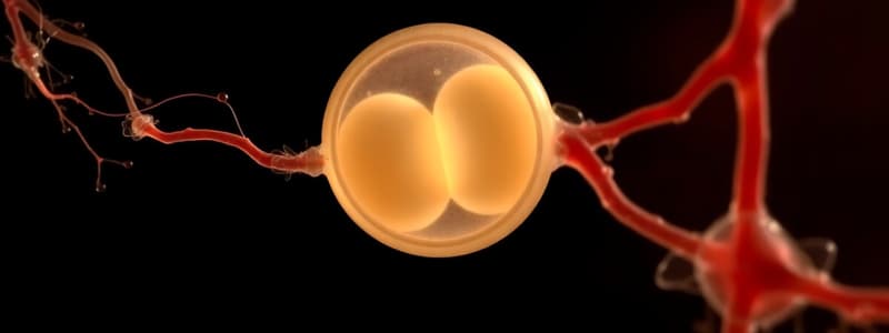
- After 3-4 days the embryo enters the morula stage, which is roughly the same size as the original zygote and consists of 16–32 blastomeres.
- The developing embryo travels to the uterine cavity during the transition from zygote to morula.
- By days 4-5, the dividing embryonic cells reorganize into a blastocyst, which contains a bastocoal, which is a fluid-filled cavity.
- Blastocysts contains Inner cell mass cells, which develop in embryo and trophoblast, of the outer cell that develops extraembryonic tissues.
Implantation and Placental Development
- The blastocyst must implant in the uterine lining for development to continue.
- Progesterone and estrogen surges trigger the secretory phase of the uterine cycle by 6–7 days after fertilization.
- The endometrial lining is primed to support blastocyst implantation.
- Developing trophoblastic cells adhere to the endometrial surface causing the blastocyst to burrow into the endometrial lining resulting in endometrial cells that proliferate and surround the embryo.
- Until the placenta is completely formed, the endometrium supports and nourishes the embryo.
- Implantation is usually complete by day 12.
- The corpus luteum typically degenerates and progesterone and estrogen levels decline if fertilization does not occur allowing for the triggering of menstruation.
- The developing trophoblastic cells secrete human chorionic gonadotropin (hCG) with successful implantation, and the corpus luteum is maintained while the placenta develops.
- The placenta forms over the first 3 months of pregnancy to facilitate nutrient, gas, and waste exchange between the maternal circulation and the developing embryo.
- The chorion is an extraembryonic membrane derived from the trophoblast after implantation as chorionic villi that invade the endometrium and form from the chorion and become vascularized.
- The chorion and chorionic villi form the bulk of the placenta.
- The corpus luteum continues to secrete estrogen and progesterone for approximately the first 9 weeks of pregnancy, effectively inhibiting the initiation of another menstrual cycle.
- The developing placenta gradually takes over secretion of hCG, estrogens, and progesterone to support the pregnancy, leading to corpus luteum degeneration.
- Yolk sac: exchange occurs and embryonic blood cell production takes place.
- Allantois: Fluid and waste exchange structure.
- Amnion: A tough membrane filled with aminiotic fluid surrounding the embryo in which placenta recieves blood.
Post-Implantation Development
- Successful blastocyst implantation signals the beginning of the embryonic period of development.
- Organogenesis: This (development of embryonic tissues and organs) begins with gastrulation, a period of rapid growth and embryonic cells.
- The three primary germ layers are established by the end of gastrulation, and mesodermal tissue induces the overlying ectoderm to become neural tissue in neurulation.
Gastrulation
- At the implantation point, the blastocyst becomes formed through 2 major cells called.
- The trophoblast cell and inner cell mass.
- 2 major cell types are called epiblast and hypoblast.
- The epiblast cells form the amnion and embryo, and that of the hypoblast form a certain amount of chorion and yolk sac.
- The primary germ layers arise from which all embryonic tissues and organs:
- Ectoderm
- Mesoderm - endoderm.
- The formation of a groove makes a primitive streak which has a posterior-anterior of the embryo.
- The blastopore or the first point of invangination forms endodermal cells.
Germ Layer Derivatives
- The ectoderm develops into the skin and nervous system.
- The mesoderm gives rise to muscle, bone, and other connective tissues.
- The endoderm forms the lining of the digestive tract and other internal organs.
Organogenesis and Neurulation
- The formation of specific and organ systems begins after primary germ layer estabishment.
- Ectodermal cells form neural tissues neurulation
- Three ectodermal cell types: -Epidermal -Neural Crest -Neural plate
- Embryos at this stage are neurulas.
- The thickening and elogation of the neural plate shows in the response by undermesodermal to show signs.
- Movement of the lateral edges of the neural folds towards the midline.
- Detatch and mirgation of neural crest cells for neurulation
Studying That Suits You
Use AI to generate personalized quizzes and flashcards to suit your learning preferences.


