Podcast
Questions and Answers
What does the neural tube give rise to?
What does the neural tube give rise to?
- Notochord
- Central nervous system (correct)
- Ganglion
- Peripheral nervous system
Which structure provides support and induces differentiation of the neuroectoderm?
Which structure provides support and induces differentiation of the neuroectoderm?
- Neural tube
- Neural crest
- Notochord (correct)
- Ectoderm
What type of fibers are myelinated at a speed of 100 m/s?
What type of fibers are myelinated at a speed of 100 m/s?
- D fibers
- C fibers
- A fibers (correct)
- B fibers
What type of glial cell produces myelin in the central nervous system?
What type of glial cell produces myelin in the central nervous system?
In which layer of embryonic development does the nervous system arise?
In which layer of embryonic development does the nervous system arise?
Which of the following statements is true regarding myelinated and unmyelinated fibers?
Which of the following statements is true regarding myelinated and unmyelinated fibers?
What can occur when the nucleus pulposus bulges?
What can occur when the nucleus pulposus bulges?
What characteristic distinguishes the structure of the spinal cord?
What characteristic distinguishes the structure of the spinal cord?
What is the primary function of the vitreous body?
What is the primary function of the vitreous body?
Which component supports the structure of the lens?
Which component supports the structure of the lens?
What are the two types of photoreceptors in the retina?
What are the two types of photoreceptors in the retina?
How does the lens change shape?
How does the lens change shape?
Which structure is responsible for converting light into electrical signals in the retina?
Which structure is responsible for converting light into electrical signals in the retina?
What role does the choroid layer of the eye serve?
What role does the choroid layer of the eye serve?
Which layer of the eye is responsible for generating visual information sent to the brain?
Which layer of the eye is responsible for generating visual information sent to the brain?
What is the primary purpose of the lens in the eye?
What is the primary purpose of the lens in the eye?
What is the primary role of the hippocampus in memory?
What is the primary role of the hippocampus in memory?
How does long-term potentiation contribute to memory?
How does long-term potentiation contribute to memory?
Which part of the brain is involved in emotional memory formation?
Which part of the brain is involved in emotional memory formation?
What is the main function of the ventral tegmental area (VTA)?
What is the main function of the ventral tegmental area (VTA)?
What is the process of memory formation?
What is the process of memory formation?
Which neurotransmitter is primarily involved in pleasure and reward?
Which neurotransmitter is primarily involved in pleasure and reward?
What role does the hypothalamus play in relation to sexual motivation?
What role does the hypothalamus play in relation to sexual motivation?
Which of the following is NOT a function of the limbic system?
Which of the following is NOT a function of the limbic system?
What is the primary function of the cochlea?
What is the primary function of the cochlea?
Which of the following accurately describes the scala media?
Which of the following accurately describes the scala media?
What does the sympathetic nervous system primarily prepare the body for?
What does the sympathetic nervous system primarily prepare the body for?
What type of signals do afferent neurons carry?
What type of signals do afferent neurons carry?
Which part of the autonomic nervous system promotes relaxation and restoration?
Which part of the autonomic nervous system promotes relaxation and restoration?
What is the role of the postganglionic neuron?
What is the role of the postganglionic neuron?
What neurotransmitter is primarily used by the parasympathetic nervous system?
What neurotransmitter is primarily used by the parasympathetic nervous system?
Where does the parasympathetic nervous system originate?
Where does the parasympathetic nervous system originate?
What is the primary effect of sympathetic stimulation on the heart?
What is the primary effect of sympathetic stimulation on the heart?
Which of the following is a characteristic of the parasympathetic nervous system?
Which of the following is a characteristic of the parasympathetic nervous system?
What neurotransmitters are used in the sympathetic nervous system?
What neurotransmitters are used in the sympathetic nervous system?
Which of the following effects is associated with parasympathetic stimulation of the digestive tract?
Which of the following effects is associated with parasympathetic stimulation of the digestive tract?
How do nicotinic and muscarinic receptors differ in the parasympathetic nervous system?
How do nicotinic and muscarinic receptors differ in the parasympathetic nervous system?
Which organs primarily experience increased respiration as a result of sympathetic stimulation?
Which organs primarily experience increased respiration as a result of sympathetic stimulation?
Where does the parasympathetic nervous system primarily originate?
Where does the parasympathetic nervous system primarily originate?
What is the effect of sympathetic stimulation on the pupils of the eyes?
What is the effect of sympathetic stimulation on the pupils of the eyes?
Flashcards are hidden until you start studying
Study Notes
Key Structures
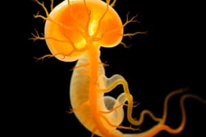
- Neural tube: Develops from neuroectoderm and gives rise to the central nervous system (CNS).
- Neural crest: Also originates from neuroectoderm and forms the peripheral nervous system (PNS).
- Notochord: Formed from mesoderm, providing support and inducing differentiation of neuroectoderm.
- Ganglion: A cluster of nerve cell bodies in the PNS.
- Nerve: A bundle of axons in the PNS.
- Tract: A bundle of axons in the CNS.
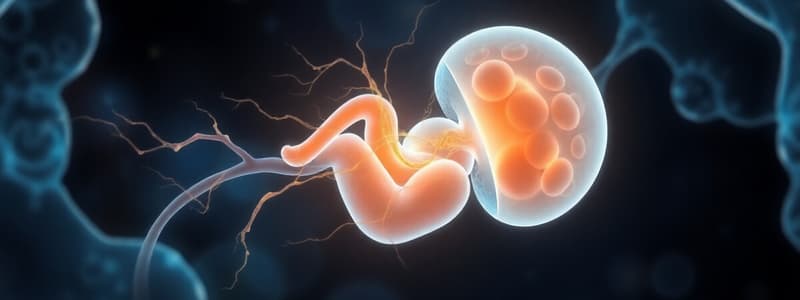
Embryonic Development of the Nervous System
- The ectoderm gives rise to the nervous system.
- The mesoderm forms the notochord, bones, and cartilage.
- The endoderm develops into the lining of the digestive tract and other internal organs.
Intervertebral Discs and Herniation
- The nucleus pulposus, a gel-like substance, is located at the center of intervertebral discs.
- Disc herniation occurs when the nucleus pulposus bulges, potentially causing neuropathic pain.
- Sciatica can be a result of disc herniation.
Neurons and Myelination
- All neurons possess a neural sheath enabling regeneration.
- Some neurons are myelinated, while others are not.
- Myelinated fibers include A and B fibers with faster conduction speeds.
- Unmyelinated fibers are C fibers with slower conduction speeds.
Myelin Production
- Oligodendrocytes produce myelin in the CNS.
- Schwann cells produce myelin in the PNS.
Spinal Cord Structure
- The spinal cord's white matter, containing myelinated axons, is located externally.
- Grey matter, composed of neuronal cell bodies, is found internally in a H-shape.
The Hippocampus and Memory
- Plays a crucial role in forming memories, especially emotionally charged ones.
- Consolidates short-term memories into long-term storage.
- Communicates with the fornix and cingulate gyrus for memory encoding and storage.
Memory Formation and Consolidation
- Memory formation involves creating new memories, primarily explicit ones.
- Memory consolidation strengthens and stabilizes memories over time, making them easier to recall.
Limbic System Functions
- The limbic system is a network of brain structures responsible for memory, emotions, motivation, and homeostasis.
Memory and Learning
- Long-term potentiation strengthens neural connections, allowing for memory formation.
- The hippocampus and amygdala work together to create emotional memories.
- The olfactory bulb plays a role in emotional memories connected to smells.
Motivation and Reward
- The ventral tegmental area (VTA) contains dopaminergic neurons that release dopamine, a neurotransmitter associated with pleasure, reward, and motor function.
- The VTA projects to the nucleus accumbens, involved in processing pleasure.
- Dopamine plays a role in controlling pleasure, reward, and motor function.
Sex Motivation
- The hypothalamus controls sex motivation and releases hormones like oxytocin and testosterone.
- The hypothalamus is often referred to as the "king" or "queen" of the brain.
The Vitreous Body
- The vitreous body is a gelatinous substance that fills the space between the lens and the retina.
- It maintains the eye’s shape and pressure.
- Composed of gelatinous material and fibers.
The Lens
- The lens is a clear, flexible structure that focuses light on the retina.
- It's made up of lens fibers, lens capsule, and epithelium.
- The lens changes shape by constricting or relaxing the ciliary muscles, which affects the compactness of lens fibers, ultimately adjusting focus.
The Retina
- The innermost layer of the eye, responsible for detecting light and transmitting visual information to the brain.
- Contains photoreceptors, specifically rods and cones, which convert light into electrical signals.
- Rods detect black and white vision.
- Cones are responsible for color vision.
- Photoreceptors connect to bipolar cells, the first sensory cells in the retina.
The Eye Structure
- Vascular layer (choroid): Contains dense, irregular connective tissue and is highly vascularized.
- Sclera: Provides protection to the eye.
- Retina: Composed of layers, including the photoreceptor layer which contains light-sensitive cells: rods and cones.
- Optic nerve: Transmits visual information from the retina to the brain.
Cochlea
- A spiral-shaped structure responsible for converting sound waves into electrical signals.
- Contains three sections:
- Scala vestibuli: Filled with fluid, transmitting sound waves.
- Scala media: Also contains fluid and helps with sound wave transmission.
- Scala tympani: Filled with fluid and involved in sound wave transmission.
- Hair cells within the cochlea are connected to afferent neurons and transmit sound information to the brain.
Understanding Sensory Receptors and Functions
- Efferent Nervous System: Carries signals away from the CNS to the body.
- Afferent Nervous System: Carries sensory information from the body toward the CNS.
- Autonomic Nervous System (ANS): Part of the efferent system, controls involuntary actions such as heart rate, digestion, and breathing. Divided into two branches:
Autonomic Nervous System Overview
- Sympathetic Nervous System: Responsible for the "fight or flight" response, preparing the body for immediate action.
- Parasympathetic Nervous System: Responsible for the "rest and digest" response, promoting relaxation and restoration.
How the Autonomic Nervous System Works
- The ANS collects sensory information and sends it to the CNS for processing.
- The CNS then transmits signals to the body through two neurons:
- Preganglionic Neuron: Connects the CNS to a ganglion.
- Postganglionic Neuron: Connects the ganglion to the target organ.
Parasympathetic Nervous System
- Location: Starts in the brain stem and sacral area.
- Effect: Promotes relaxation and restoration.
- Transmitters: Utilizes acetylcholine as a neurotransmitter.
- Receptors: Utilizes nicotinic receptors between pre- and postganglionic neurons and muscarinic receptors between postganglionic neurons and target organs.
| Organ | Effect of Parasympathetic Stimulation |
|---|---|
| Heart | Decreased heart rate |
| Digestive Tract | Increased digestion |
| Lungs | Increased respiration |
| Eyes | Increased tear production |
Sympathetic Nervous System
- Location: Starts in the thoracic and lumbar areas.
- Effect: Prepares the body for immediate action.
- Transmitters: Utilizes acetylcholine between pre- and postganglionic neurons and noradrenaline (norepinephrine) between postganglionic neurons and target organs.
- Receptors: Utilizes nicotinic receptors between pre- and postganglionic neurons and alpha and beta receptors between postganglionic neurons and target organs.
| Organ | Effect of Sympathetic Stimulation |
|---|---|
| Heart | Increased heart rate |
| Digestive Tract | Decreased digestion |
| Lungs | Increased respiration |
| Eyes | Increased pupil dilation |
Comparison of Parasympathetic and Sympathetic Nervous Systems
| Feature | Parasympathetic | Sympathetic |
|---|---|---|
| Location | Brain stem and sacral area | Thoracic and lumbar areas |
| Effect | Relaxation and restoration | Preparation for immediate action |
| Transmitters | Acetylcholine | Acetylcholine and noradrenaline |
| Receptors | Nicotinic and muscarinic | Nicotinic, alpha, and beta |
Autonomic Nervous System Receptors
- The sympathetic nervous system uses different receptors to transmit information to effector organs.
- Nicotinic receptors are always excitatory, while muscarinic receptors can be either excitatory or inhibitory, allowing for more nuanced control over the body's responses.
Studying That Suits You
Use AI to generate personalized quizzes and flashcards to suit your learning preferences.