Podcast
Questions and Answers
What two parts does the pontine flexure divide the hindbrain into?
What two parts does the pontine flexure divide the hindbrain into?
- Anterior and posterior
- Dorsal and ventral
- Rostral and caudal (correct)
- Superior and inferior
Which structure develops from the myelencephalon?
Which structure develops from the myelencephalon?
- Thalamus
- Pons
- Cerebellum
- Medulla oblongata (correct)
During the development of the myelencephalon, what major structures form from neuroblasts in the alar plates?
During the development of the myelencephalon, what major structures form from neuroblasts in the alar plates?
- Dorsal horns of the spinal cord
- Motor nuclei
- Gracile and cuneate nuclei (correct)
- Corticospinal fibers
What is the shape of the cavity in the myelencephalon as a result of the pontine flexure?
What is the shape of the cavity in the myelencephalon as a result of the pontine flexure?
What is the role of the metencephalon in hindbrain development?
What is the role of the metencephalon in hindbrain development?
Which part of the cerebellum is the oldest phylogenetically?
Which part of the cerebellum is the oldest phylogenetically?
Which part of the cerebellum is primarily associated with sensory data from the limbs?
Which part of the cerebellum is primarily associated with sensory data from the limbs?
What happens to the gray matter in the pons due to the pontine flexure?
What happens to the gray matter in the pons due to the pontine flexure?
What developmental feature allows for an increase in the surface area of the cerebral cortex?
What developmental feature allows for an increase in the surface area of the cerebral cortex?
What common abnormal development can result from defective closure of the rostral neuropore?
What common abnormal development can result from defective closure of the rostral neuropore?
Which condition is characterized by the protrusion of both nerve tissue and the meninges?
Which condition is characterized by the protrusion of both nerve tissue and the meninges?
What is the primary cause of most cases of cerebral palsy?
What is the primary cause of most cases of cerebral palsy?
Which embryonic structures are primarily involved in alterations leading to brain birth defects?
Which embryonic structures are primarily involved in alterations leading to brain birth defects?
What prenatal factor can contribute to the risk of abnormal brain development?
What prenatal factor can contribute to the risk of abnormal brain development?
Which structure primarily covers the protrusion in meningoencephalocele?
Which structure primarily covers the protrusion in meningoencephalocele?
Which brain region is most commonly associated with posterior fonticle openings?
Which brain region is most commonly associated with posterior fonticle openings?
What is the primary role of the pons in the brainstem?
What is the primary role of the pons in the brainstem?
Which structure is primarily responsible for processing visual and auditory information in the midbrain?
Which structure is primarily responsible for processing visual and auditory information in the midbrain?
What is the significance of the cerebellum within the brain?
What is the significance of the cerebellum within the brain?
How do the telencephalon and diencephalon differ?
How do the telencephalon and diencephalon differ?
What connects the cerebral vesicles to the third ventricular cavity?
What connects the cerebral vesicles to the third ventricular cavity?
Which layer provides a significant number of dopaminergic neurons associated with Parkinson's disease?
Which layer provides a significant number of dopaminergic neurons associated with Parkinson's disease?
What is the main function of the cerebral aqueduct in the midbrain?
What is the main function of the cerebral aqueduct in the midbrain?
What type of developmental changes does the midbrain undergo compared to other brain regions?
What type of developmental changes does the midbrain undergo compared to other brain regions?
What is the primary role of oligodendrocytes in the central nervous system?
What is the primary role of oligodendrocytes in the central nervous system?
At what level does the spinal cord typically terminate in adults?
At what level does the spinal cord typically terminate in adults?
Which layer of the meninges is derived from neural crest cells?
Which layer of the meninges is derived from neural crest cells?
What happens to the pia mater distal to the caudal end of the spinal cord?
What happens to the pia mater distal to the caudal end of the spinal cord?
Which process occurs first during the development of spinal ganglion cells?
Which process occurs first during the development of spinal ganglion cells?
What is the primary reason for the spinal cord's position in relation to the vertebral canal during development?
What is the primary reason for the spinal cord's position in relation to the vertebral canal during development?
Which component forms the outermost layer of the meninges?
Which component forms the outermost layer of the meninges?
Which type of myelination occurs first according to the given developmental process?
Which type of myelination occurs first according to the given developmental process?
From which embryonic layer is the nervous system primarily derived?
From which embryonic layer is the nervous system primarily derived?
What is the initial structure formed during the third week of embryonic development that precedes the neural tube?
What is the initial structure formed during the third week of embryonic development that precedes the neural tube?
During which week does the neurulation process begin?
During which week does the neurulation process begin?
What structure induces the formation of the neural plate?
What structure induces the formation of the neural plate?
What does the lumen of the neural tube become?
What does the lumen of the neural tube become?
What part of the neural structure represents the future spinal cord?
What part of the neural structure represents the future spinal cord?
What are the openings at either end of the neural tube called?
What are the openings at either end of the neural tube called?
What develops from the caudal part of the neural plate?
What develops from the caudal part of the neural plate?
What is the role of the ventricular zone in the developing spinal cord?
What is the role of the ventricular zone in the developing spinal cord?
What structures develop from the intermediate zone of the spinal cord?
What structures develop from the intermediate zone of the spinal cord?
Which zone of the spinal cord contains cell bodies that form the dorsal gray horns?
Which zone of the spinal cord contains cell bodies that form the dorsal gray horns?
What initiates the separation of the dorsal part from the ventral part of the spinal cord?
What initiates the separation of the dorsal part from the ventral part of the spinal cord?
What type of cells are derived from neural crest cells in the development of spinal ganglia?
What type of cells are derived from neural crest cells in the development of spinal ganglia?
What becomes of neuroepithelial cells once they stop producing neuroblasts and glioblasts?
What becomes of neuroepithelial cells once they stop producing neuroblasts and glioblasts?
Which structure forms from the bulging of the basal plates?
Which structure forms from the bulging of the basal plates?
What is the primary function of glioblasts in the early development of the CNS?
What is the primary function of glioblasts in the early development of the CNS?
What is the main function of the cerebellum beyond motor coordination?
What is the main function of the cerebellum beyond motor coordination?
Which structure forms the connection between the third and fourth ventricles in the midbrain?
Which structure forms the connection between the third and fourth ventricles in the midbrain?
Which part of the forebrain comprises the primordia of the cerebral hemispheres?
Which part of the forebrain comprises the primordia of the cerebral hemispheres?
What do the optic vesicles in the forebrain eventually develop into?
What do the optic vesicles in the forebrain eventually develop into?
What is the primary role of the Crus cerebri in the midbrain?
What is the primary role of the Crus cerebri in the midbrain?
How does the volume of the cerebellum compare to that of the entire brain?
How does the volume of the cerebellum compare to that of the entire brain?
What forms the lateral ventricles in the developing brain?
What forms the lateral ventricles in the developing brain?
Which layer in the midbrain is associated with the production of dopaminergic neurons?
Which layer in the midbrain is associated with the production of dopaminergic neurons?
What major structures develop from the metencephalon during hindbrain development?
What major structures develop from the metencephalon during hindbrain development?
Which part of the cerebellum is primarily responsible for the selective control of limb movements?
Which part of the cerebellum is primarily responsible for the selective control of limb movements?
What determines the diamond shape of the cavity in the myelencephalon?
What determines the diamond shape of the cavity in the myelencephalon?
What is the result of neuroblast development in the basal plate of the myelencephalon?
What is the result of neuroblast development in the basal plate of the myelencephalon?
How do the lateral walls of the pons change as a result of the pontine flexure?
How do the lateral walls of the pons change as a result of the pontine flexure?
Which nuclei are formed by neuroblasts migrating from the alar plates in the myelencephalon?
Which nuclei are formed by neuroblasts migrating from the alar plates in the myelencephalon?
What role does the archicerebellum play in the cerebellum's structure?
What role does the archicerebellum play in the cerebellum's structure?
What happens to the gray matter in the pons as a consequence of the developmental changes during the pontine flexure?
What happens to the gray matter in the pons as a consequence of the developmental changes during the pontine flexure?
What is the primary component formed by the elongation of the thalamus that bulges into the lateral ventricle?
What is the primary component formed by the elongation of the thalamus that bulges into the lateral ventricle?
Which region remains thin due to a low increase in neuroblasts adjacent to the diencephalon?
Which region remains thin due to a low increase in neuroblasts adjacent to the diencephalon?
The largest cerebral commissure, which connects neocortical areas, is known as what?
The largest cerebral commissure, which connects neocortical areas, is known as what?
What separates the thalamus from the hypothalamus?
What separates the thalamus from the hypothalamus?
Which layer of the neural tube gives rise to the cortical layers and is located peripherally?
Which layer of the neural tube gives rise to the cortical layers and is located peripherally?
Which commissure connects the olfactory bulbs and related areas of the hemispheres?
Which commissure connects the olfactory bulbs and related areas of the hemispheres?
Which structure develops as a median diverticulum from the caudal part of the diencephalon?
Which structure develops as a median diverticulum from the caudal part of the diencephalon?
Which of the following zones of the neural tube is innermost?
Which of the following zones of the neural tube is innermost?
Flashcards
Pontine Flexure
Pontine Flexure
A bend in the brainstem that divides the hindbrain into myelencephalon and metencephalon.
Myelencephalon
Myelencephalon
The caudal part of the hindbrain, developing into the medulla oblongata.
Medulla Oblongata
Medulla Oblongata
The part of the brain stem that is formed from the myelencephalon.
Metencephalon
Metencephalon
Signup and view all the flashcards
Fourth Ventricle
Fourth Ventricle
Signup and view all the flashcards
Gracile Nuclei
Gracile Nuclei
Signup and view all the flashcards
Cuneate Nuclei
Cuneate Nuclei
Signup and view all the flashcards
Corticospinal Fibers
Corticospinal Fibers
Signup and view all the flashcards
Archicerebellum
Archicerebellum
Signup and view all the flashcards
Paleocerebellum
Paleocerebellum
Signup and view all the flashcards
Neocerebellum
Neocerebellum
Signup and view all the flashcards
Cerebral Hemisphere Growth
Cerebral Hemisphere Growth
Signup and view all the flashcards
Insula Location
Insula Location
Signup and view all the flashcards
Gyri
Gyri
Signup and view all the flashcards
Sulci
Sulci
Signup and view all the flashcards
Brain Surface Area Increase
Brain Surface Area Increase
Signup and view all the flashcards
Birth Defects: Cause
Birth Defects: Cause
Signup and view all the flashcards
Birth Defects: Rostral Neuropore
Birth Defects: Rostral Neuropore
Signup and view all the flashcards
Meningocele
Meningocele
Signup and view all the flashcards
Meningoencephalocele
Meningoencephalocele
Signup and view all the flashcards
Meningo-hydroencephalocele
Meningo-hydroencephalocele
Signup and view all the flashcards
Pons
Pons
Signup and view all the flashcards
Cerebellum
Cerebellum
Signup and view all the flashcards
Midbrain (mesencephalon)
Midbrain (mesencephalon)
Signup and view all the flashcards
Cerebral Aqueduct
Cerebral Aqueduct
Signup and view all the flashcards
Colliculi (superior and inferior)
Colliculi (superior and inferior)
Signup and view all the flashcards
Substantia Nigra
Substantia Nigra
Signup and view all the flashcards
Crus Cerebri
Crus Cerebri
Signup and view all the flashcards
Telencephalon
Telencephalon
Signup and view all the flashcards
Diencephalon
Diencephalon
Signup and view all the flashcards
Lateral Ventricles
Lateral Ventricles
Signup and view all the flashcards
Optic Vesicles
Optic Vesicles
Signup and view all the flashcards
Telencephalic Vesicles
Telencephalic Vesicles
Signup and view all the flashcards
Neural Tube Formation
Neural Tube Formation
Signup and view all the flashcards
Neural Crest Cells
Neural Crest Cells
Signup and view all the flashcards
Neuroectoderm
Neuroectoderm
Signup and view all the flashcards
Neural Plate
Neural Plate
Signup and view all the flashcards
Neurulation
Neurulation
Signup and view all the flashcards
CNS Development
CNS Development
Signup and view all the flashcards
PNS Development
PNS Development
Signup and view all the flashcards
Notochord Role
Notochord Role
Signup and view all the flashcards
Rostral Neuropore
Rostral Neuropore
Signup and view all the flashcards
Caudal Neuropore
Caudal Neuropore
Signup and view all the flashcards
Neuroepithelial cells
Neuroepithelial cells
Signup and view all the flashcards
Ventricular zone
Ventricular zone
Signup and view all the flashcards
Marginal zone
Marginal zone
Signup and view all the flashcards
Mantle layer
Mantle layer
Signup and view all the flashcards
Neuroblasts
Neuroblasts
Signup and view all the flashcards
Glioblasts
Glioblasts
Signup and view all the flashcards
Ependymal cells
Ependymal cells
Signup and view all the flashcards
Alar plate
Alar plate
Signup and view all the flashcards
Basal plate
Basal plate
Signup and view all the flashcards
Spinal ganglia (dorsal root ganglia)
Spinal ganglia (dorsal root ganglia)
Signup and view all the flashcards
Sulcus limitans
Sulcus limitans
Signup and view all the flashcards
Spinal Ganglion Axons
Spinal Ganglion Axons
Signup and view all the flashcards
Peripheral Process Function
Peripheral Process Function
Signup and view all the flashcards
Central Process Role
Central Process Role
Signup and view all the flashcards
Meninges Development
Meninges Development
Signup and view all the flashcards
Dura Mater
Dura Mater
Signup and view all the flashcards
Pia Arachnoid
Pia Arachnoid
Signup and view all the flashcards
Myelination in CNS
Myelination in CNS
Signup and view all the flashcards
Myelination in PNS
Myelination in PNS
Signup and view all the flashcards
Spinal Cord Length (Embryonic)
Spinal Cord Length (Embryonic)
Signup and view all the flashcards
Spinal Cord Descent
Spinal Cord Descent
Signup and view all the flashcards
Conus Medullaris
Conus Medullaris
Signup and view all the flashcards
Filum Terminale
Filum Terminale
Signup and view all the flashcards
Pontine Flexure
Pontine Flexure
Signup and view all the flashcards
Myelencephalon
Myelencephalon
Signup and view all the flashcards
Medulla Oblongata
Medulla Oblongata
Signup and view all the flashcards
Metencephalon
Metencephalon
Signup and view all the flashcards
Fourth Ventricle
Fourth Ventricle
Signup and view all the flashcards
Gracile Nuclei
Gracile Nuclei
Signup and view all the flashcards
Cuneate Nuclei
Cuneate Nuclei
Signup and view all the flashcards
Corticospinal Fibers
Corticospinal Fibers
Signup and view all the flashcards
Cerebellum
Cerebellum
Signup and view all the flashcards
Archicerebellum
Archicerebellum
Signup and view all the flashcards
Paleocerebellum
Paleocerebellum
Signup and view all the flashcards
Neocerebellum
Neocerebellum
Signup and view all the flashcards
Corpus Striatum Formation
Corpus Striatum Formation
Signup and view all the flashcards
Hippocampus Location
Hippocampus Location
Signup and view all the flashcards
Cerebral Hemisphere Zones
Cerebral Hemisphere Zones
Signup and view all the flashcards
Gray Matter Location
Gray Matter Location
Signup and view all the flashcards
White Matter Formation
White Matter Formation
Signup and view all the flashcards
Diencephalon Swellings
Diencephalon Swellings
Signup and view all the flashcards
Cerebral Commissures
Cerebral Commissures
Signup and view all the flashcards
Lamina Terminalis
Lamina Terminalis
Signup and view all the flashcards
Anterior Commissure
Anterior Commissure
Signup and view all the flashcards
Corpus Callosum
Corpus Callosum
Signup and view all the flashcards
Pons Function
Pons Function
Signup and view all the flashcards
Cerebellum Structure
Cerebellum Structure
Signup and view all the flashcards
Midbrain (Mesencephalon) Change
Midbrain (Mesencephalon) Change
Signup and view all the flashcards
Cerebral Aqueduct Function
Cerebral Aqueduct Function
Signup and view all the flashcards
Colliculi Formation
Colliculi Formation
Signup and view all the flashcards
Substantia Nigra Function
Substantia Nigra Function
Signup and view all the flashcards
Crus Cerebri Function
Crus Cerebri Function
Signup and view all the flashcards
Telencephalon Definition
Telencephalon Definition
Signup and view all the flashcards
Diencephalon Definition
Diencephalon Definition
Signup and view all the flashcards
Lateral Ventricles' Position
Lateral Ventricles' Position
Signup and view all the flashcards
Optic Vesicles Origin
Optic Vesicles Origin
Signup and view all the flashcards
Telencephalic Vesicles
Telencephalic Vesicles
Signup and view all the flashcards
Study Notes
Nervous System Development
- The nervous system develops from the ectoderm, the outermost layer of the embryonic disc.
- Cells differentiate at the primitive streak and form the mesodermal and endodermal layers.
- The ectoderm originates from the epiblast.
Neural Plate and CNS Development
- The developing nervous system appears in the third week of embryonic development.
- The neural plate, a thickening of the ectoderm, forms on the posterior aspect of the trilaminar embryo.
- The notochord induces the formation of the neural plate.
- The neural plate differentiates to form neural folds with a neural groove in between.
- Neurulation occurs, forming the neural tube.
- The neural tube gives rise to the central nervous system (CNS).
- The neural crest gives rise to the peripheral nervous system (PNS) and autonomic nervous system (ANS).
- Neural tube formation starts during the fourth week (22-23 days) in the region of the fourth to sixth pairs of somites.
- The cranial two-thirds of the neural plate and tube form the future brain, and the caudal one-third forms the future spinal cord.
- The neural folds fuse to form the neural tube, occurring from the fifth somite cranially and caudally.
- The neural tube's lumen becomes the neural canal, communicating with the amniotic cavity.
- Cranial (rostral) neuropore closure occurs around day 25; caudal (caudal) neuropore closure occurs around day 27.
Spinal Cord Development
- The primordial spinal cord develops from the caudal part of the neural plate and caudal eminence.
- The neural tube develops into the spinal cord caudally of the fourth pair of somites.
- Initially, the wall of the neural tube is composed of a thick, pseudostratified columnar neuroepithelium.
- Neuroepithelial cells form the ventricular zone (ependymal layer), giving rise to neurons and macroglial cells.
- The outer parts of the neuroepithelial cells form the marginal zone, which becomes the white matter of the spinal cord.
- The middle layer (mantle) becomes the gray matter.
- Nerve fibers (axons) grow into the marginal zone from the spinal cord, spinal ganglia, and brain.
Spinal Ganglia Development
- Unipolar neurons in the spinal ganglia develop from neural crest cells.
- Initial bipolar neurons' axons unite to form a T-shaped structure.
- Peripheral processes act as dendrites, while central processes form the dorsal roots of spinal nerves.
Meninges Development
- Meninges (spinal cord membranes) develop from neural crest cells and mesenchyme between days 20 and 35.
- These cells surround the neural tube.
- The external layer thickens into the dura mater.
- The internal layer comprises pia mater and arachnoid mater (leptomeninges).
- The dura mater is a tough and durable membrane.
- Arachnoid mater and pia mater are a single layer.
- Pia mater intimately covers the CNS.
Myelination
- Myelination begins at the end of the fetal period and continues postnatally.
- Oligodendrocytes are responsible for myelination in the CNS.
- Schwann cells are responsible for myelination in the periphery.
- Myelin is observed in peripheral nerves by the 20th week.
- Motor roots myelinate before sensory roots.
Spinal Cord Positional Changes
- The spinal cord initially extends the entire length of the vertebral canal.
- The cord's inferior end (conus medullaris) remains elevated in the embryo.
- At birth, the spinal cord terminates at the first lumbar vertebra.
- In adults, the spinal cord typically terminates at the lower border of the first lumbar vertebra.
- Dura mater and arachnoid mater typically end at S2, but pia mater extends into filum terminale.
- The filum terminale attaches to the first coccygeal vertebra.
- Spinal nerves exit intervertebral foramina opposite their points of origin.
Brain Development Overview
- The neural tube cranial to the fourth somite forms the brain.
- Neuroprogenitor cells differentiate to form specific brain areas.
- Neural folds close, forming three primary brain vesicles (forebrain, midbrain, hindbrain).
- The forebrain divides into telencephalon and diencephalon; the midbrain does not divide; the hindbrain divides into metencephalon and myelencephalon, forming five secondary brain vesicles.
Midbrain Development
- The midbrain (mesencephalon) undergoes minimal change.
- The neural canal narrows into the cerebral aqueduct, connecting the third and fourth ventricles.
Forebrain Development
- The rostral (anterior) part of the forebrain forms the telencephalon.
- The caudal (posterior) part of the forebrain forms the diencephalon.
- The cavities of the telencephalon and diencephalon contribute to the formation of the third ventricle.
- The optic vesicles originate from the forebrain.
- The telencephalic vesicles form the cerebral hemispheres.
- The lateral ventricles originate from these vesicles.
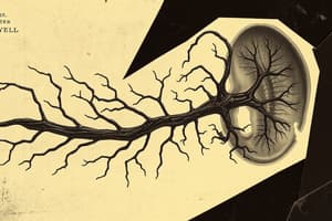
Hindbrain Development
- The cervical flexure separates the hindbrain from the spinal cord.
- The pontine flexure divides the hindbrain into the rostral (metencephalon) and caudal (myelencephalon) portions.
- The myelencephalon becomes the medulla oblongata.
- The metencephalon becomes the pons and the cerebellum.
- The cavity of the hindbrain becomes the fourth ventricle and central canal of the medulla.
Metencephalon Development
- The metencephalon forms the pons and cerebellum.
- The pontine flexure causes lateral expansion of pons.
- Three columns of motor nuclei are present on the basal plates.
- The cerebellum forms from the dorsal parts of the alar plates.
Myelencephalon Development
- Neuroblasts in the alar plates migrate to the marginal zone.
- The medulla forms the gracile and cuneate nuclei.
- The pyramids consist of corticospinal fibers.
- The roof plate thins during pontine flexure.
- The cavity of the myelencephalon becomes diamond-shaped.
Cerebellum Development
- The cerebellum structure reflects its evolutionary development.
- The archicerebellum (flocculonodular lobe) is the oldest part, with connections to the vestibular apparatus.
- The paleocerebellum (vermis and anterior lobe) is newer, involved in sensory data from the limbs.
- The neocerebellum (posterior lobe) is the newest, involved in selective limb movements.
Cerebral Commissures
- Groups of nerve fibers (commissures) connect corresponding areas of cerebral hemispheres as the cortex develops.
- The lamina terminalis is the natural pathway between the hemispheres.
- The first commissures to form are the anterior commissure and hippocampal commissure.
- The corpus callosum is a large commissure connecting neocortical areas.
- The corpus callosum initially develops within the lamina terminalis, then arches over the diencephalon.
Cerebral Hemisphere Development
- Hemispheres grow in anterior, dorsal, and inferior directions.
- Frontal, temporal, and occipital lobes develop.
- The insula is the area between the frontal and temporal lobes.
- Surface gyri and sulci create increased surface area in the cortex.
Birth Defects of the Brain
- Abnormal brain development is relatively common (approximately 3 per 1,000 births).
- Birth defects can stem from:
- Embryologic history complexity
- Defective closure of the rostral neuropore.
- Involvement of overlying tissues (meninges and calvaria)
- Genetic, nutritional, and environmental factors
Other Brain Development Note
- Prenatal risk factors (maternal infection/thyroid disorder, Rh incompatibility, genetic conditions) contribute to cerebral palsy.
- Central motor deficits sometimes result from birth events.
- Holoprosencephaly (incomplete cerebral hemisphere separation) often includes facial abnormalities. Genetic and environmental factors are implicated and may arise from destruction of embryonic midline cells.
- Hydranencephaly (absence of cerebral hemispheres) is rare with little to no cognitive development.
- Microcephaly is a neurodevelopmental disorder characterized by a small calvaria and brain, but normal-sized face. Factors such as genetic origin, exposure to ionizing radiation, infectious agents, certain drugs, and premature synostosis can contribute to this disorder.
Studying That Suits You
Use AI to generate personalized quizzes and flashcards to suit your learning preferences.